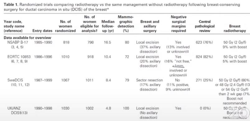
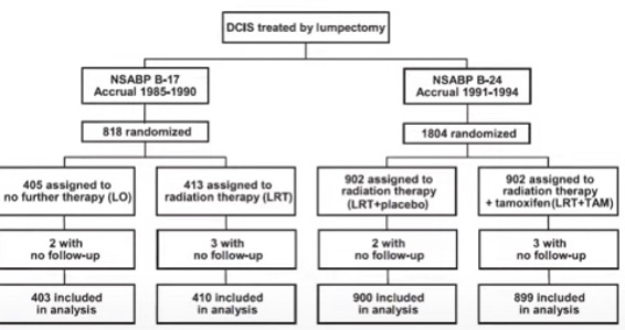
Seminal first generation trials of radiation therapy after breast conserving surgery (BCS) for DCIS:
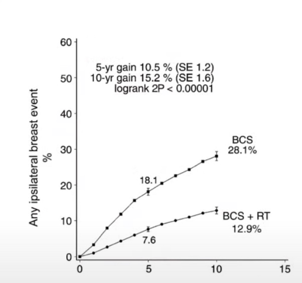
Meta-Analysis of DCIS Trials. These trials showed that radiation therapy in DICS significantly decreased local recurrence rates of both DCIS and IBC.
Radiation therapy is associated with a roughly 50% relative risk reduction in local recurrence and absolute risk reduction of 15%:
In these studies most patients did not receive tamoxifen
The first generation seminal trials:
Were conducted in the 1980’s and 1990’s
Many patients had symptomatic DCIS:
With palpable tumors of bloody nipple discharge:
Screening mammogram was uncommon
10% to 20% of patients had a positive or close margins
Greater than 1/3 of the patients had high grade DCIS
The DCIS patients that we see in our clinics today are:
Smaller, low grade, and are diagnosed earlier:
Because of the screening mammograms
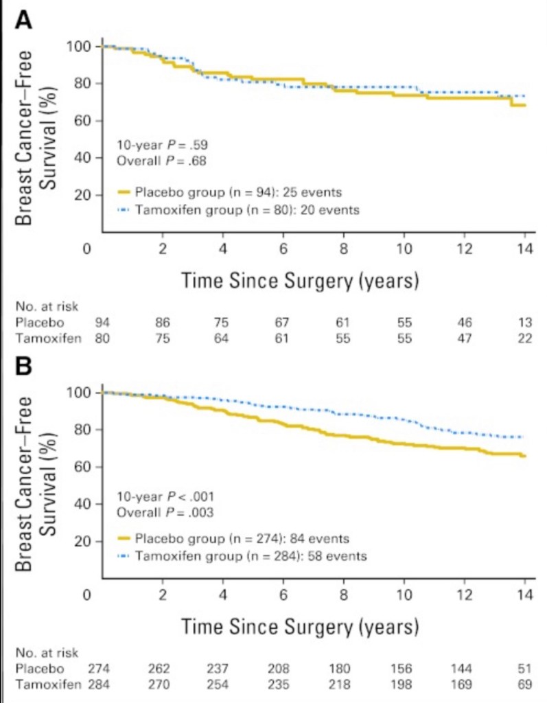
The effect of tamoxifen on local control:
NSABP B 24. Lumpectomy + RT vs. Lumpectomy + RT + Tamoxifen. A. Estrogen receptor negative patients B. Estrogen receptor positive patients
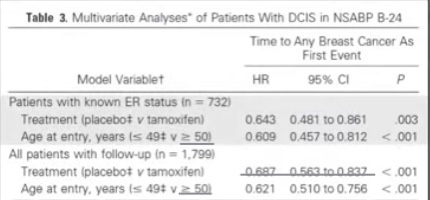
In the NSABP B 24 trial in multivariable analysis:
Young age and no tamoxifen were adverse prognostic factors
Long-term outcomes of invasive ipsilateral breast cancer recurrences after lumpectomy in the NSABP B-17 and B-24 randomized trials for DCIS:
The cumulative analysis of these trials showed that radiation therapy has a relatively greater impact on preventing local recurrence than tamoxifen. Tamoxifen plays a role in significantly decreasing the risk of contralateral breast cancer.
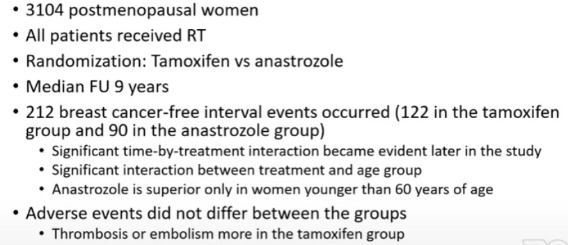
Anastrozole vs Tamoxifen in DCIS (IBIS-II DCIS Trial):
2980 postmenopausal women
71% of the patients received radiation therapy
Randomization:
Anastrozole vs tamoxifen
Median follow-up:
7.2 years
144 breast cancer recurrences
The non-inferiority of anastrozole was established:
But its superiority to tamoxifen was not
Adverse effects:
Anastrozole:
More fractures, musculoskeletal events, hypercholesterolemia, and strokes
Tamoxifen:
More muscle spasms, gynecological cancers and symptoms, vasomotor symptoms, and deep vein thromboses
NSABP B-35 Trial
Summary of radiation therapy in DCIS:
If prevention of ipsilateral breast tumor recurrence (IBTR) is the primary goal:
Tamoxifen cannot replace the impact of radiation therapy on local control after BCS
Tamoxifen may further decrease local regional recurrence risk after BCS therapy with radiation and significantly reduce contralateral breast cancer
In postmenopausal women less than 60 years of age:
Instead of trying to identify patients with underlying invasion:
Let’s find a way to mark the sentinel lymph node (SLN) in advance
Senti nel Lymph Node Dissection in DCIS and how to
Not to do it
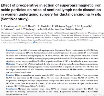
The SentiNot Trial
Concept:
Inject supraparamagnetic iron oxide particles (SPIO) at the primary operation:
Mark the SLN but do not remove it
If pathology report shows invasion:
Then we can go back and perform the SLNB
A multicenter cohort study:
Has been completed
Multicenter randomized trial:
Is ongoing
Inclusion criteria: Grade 2, > 20 mm, any grade 3, mass forming DCIS, Andy DCIS planned for mastectomyThe detection rate in the WLE were not different but in the setting of OPBCT and mastectomy the detection rate was inferior to standard technique The procedure was not accurate in any setting others than WLE
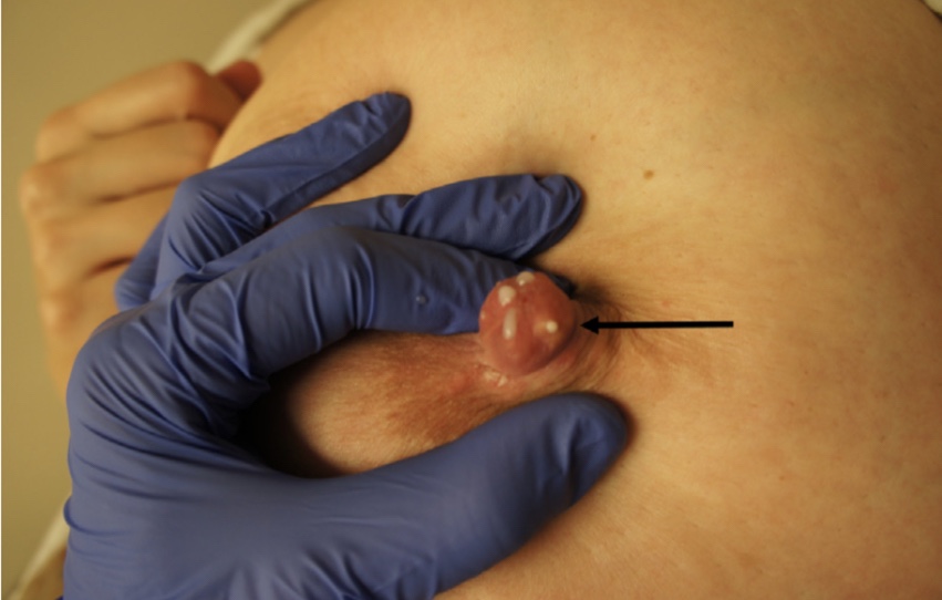
Nipple blebs are caused by trauma from shallow infant latch
Science:
Blebs appear as:
Small white, yellow, or red blisterlike lesions on the surface of a nipple
They are inflammatory lesions that may occlude a nipple orifice:
They reflect underlying ductal inflammation and microbiome disruption with biofilm formation
Blebs are associated with:
Hyperlactation (oversupply)
Pumping (which alters the breast microbiome)
C-section births (which also alter the breast microbiome),
Other characteristics of individual variation in microbiome expression
Blebs are not related to infant trauma or latch in any way
Because blebs are very painful:
Moms often believe the infant has a poor latch or otherwise has contributed to the problem:
However, this represents an association rather than causation
Treatment:
Asymptomatic blebs do not require any specific treatment
Blebs causing milk obstruction warrant treatment:
To reduce underlying ductal inflammation and decrease the viscosity of milk
Sunflower lecithin by mouth:
Is effective for breastmilk emulsification and can help to both treat and prevent blebs
Therapeutic ultrasound can also be used to reduce breast inflammation
Symptomatic blebs occluding an orifice:
Should be treated with oral lecithin as well as a topical medium-potency steroid:
Such as 0.1% triamcinolone cream
Blebs should not be routinely unroofed with a sterile needle or other means:
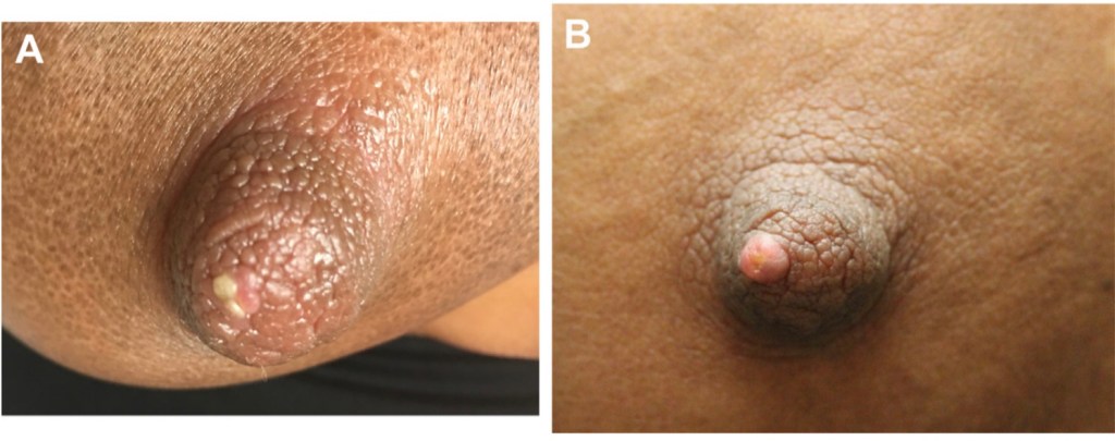
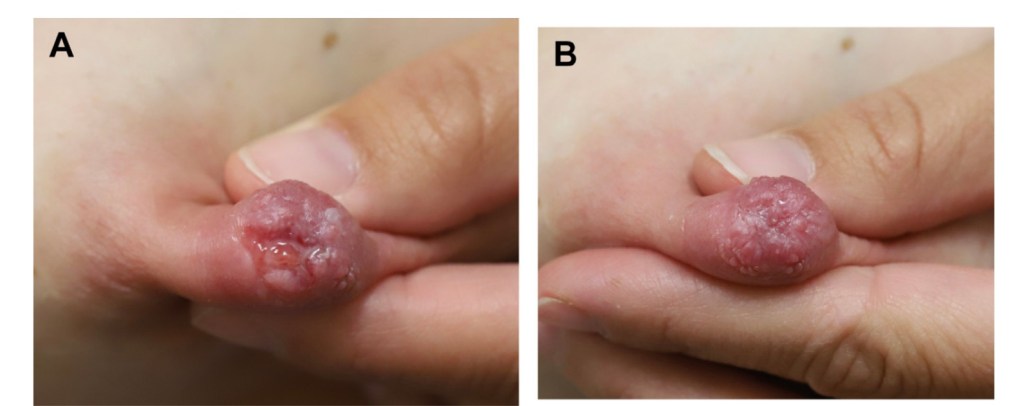
As this may transiently relieve milk obstruction in an associated ductal orifice but will also cause local tissue trauma and can lead to scarring (Figure):
This scarring can result in permanent occlusion of the nipple orifice
Patients should be instructed not to attempt to squeeze out a bleb or pick at it with their fingernails, as this can cause bleeding and further trauma
Nipple bleb at presentation (A) and after chronic tissue trauma from frequent un- roofing (B).
Oncocytic follicular cell–derived thyroid carcinomas as a group can include many different entities:
Oncocytic papillary thyroid carcinomas (PTC)
Oncocytic encapsulated follicular subtype of PTC
Oncocytic poorly differentiated carcinoma
Oncocytic medullary thyroid carcinoma
The term “oncocytic carcinoma of the thyroid”:
Is used in the new World Health Organization (WHO) to refer to:
Invasive malignant follicular cell neoplasms:
Composed of at least 75% oncocytic cells:
In which the nuclear features of PTC and high-grade features are absent
This term replaces Hürthle cell carcinoma:
A misnomer given that Hürthle actually described parafollicular C cells
Oncocytic cells:
Have abundant granular eosinophilic cytoplasm:
Secondary to a marked accumulation of dysfunctional mitochondria
Oncocytic carcinoma of the thyroid (OCA):
Represents the malignant counterpart of oncocytic adenoma
Accounts for approaching 2% to 5% of differentiated thyroid carcinomas in the USA
Can occur anywhere in the thyroid
Usually presents as a slowly enlarging painless solitary thyroid nodule
Thyroid ultrasound cannot distinguish between oncocytic adenoma and OCA:
Though larger tumors have a higher rate of malignancy
There are no known risk factors for developing OCA
The mean age at diagnosis is approaching 60 years:
Which is roughly 10 years later than the mean age of diagnosis for patient with follicular thyroid carcinoma
OCA, although more common in women (with a 1.6 to 1 female-to-male ratio):
Has a lower female-to-male ratio than is seen with follicular thyroid carcinoma
Histologically:
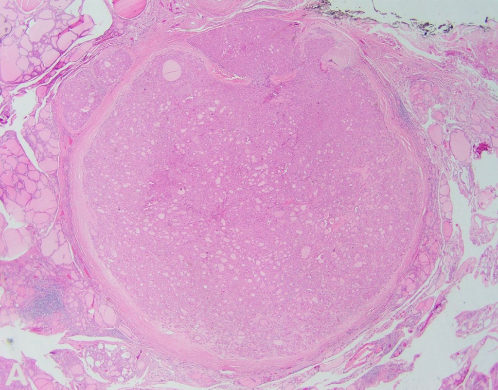
OCAs are encapsulated tumors with capsular and / or vascular invasion and at least 75% oncocytic cells (Figures)
OCAs are subclassified into:
Minimally invasive:
Those with capsular invasion only
Encapsulated angioinvasive
Widely invasive:
Those with gross invasion through the gland
When evaluating OCA, it is important not only to document extent of invasion, but also to evaluate for progression to oncocytic poorly differentiated thyroid carcinoma:
Thus, all tumors should be assessed for increased mitotic activity (3 or more mitoses per 10 high-power fields / ~ 2 mm2) and tumor necrosis
OCA can metastasize to lymph nodes:
However, some authors have shown that most of the so-called lymph nodes metastasis of OCA represent tumor plugs in veins in the neck and not lymph nodes involved by tumor
The important clue is the almost perfect roundness of these tumor plugs compared to oval or somewhat irregular outline for nodal metastases
OCA (like follicular thyroid carcinoma) usually spreads to distant sites via blood vessels:
Distant metastasis at presentation are seen in 15% to 27% of patients with OCA:
In up to 40% of tumors with extensive vascular invasion
Prognostic parameters for OCA include:
Patient age, tumor size, vascular invasion, extrathyroidal extension, and the presence of distant metastases:
Distant metastases at diagnosis are the most important prognostic factor for OCA
For OCA, the 5-year overall survival has been reported to be 85%:
But only 24% among patients with distant metastases at diagnosis compared to 91% for patients with M0 disease at diagnosis
Although it is not clear that OCA is more aggressive than follicular thyroid carcinoma after adjusting for variables such as patient age, gender, and tumor stage:
Due to decreased efficacy of radioactive iodine with OCA compared to follicular thyroid carcinoma:
Treating OCA is currently more difficult once there is disease recurrence
Benign and malignant oncocytic thyroid tumors:
Have both been shown to harbor homoplasmic or highly heteroplasmic (> 70%) mitochondrial DNA mutations in complex I subunit genes of the electron transport chain
Additionally, OCAs demonstrate widespread chromosome losses that result in near-genome-wide haploidization with or without subsequent genome endoreduplication:
Chromosomal changes have been found to be associated with extent of invasion:
Most OCAs with capsular invasion only or focal vascular invasion have been shown to be diploid
Whereas tumors with extensive vascular invasion and widely invasive tumors:
Are usually polysomic and nearly always demonstrate chromosome 7 amplification
Additionally, the near-haploid state has been shown to be maintained in metastases, implying selection during tumor evolution
OCAs have also been shown to have recurrent DNA mutations, including RAS mutations (though at a lower rate than is seen with follicular thyroid carcinoma), EIF1AX, TERT, TP53, NF1, and CDKN1A, among others
Oncocytic carcinoma of the thyroid – Invasive growth through the capsule is evident at low powerOncocytic carcinoma of the thyroid – At high power, the cells have abundant granular cytoplasm and prominent nucleoli
Women with nipple / areolar lesions and nipple piercings should be discouraged from breastfeeding
Science:
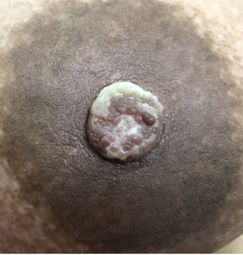
Several lesions can occur on the nipple / areolar complex including:
Nipple adenomas
Skin tags
Eczema
Hyperkeratosis
Patients with these conditions are often advised to avoid breastfeeding:
Due to concerns about latch and milk extraction, as well as theoretic risks of an infant choking on a protruding lesion or suffering toxicity from medications used to treat dermatologic conditions
Patients with nipple piercings may be advised to avoid breastfeeding altogether due to concerns about milk fistulae:
Milk will indeed pass through the piercing sites (Figure):
However, this does not negatively affect milk production or extraction, nor does it pose a risk to the mother’s health in any way
Treatment:
Women with nipple / areolar lesions should be evaluated by a breastfeeding medicine physician prenatally
These physicians may recommend removal of larger lesions if they are concerned about interference with latch or potential for tissue trauma
Surgical excision can be performed under local anesthesia during pregnancy or lactation with minimal risk
In most of the cases, prenatal evaluation of nipple / areolar lesions will consist of review of the lactational safety of medications and reassurance
Topical steroids, keratolytic ointments, and most immunomodulators used for conditions such as eczema, psoriasis, and hyperkeratosis are safe in lactation, with the exception of methotrexate
Nipple shields should not be recommended to cover nipple /areolar complex lesions, as there is no benefit to this practice and nipple shields are associated with decreased physiologic milk transfer and increased risk of microbiome disruption and mastitis
Ideally, patients should remove nipple piercings during early pregnancy:
As the nipple is expected to hypertrophy and can make later removal more difficult
Nipple piercings are a choking hazard, and patients should not breastfeed with them
When consenting for piercings, women should be counseled that the procedure may result in ductal trauma or scarring that could impair lactation
Nipple piercing orifice with milk emanating from it.
Nipple wounds in lactating women should be treated with drying agents and topical antibiotics to prevent progression to mastitis
Science:
Surgical training provides strong education in wound care techniques and teaches the difference between traumatized versus infected tissue
Surgeons debride devitalized tissue and understand the need to provide absorption for serous fluid and fibrinous exudate
However, traditional lactation recommendations have contradicted principles of closed, moist healing for wound care
Patients are often counseled to express a small amount of breastmilk onto nipple wounds and allow it to air dry
They also may be recommended to soak nipples in Epsom salt or salt water and to use a hair dryer to prevent moisture build-up
They are also often instructed to avoid wearing a bra or allow anything touch the nipples
In addition, breastfeeding patients with nipple wounds are often warned they are at risk for ascending intraductal infection, that is, bacterial mastitis
They are therefore encouraged to apply topical antibiotics to their wounds
However, it is very uncommon for open, vascularized wounds to become secondarily infected in immunocompetent hosts
Overutilization of antibiotics contributes to disruption of the microbiome and development of multidrug-resistant pathogens
Routine use of topical antibiotics for open wounds is not recommended
Furthermore, the hypothesis that bacterial mastitis is a result of ascending infection from nipple wounds is not supported by breast anatomy and physiology
The highly vascular nature of the lactating breast and the multiple immune components of breastmilk prevent such infection
Treatment:
Care of the nipples include:
Moist, closed wound healing principles should be followed, with the use of nonallergenic ointments / balms and sterile, absorbent dressings (Figure)
Avoid ointments / balms containing potentially allergenic ingredients such as lanolin and petroleum
APNO (All Purpose Nipple Ointment) should be avoided:
This compounded prescription ointment contains an antifungal, antibacterial, and a steroid
Although often recommended by lactation consultants and readily prescribed by the physicians to whom patients are referred, this nonspecific medication can cause additional complications
Although patients may achieve some pain relief due to steroids and antiinflammatory properties of the antifungal, this potential benefit is outweighed by the risks of impaired wound healing from steroids and of microbiome disruption from nonselective elimination of normal flora
Furthermore, other ingredients in this ointment may cause dermatitis
This medication is generally expensive, even for patients with insurance
Breast shells designed to “keep the nipple dry” or “protect the nipple from the bra” worsen swelling in the nipple, cause areola compression, and subsequently worsen pain
Do not use drying agents such as antiseptics, alcohol, or Epsom salt soaks
Similarly, do not use a hair dryer to blow hot, dehumidified air on nipples:
These practices cause tissue desiccation, which is counterproductive for wound healing and increase the risk of skin breakdown
Nipple previously air dried (A), with resolution of tissue defect with PolyMem therapy (B).Blistering from small pump flanges worsened with Epsom salt soaks.
“Plugging” represents occlusion of ducts by stagnant milk, and “plugs” should be extruded through massage
Science:
The sensation of “plugging” does not represent discrete collections of breastmilk
More accurately, a “plug” represents a focal area of congested capillaries, distended alveolar cells, and tissue edema:
The root cause of “plugging” relates to:
Tissue hypervascularity
Edema
Ductal narrowing from microbiome changes and luminal inflammation
Lactation consultants often recommend massage for “plugs”:
However, this recommendation lacks scientific validity and causes tissue trauma that can result in significant complications
As surgeons are well aware, lactating breasts have robust blood supply, lymphatic vessels to drain increased interstitial fluid during lactation, nerves, fibroadipose tissue, and functional glandular tissue with a complex network of interlacing ducts:
Attempts to extrude a milk “plug” from a duct with aggressive massage will result in tissue trauma, edema, collapse of ducts, and capillary damage
All patients report worsened pain with massage
As we would injure a thyroid, pancreas, or other functional gland with massage or tissue mishandling during a surgical procedure, it similarly must be avoided in the lactating breast
Massage is associated with development of lactational phlegmon (Figure), particularly in the setting of hyperlacta- tion or excessive pumping
Treatment:
Deep manual massage, vibrators, electric toothbrushes, or any commercial breast massage products designed to extrude a “plug” should be strictly avoided
Patients with symptomatic “plugging” should be evaluated for:
Proinflammatory conditions such as:
Hyperlactation and subacute mastitis
Patients can use ice for both pain relief and vasoconstriction
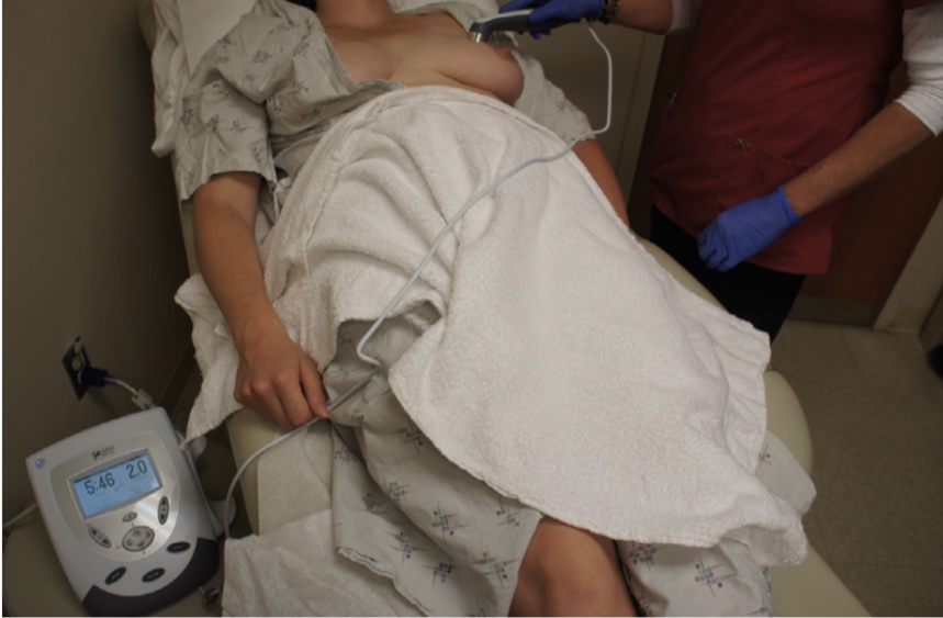
Therapeutic ultrasound, also used to treat conditions such as radiation fibrosis, can reduce inflammation and pain through application of thermal energy (Figure)
If a mass or erythema persists, diagnostic breast imaging should be performed
Mastitis represents a bacterial infection resulting from milk stasis, engorgement, and “plugging”
Science:
When an infant sleeps through the night or mothers do not express their milk at work as regularly as the infant breastfeeds at home:
Patients experience transient engorgement and pain
Women may also develop breast erythema and edema:
From congested capillaries and interstitial fluid (Figure):
Which can cause sweating, fever, and chills:
As it is an inflammatory process in a body organ with robust blood supply
This systemic inflammatory response syndrome may be mistaken for signs and symptoms of infection:
Raising alarm for impending development of bacterial mastitis
However, unless a person has developed a very rare rapidly progressive soft tissue infection:
It is otherwise very unusual for average bacterial mastitis to present this quickly
Lactation literature commonly describes a theory that mastitis results from milk stasis:
Mothers are warned to avoid long stretches without breastfeeding or pumping to avoid build-up of stagnant milk and progression to “plugging” and infectious mastitis:
However, there is no scientific evidence to support the idea that mastitis results from milk stasis
In contrast to a passive repository such as a bladder, the breast is a gland with production regulated by Feedback Inhibitor of Lactation (FIL):
Therefore, continued removal of milk increases production and worsens tissue edema and inflammation:
Reduced removal of milk will allow for FIL to downregulate production and enable resorption of milk not used
Mastitis results from underlying microbiome disruption and ductal inflammation and is therefore often observed in patients with hyperlactation and/or blebs
Most cases of lactational mastitis are purely inflammatory and can be resolved with conservative measures and appropriate management of lactation
Breastmilk contains numerous immunologic cells and substances:
That counteract infection
In similar fashion, it is uncommon for stagnant milk contained in a galactocele to become infected without an inciting event such as instrumentation
It also should be noted that external compression by a bra or tight clothing can obstruct ducts:
Is not scientifically founded.
Treatment:
With early inflammatory mastitis:
Patients should feed physiologically (eg, eliminate breast pump usage if possible, and do not continue “overfeeding” on the affected breast):
Reducing overstimulation of an engorged or inflamed breast will allow down-regulation of milk production through the FIL receptor
Patients should never be counseled to “pump to relieve engorgement” as this prevents FIL from activating and perpetuates hyperlactation
Patients can use ice and antiinflammatory medication by mouth such as nonsteroidal antiinflammatory drugs and/or obtain pain relief from acetaminophen:
Ice is generally the most helpful, but some people also prefer heat for comfort
Therapeutic ultrasound:
Can use thermal energy to reduce inflammation and pain, as can lymphatic drainage
A supportive bra is necessary during lactation:
To prevent dependent lymphedema and back pain
Massage should be strictly avoided
If symptoms persist or worsen:
Antibiotics should be considered (Table)
Women should be counseled that there is no medical indication to “pump and dump” while taking these antibiotics:
The Relative Infant Dose (RID) estimates an infant’s exposure to a medication through breastmilk and depends on multiple factors including the medication’s plasma concentration, half-life, solubility, and oral bioavailability:
In general, medications with RID less than 10% are considered safe
Acute inflammatory mastitis (A) managed with decreased removal of breastmilk, ice, and antiinflammatory medication, with resolution of erythema (B).
Fluid collections in the lactating breast require operative incision and drainage or aspiration alone
Science:
As surgeons have moved away from large incision and drainage procedures performed on the lactating breast in the operating room setting:
They have turned to minimally invasive aspiration approach:
However, aspiration alone can result in incomplete drainage
Unlike simple breast cysts, abscesses and galactoceles in the lactating breast contain breastmilk:
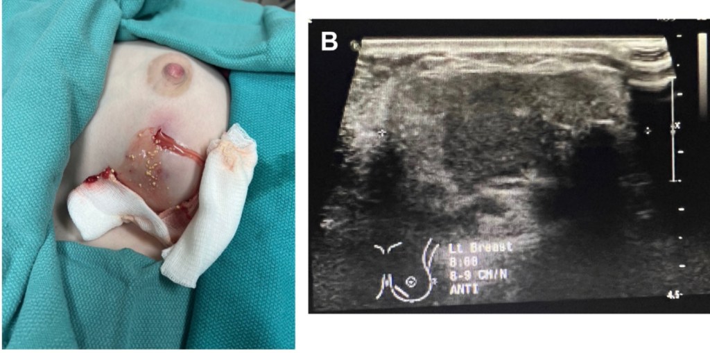
Which is highly viscous and loculated (Figure):
Therefore, a needle aspiration alone will likely remove only part of the fluid collection, particularly if it is chronic
If a needle aspiration is successful in removing the entire volume of an acute collection:
The area can refill with milk very quickly and require repeated procedures
Treatment:
Lactational abscess and infected galactocele:
Require drainage for source control
Drainage may also be appropriate for symptomatic noninfected galactoceles:
Small stab incision and drain placement will definitively resolve fluid collections in the lactating breast:
The small stab incision allows for access to the cavity with an instrument that can be used to disrupt loculations and provide complete drainage, such as a hemostat
A stent or drain can be placed to allow passive decompression of the area for 3 to 5 days:
This could involve a Penrose drain, Seromacath, Blake drain, or other wicks such as a small foley catheter:
Drains should be placed to gravity rather than suction
In addition to the surgical management, many patients developing fluid collections during lactation require treatment of idiopathic or iatrogenic hyperlactation:
Patients should not be instructed to massage their breast:
As this results in tissue necrosis and phlegmon development
Ice and antiinflammatory medication by mouth should be recommended for symptomatic relief
Antibiotics may be indicated if significant surrounding cellulitis exists
Chronic, loculated fluid collection demonstrating thick milk chunks at definitive drainage after needle aspiration failed to resolve. Ultrasound image corresponding to semisolid appearance of the collection.
Previously, there was no consensus on the optimal time intervals for imaging evaluation of patients following breast-conservation surgery:
Many practices performed 6-month interval mammography of the treated breast for a variable number of years in an effort to monitor post-procedure changes:
The usefulness of short-term follow-up mammography in women undergoing breast conservation has been challenged:
Local recurrence in the ipsilateral breast following breast conservation and radiotherapy occurs at an estimated rate of 1% in the first year, 3% to 9% at 5 years, and 14% to 20% at 20 years
Retrospective studies have verified that the yield from a 6-month follow-up mammogram on the affected side is low (≤1%)
Therefore, the ACR and ASCO recommendations are:
To perform a bilateral mammogram at 12 monthsfrom the initial mammogram:
Which should be at least 6 months following completion of radiotherapy and should include a diagnostic mammogram on the affected side and a screening mammogram on the nontreated side
Bilateral annual mammography is recommended thereafter
Elimination of the 6-month and 18-month interval diagnostic mammograms after breast conservation should spare women unnecessary anxiety, discomfort, and inconvenience while lowering costs and improving efficiency
Monticciolo DL, Newell MS, Moy L, Niell B, Monsees B, Sickles EA. Breast cancer screening in women at higher-than-average risk: recommendations from the ACR. J Am Coll Radiol. 2018;15(3 Pt A):408-414.
Khatcheressian JL, Hurley P, Bantug E, Esserman LJ, Grunfeld E, Halberg F, et al. Breast cancer follow-up and management after primary treatment: American Society of Clinical Oncology clinical practice guideline update. J Clin Oncol. 2013;31(7):961-965.