Blog

Management of Common Complications of Lactation – Milk Fistula

  • Milk Fistula:
    • Myth:
      • Procedures should be avoided on the lactating breast due to the risk of milk fistula
    • Science:
      • Milk fistula is rare if lactation and surgical interventions are managed appropriately
      • After a procedure:
        • Patients should not avoid breastfeeding:
          • In fact, the preferential flow of breastmilk through the nipple will decrease the flow through a needle or incision tract
        • On the other hand, patients should not be counseled to “pump to empty” their breasts or breastfeed more frequently on the affected breast:
          • As this will cause increase in milk production:
            • Which will promote fistula persistence
    • Treatment:
      • Large surgical incision and drainage:
        • Should be avoided in lactation patients
      • Any incision made should be as small as possible, and as distant from the nipple areolar complex as possible
      • If a distal incision is not possible:
        • It should be made outside the region where an infant latches or pump flanges contact the skin:
          • Periareolar incisions, although cosmetic, are particularly high risk due to the potential for latch or pump trauma.
    • Patients should feed physiologically after a procedure:
      • Local anesthetic agents are not absorbed orally by the infant, and blood is safe for the infant to ingest
    • A transient fistula will form after any procedure but is expected to close within a week if lactation is managed appropriately:
      • Should a persistent, high-output fistula develop:
        • A distal diverting drain can be placed to hasten closure (Figure)
        • Milk passing through a fistula tract may be collected and is safe to feed to the infant
        • Absorbent dressings may be used to prevent skin maceration from moisture but should be removed before breastfeeding:
          • As they are potential choking hazards and/or may interfere with latch
          • Wound vacuum systems should not be used on the lactating breast, as this will promote chronic fistulization and maintain tract patency
Transient milk fistula in setting of hyperlactation and pump trauma, located near nipple areolar complex with resolution 24 hours after placement of distal diverting Interventional Radiology (IR) drain.

#Arrangoiz #BreastSurgeon #CancerSurgeon #SurgicalOncologist #Surgeon #Surgeon #Doctor #MountSinaiMedicalCenter #MSMC #Miami #Mexico #MilkFistula

When to Perform A Completion Thyroidectomy?

  • Removal of the contralateral lobe of the thyroid may be necessary after lobectomy:
    • Particularly if total thyroidectomy would have been recommended had the diagnosis been known preoperatively
  • With lobectomy being sufficient therapy for the majority of low-risk cancers, the need for completion thyroidectomy is diminishing:
    • However, in the hands of an experienced surgeon, the complication rates for completion thyroidectomy are comparable to those of total / near total thyroidectomy
  • The use of RAI therapy to ablate the remaining tissue after lobectomy is not recommended routinely but may be considered in select cases when additional surgery is not feasible
#Arrangoiz #Doctor #Surgeon #CancerSurgeon #ThyroidSurgeon #ParathyroidSurgeon #HeadandNeckSurgeon #EndocrineSurgery #MountSinaiMedicalCenter #MSMC #ThyroidCancer #Miami #Mexico

Thyroid Cancer Preoperative Imaging

  • Before removal of thyroid cancer:
    • It is critical to perform a thorough evaluation:
      • To determine the extent of disease
  • Preoperative imaging should include:
    • A comprehensive ultrasound (US) of the neck to examine the contralateral lobe of the thyroid, the central neck compartments, and the lateral neck lymph nodes (LN):
      • Such imaging may change the surgical approach in up to 40% of cases
  • The anterior neck is divided into seven contiguous compartments in which thyroid cancer metastatic LN spread occurs
  • The central neck compartment (level VI) contains the thyroid and poses the greatest challenge to clinicians when deciding the optimal surgery:
    • It is bordered laterally by the carotid arteries, inferiorly by the clavicles, and superiorly by the hyoid bone
    • Level VI is the compartment that is most frequently involved with LN metastases:
      • But sonographic identification of diseased nodes is hampered by poor preoperative sensitivity:
        • The intact thyroid gland obscures visualization of the majority of nodal metastases
  • The lateral neck is further subdivided into four compartments lateral to the carotid:
    • Level IV is bordered laterally by the sternocleidomastoid (SCM), inferiorly by the clavicle, and superiorly by the cricoid cartilage
    • Level III, located immediately cephalad to level IV, extends superiorly to the carotid bifurcation
    • Level II is located below the mandible and extends to the hyoid bone
    • Level V nodes are located in the posterior triangle, lateral to the lateral edge of the SCM.
  • The presence of malignancy in sonographically suspicious nodes:
    • Can be confirmed with FNA for cytologic analysis and measurement of thyroglobulin (Tg) in the needle washout
  • If advanced, bulky nodal disease is identified on US, or the patient has clinical signs or symptoms of advanced disease (hoarseness, hemoptysis, a nonmobile thyroid mass):
    • CT or magnetic resonance imaging (MRI) of the neck may be considered to search for additional metastases in areas that cannot be visualized sonographically, including within the mediastinum, at the skull base, and posterior to the trachea
#Arrangoiz #ThyroidSurgeon #CancerSurgeon #MountSinaiMedicalCenter #MSMC #Doctor #Surgeon #HeadandNeckSurgeon #SurgicalOncologist #Miami #Mexico

Sentinel Lymph Node Biopsy in Multicentric / Multifocal Breast Tumors

  • Patients with multiple ipsilateral breast cancers:
    • Were excluded from the initial trials of SLNB
  • A systematic review evaluated the accuracy of SLNB in multifocal and multicentric tumors:
    • This review reported:
      • Significantly higher rates of lymph node metastases
      • Higher rates of involved nonsentinel lymph nodes:
        • In patients with multifocal tumors than patients with unifocal primary tumors
  • In several studies:
    • There is no significant difference in the false negative rate or failed localizations for patients with multifocal tumors
  • In a prospective study:
    • 30 patients with multifocal tumors underwent SLNB followed by ALND:
      • In 30 of 30 patients, the sentinel lymph node was identified and the false negative rate was 0%:
        • Supporting the use of SLNB for patients with multifocal tumors

#Arrangoiz #BreastSurgeon #CancerSurgeon #SurgicalOncology #Surgeon #Teacher #BreastCancer #BreastExpert #MSMC #MountSinaiMedicalCenter #Miami #Mexico #SLNM #SLNB #SentinelLymphNodeBiopsy

Pregnancy and the Use of Sentinel Lymph Node Biopsy

  • Breast cancer:
    • Is the most common pregnancy-associated malignancy
  • The ASCO recommends that pregnant patients:
    • Should not undergo SLNB based on the lack of safety data
  • Unfortunately, approximately 11% of pregnant patients with breast cancer who undergo ALND:
    • Develop lymphedema postoperatively:
      • Which has significant consequences on quality of life in these young women
  • This has led to increased interest in the use of SLNB for pregnant patients
  • Multiple studies have attempted to estimate the exposure of the fetus to radiation when using 99mTc to perform SLNB:
    • Conservative estimates suggest that fetal doses as low as 10 to 50 mGy:
      • Could increase the risk of malignancy in the fetus
    • Estimates suggest that fetal exposure during SLNB:
      • Is minimal at 1.14 mGy to 4.3 mGy:
        • Which is close to the levels of background radiation absorbed on an average day
    • Given this information, small studies have reported sentinel lymph node mapping with 99mTc in pregnant patients:
      • There have been no reported ill effects
    • Both methylene blue and isosulfan blue:
      • Are category C drugs in pregnancy:
        • With unknown levels of teratogenicity
    • Of 30 pregnant patients treated with SLNB using methylene blue:
      • One patient electively terminated her pregnancy and 29 gave birth to healthy infants
  • Safety conclusions are limited by small numbers and a lack of long-term follow-up data:
    • Some offer SLNB to pregnant patients:
      • Whereas others routinely perform ALND

#Arrangoiz #BreastSurgeon #CancerSurgeon #SurgicalOncologist #Surgeon #Teacher #BreastCancer #BreastDiseases #AxillaryDissection #BreastSurgery #MountSinaiMedicalCenter #MSMC #Miami #Mexico #SLNM #SLNB #SentinelLymphNodeBiopsy

Natalee Trial in Early Breast Cancer

  • Adding the CDK4/6 inhibitor ribociclib to endocrine therapy resulted in a significant improvement in invasive disease–free survival for patients with hormone receptor–positive, HER2-negative early-stage breast cancer
    • Findings from this phase III NATALEE trial were presented at the 2023 ASCO Annual Meeting (Abstract LBA500)
  • Hormone receptor–positive, HER2-negative breast cancer:
    • Is the most common subtype of the disease:
      • Making up nearly 70% of all breast cancer cases in the United States
    • Roughly one-third of patients with stage II hormone receptor–positive, HER2-negative disease:
      • Experience a recurrence following standard-of-care treatment
    • More than one-half of patients with stage III hormone receptor–positive, HER2-negative disease:
      • Experience a recurrence
  • If a recurrence occurs:
    • It is often at a more advanced stage
  • Ribociclib is currently approved by the U.S. Food and Drug Administration to treat:
    • Hormone receptor–positive, HER2-negative advanced or metastatic breast cancer in combination with an aromatase inhibitor for premenopausal patients or in combination with fulvestrant for postmenopausal patients
  • While ribociclib has previously shown survival benefits in people with metastatic disease:
    • In the NATALEE study, researchers showed that it may also improve outcomes for people with earlier-stage disease, including those with cancer that has not yet spread to the lymph nodes
  • The NATALEE phase III clinical trial:
    • Included men and premenopausal or postmenopausal women from 20 different countries with stage IIA, IIB, or III hormone receptor–positive, HER2-negative breast cancer who were at risk for disease recurrence
    • Participants were randomly assigned to receive either 400 mg of adjuvant ribociclib for 3 years with hormonal therapy for at least 5 years (n = 2,549) or hormonal therapy alone for at least 5 years (n = 2,552)
    • Men and premenopausal women also received Goserelin
    • Prior hormonal therapy use was allowed if it was initiated no more than 1 year before the start of the study
    • The current recommended starting dose of ribociclib for people with metastatic disease is 600 mg:
      • However, an extended duration of treatment may help to stop cells from duplicating and dividing and destroy any remaining cancer cells:
        • Because of this, study authors chose a 3-year treatment duration of ribociclib at a dose of 400 mg to reduce side effects while maintaining efficacy
  • Key Findings:
    • At a median follow-up of 34 months:
      • 20.2% of participants in the ribociclib group had completed 3 years of treatment
      • 56.8% had completed 2 years of treatment
    • Overall, 74.7% of participants remained on study treatment at data cutoff, with 1,984 patients on ribociclib and 1,826 patients on hormonal therapy alone
    • The study found that adding ribociclib to hormonal therapy:
      • Led to a significant improvement in invasive disease–free survival compared with hormonal therapy alone:
      • Researchers evaluated invasive disease–free survival after 426 invasive disease–free survival events occurred, a number that was prespecified for the interim analysis
      • Of those events, 189 occurred in the ribociclib group (7.4% of patients) vs 237 in the hormonal therapy alone group (9.2% of patients)
    • The 3-year invasive disease–free survival rates were:
      • 90.4% in the ribociclib group compared with 87.1% in the hormonal therapy alone group
    • Overall, the addition of ribociclib reduced the risk of disease recurrence by:
      • 25%
    • The invasive disease–free survival benefit seen in the ribociclib group was generally consistent across clinically relevant patient subgroups
    • Ribociclib also showed more favorable outcomes in overall survival, recurrence-free survival, and distant disease–free survival
    • For patients receiving ribociclib, the most common adverse effects were:
      • Neutropenia and joint pain
    • Rates of gastrointestinal adverse effects and fatigue were low in patients receiving ribociclib
    • For patients receiving hormonal therapy alone, the most common adverse effects were:
      • Joint pain and hot flash.
  • Currently approved targeted treatments can only be used in a small population of patients diagnosed with hormone receptor positive, HER2-negative early breast cancer:
    • Leaving many without an effective treatment option for reducing risk of the cancer returning:
      • Thus, there is a significant unmet need for both reducing the risk of recurrence and providing a tolerable treatment option that keeps patients cancer-free without disrupting their daily life:
        • The NATALEE study investigated the addition of ribociclib to standard-of-care adjuvant endocrine therapy and was specifically designed to address these unmet needs
#Arrangoiz #CancerSurgeon #BreastSurgeon #SurgicalOncologist #Surgeon #Doctor #BreastCancer #MountSinaiMedicalCenter #MSMC #Miami #Mexico

What is the Appropriate Margin Width for a Malignant Phyllodes Tumor

👉Malignant phyllodes tumors carry a high risk of local recurrence if not excised to wide negative margins.

👉How wide these margins should be is a matter of debate with data supporting a negative margin of any width to a negative margin as wide as 2 cm.

👉Currently, the National Comprehensive Cancer Network recommends a margin width of 1cm; however, recent studies suggest margin width is not associated with local recurrence or local recurrence free survival.

👉The addition of radiation therapy when margins are closer than 1 cm or other high-risk features are present can also be a consideration but it is controversial.

👉Most data indicate unacceptably high recurrence rates if the tumor is at the inked margin of a malignant phyllodes tumor excision.

👉Mastectomy is not required unless excision cannot achieve tumor-free margins.

👉The most appropriate margin thickness for borderline or benign phyllodes tumors is even less well-defined.

👉Although achieving widely negative margins can reduce local recurrence rates, malignant phyllodes tumors also have metastatic potential regardless of negative margin status. 

👉REFERENCES

  1. Belkacémi Y, Bousquet G, Marsiglia H, et al. Phyllodes tumor of the breast. Int J Radiat Oncol Biol Phys. 2008;70:492-500.
  2. Jang JH, Choi MY, Lee SK, et al. Clinicopathologic risk factors for the local recurrence of phyllodes tumors of the breast. Ann Surg Oncol. 2012;19:2612-2617. 
  3. Mituś J, Reinfuss M, Mituś JW, et al. Malignant phyllodes tumor of the breast: treatment and prognosis. Breast J.2014;20:639-644. 
  4. Petrek J. Phyllodes tumors. In: Harris JR, Lippman ME, Morrow M, et al. eds. Diseases of the Breast, 2nd ed. Philadelphia PA: Lippincott-Raven Publishers, 2000:669-675.
  5. Telli ML, Horst KC, Guardino AE, et al. Phyllodes tumors of the breast: natural history, diagnosis, and treatment. J Natl Compr Canc Netw. 2007;5:324-330. 
  6. Yom CK, Han W, Kim SW, et al. Reappraisal of conventional risk stratification for local recurrence based on clinical outcomes in 285 resected phyllodes tumors of the breast. Ann Surg Oncol. 2015;22:2912-2918.

#Arrangoiz #PhyllodesTumors

#Surgeon #MountSinaiMedicalCenter #MSMC #Miami #Mexico

#Cirujano

#SurgicalOncologist

#CirujanoOncologo

#BreastSurgeon

#CirujanodeMama

#CancerSurgeon

#CirujanodeCancer

Phyllodes Tumors Part 2

Clinical Presentation

Phyllodes tumors generally present as a palpable mass or, in women being screened, as an abnormal finding on imaging:

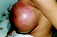
  • In one third of patients, these tumors grow rapidly and may fill a substantial portion of the breast, causing thinning of the overlying skin and prominent vascularity

angiosarc

Clinical concern for a phyllodes tumor versus a fibroadenoma usually hinges on a history of rapid growth, large tumor size (> 3 cm), and / or older patient age:

  • However, with increased adoption of breast screening and breast awareness, smaller and asymptomatic lesions are being detected more often

Imaging Findings

Most phyllodes tumors appear as lobulated masses on mammography and ultrasonography:

Phyllodes tumors may, however, have irregular margins on ultrasonography, a finding seen more frequently in borderline or malignant tumors than in benign tumors

Magnetic resonance imaging (MRI) has been used to evaluate patients with phyllodes tumors, but the clinical benefit of this approach is unclear as these tumors are generally well delineated by physical examination and / or sonography, and correlation of tumor size among mammography, ultrasonography, and MRI is high

Papillary Lesions of the Breast

  • Papillary lesions of the breast:
    • Are common
  • They are highly vascular lesions that are intraductal and may transform into malignant variants
  • In benign papillary lesions:
    • A vascular stalk may be demonstrated on color Doppler scanning:
      • While multiple feeding vessels may be seen when imaging malignant papillary lesions
  • When papillary lesions infarct:
    • The vascular stalk will not be demonstrated
  • The ability to reliably distinguish papilloma, in-situ papillary carcinoma, and invasive papillary carcinoma:
    • Is not possible with ultrasound and is quite challenging even on core biopsy:
      • Open surgical biopsy may need to be performed to distinguish malignant from benign papillary lesions
Acorn Cyst
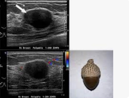
  • An “acorn” cyst is lined with papillary apocrine metaplasia:
    • Which can form a mural nodule:
      • The nodule in an acorn cyst is less echogenic than papillomas or papillary carcinomas:
        • Is usually concave, following the contour of the cyst (thus the appearance of a cap on an acorn) instead of convex, and does not have a vascular stalk
          • The mural nodule caused by papillary apocrine metaplasia:
            • Also would not extend into the duct as the papillary lesion shown in the image does
  • Tubular adenomas and fibroadenomas:
    • Have a similar sonographic appearance and are frequently round or oval, although tubular adenomas can be fusiform or spindle shaped:
      • Both lesions occur during reproductive years and would not commonly present as a new finding in a postmenopausal patient
  • References
    • Jagmohan P, Pool FJ, Putti TC, Wong J. Papillary lesions of the breast: imaging findings and diagnostic challenges. Diagn Interv Radiol. 2013;19(6):471-478.
    • Wyss P, Varga Z, Rössle M, Rageth CJ. Papillary lesions of the breast: outcomes of 156 patients managed without excisional biopsy. Breast J. 2014;20(4):394-401.
    • Stavros AT. Breast Ultrasound. Philadelphia, PA: Lippincott Williams & Wilkins; 2004.
#Arrangoiz #BreastSurgeon #CancerSurgeon #SurgicalOncologist #MountSinaiMedicalCenter #MSMC #Miami #Mexico

Phyllodes Tumors of the Breast

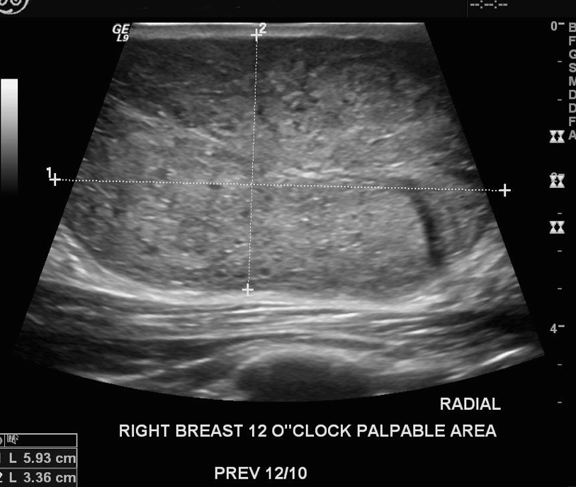
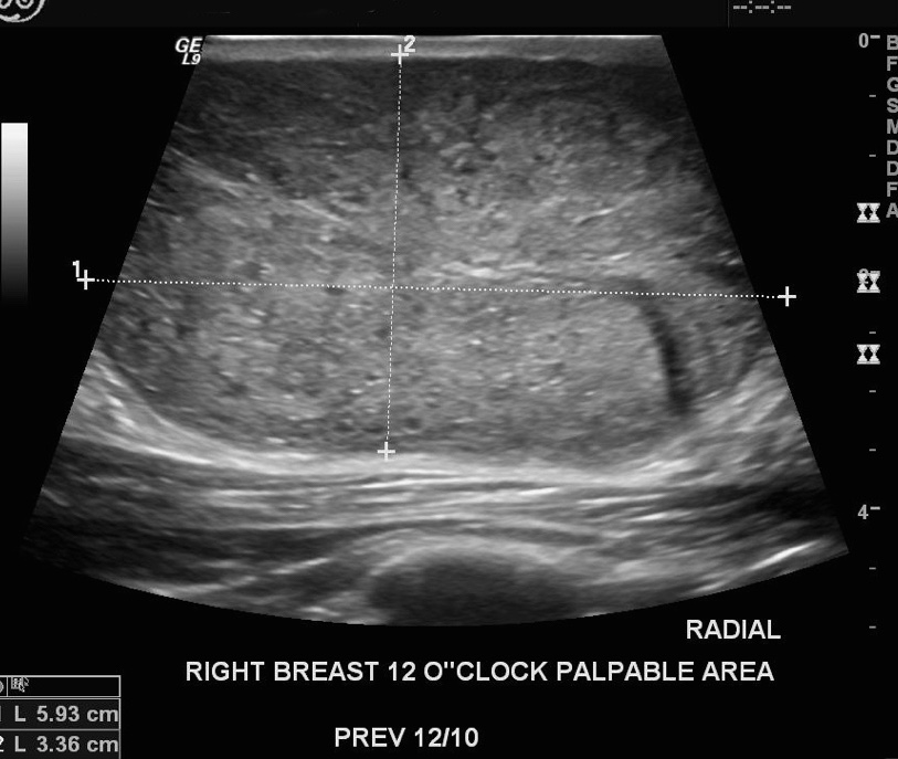
Palpable right subareolar mass
Ultrasound from 6-month follow-up
  • The rapid growth of this lesion:
    • Suggests that it is a phyllodes tumor:
      • Although a giant fibroadenoma is another possibility
  • The most recent ultrasound image shows:
    • An isoechoic, heterogeneous mass that contains cystic, fluid filled spaces, and is vascular on Doppler examination
  • In most cases, benign phyllodes tumors:
    • Have margins that are well-circumscribed, and a thin, echogenic capsule is demonstrable
    • The doubling time for a:
      • Benign phyllodes tumor:
        • Is about four months
      • Malignant phyllodes tumor:
        • Is a little over a month
    • Rapidly growing phyllodes tumors:
      • Whether benign or malignant:
        • Often cause prominent veins on the skin:
          • From the developing vascularity
    • Phyllodes tumors are more common in women of Mexican descent:
      • Latin American women with phyllodes tumors tend to be diagnosed at an earlier age than other women
  • A malignant phyllodes tumor:
    • Would in all likelihood have a less distinct border than the lesion shown above
  • A core needle biopsy:
    • Cannot reliably distinguish phyllodes tumors from fibroadenomas:
      • It is therefore not sufficient for a definitive diagnosis
  • The correct management is excision:
    • With or without a pre-operative core biopsy:
      • With care to completely excise the tumor
  • Phyllodes tumors can also be difficult to distinguish from giant juvenile fibroadenomas:
    • But they should also be treated by surgical excision
  • The rapid growth of this lesion suggests a phyllodes tumor, not a fibroadenoma
  • References:
    • Guillot E, Couturaud B, Reyal F, Curnier A, Ravinet J, Lae M, et al. Management of phyllodes breast tumors. Breast J. 2011;17(2):129-137.
    • Plaza MJ, Swintelski C, Yaziji H, Torres-Salichs M, Esserman LE. Phyllodes tumor: review of key imaging characteristics. Breast Dis. 2015;35(2):79-86.
    • Rajan PB, Cranor ML, Rosen PP. Cystosarcoma phyllodes in adolescent girls and young women: a study of 45 patients. Am J Surg Pathol. 1998;22(1):64-69.
    • Sosin M, Pulcrano M, Feldman ED, Patel KM, Nahabedian MY, Weissler JM, et al. Giant juvenile fibroadenoma: a systematic review with diagnostic and treatment recommendations. Gland Surg. 2015;4(4):312-321.
    • Stavros AT. Atypical, high-risk, premalignant, and locally aggressive lesions. In: Stavros AT. Breast Ultrasound. Philadelphia, PA: Lippincott Williams & Wilkins; 2004:689-711.
#Arrangoiz #BreastSurgeon #CancerSurgeon #Surgeon #SurgicalOncologist #Doctor #Miami #MountSiniaMedicalCenter #MSMC #BreastCancer #Phyllodes Tumor