Blog

Wharton Duct Ligation During Level 1B Dissection

  • Ligation of Whartons duct in the mylohyoid-hyoglossal hiatus during submaxillectomy requires prior exposure of the lingual and hypoglossal nerves
  • Landmarks:
    • 1. Submandibular canal
    • 2. Hypoglossal nerve
    • 3. Lingual nerve
    • 4. Eardrum cord
    • 5. Mylohyoid nerve
    • 6. Submental artery
    • 7. Frog veins (“of the hypoglossus”)
    • 8. Hyoglossal muscle (floor of the submandibular triangle)
    • 9. Anterior belly of the digastric muscle
    • 10. Facial artery
    • 11. Facial vein
    • 12. Posterior belly of the digastric muscle
    • 13. Projection of Jaffe’s mandibular marginal nerve
    • 14. Hayes-Martin manoeuvre
    • 15. Platysma muscle
    • 16. Sector of the venous vascular lamina over the hyoglossal muscle

Omitting Surgery May Be Safe in Early Breast Cancer (BC) After Neoadjuvant Pathologic Complete Response (pCR)

  • A small trial headed by MD Anderson Cancer Center, Houston, has helped to further identify women who can safely skip surgery after neoadjuvant therapy for early breast cancer.
  • Among 50 women in the study with cT1, cT2, cN0, cN1, M0 triple negative or HER2-positive disease:
    • 31 (62%) had a complete pathologic response (pCR) to neoadjuvant therapy on image-guided vacuum-assisted core biopsy (VACB)
  • They went onto whole breast radiation with a boost, but given their response to neoadjuvant treatment and the accuracy of VACB, the women did not have surgery
  • So far, it seems to have been the right call:
    • At 3 years, there’s been no tumor recurrences and disease-free and overall survival are both 100%
  • Eliminating breast surgery in highly-selected patients with image-guided VACB-determined pCR following” neoadjuvant systemic therapy has “very promising 3-year results:
    • This was presented by Henry M. Kuerer, MD, PhD, a breast cancer surgeon at MD Anderson, who presented the findings at the European Society for Medical Oncology (ESMO) 2023 annual meeting.
  • The study speaks to a trend in breast cancer toward deescalation of treatment:
    • Particularly surgery:
      • To save women from the side effects of treatments they don’t need
  • With the success of modern systemic therapy, it’s only natural that we think this way:
    • This study is really important:
      • It’s addressing a very important question whether we can omit surgery in certain groups of patients:
        • We do want to deescalate surgery, and the study results are “very good:
        • However, larger trials with longer follow-up are needed to draw any firm conclusions.

Study details

  • Women in the trial were a median of 60.4 years old
  • 58% had HER2-positive and the rest triple-negative unicentric breast cancer
  • Mean baseline tumor size was 2.8 cm
  • Just 12% of the participants had lymph node involvement
  • Neoadjuvant systemic therapy was clinician’s choice
  • Breast lesions had to shrink to less than 2 cm on imaging after systemic therapy to be eligible for the study
  • A minimum of 12 cores had to be obtained on VACB
  • The 38% of women in the study with residual disease after systemic treatment went on to surgery
  • Two patients were circulating tumor cell (CTC)-positive at baseline, two were positive at 6 months, and one at 12 months
  • No patients had CTCs detected at more than one timepoint
  • The work was funded by the National Cancer Institute
#Arrangoiz #BreastSurgeon #CancerSurgeon #SurgicalOncologist #Surgeon #Doctor #MountSianiMedicalCenter #MSMC #Miami #Mexico

What proportion of patients with inherited variants in high-risk cancer genes had changes to medical management based on their genetic test results?

Samadder NJ, et al. JAMA Oncol. 2021;7(2):230‑237. doi:10.1001/jamaoncol.2020.6252

More than 1 in 4 patients with inherited variants in high‑risk cancer genes had changes to medical management based on their test results, including chemotherapy and surgical decisions.6

If a patient has cancer, a positive result6‑8:

  • May open the door for more personalized care options that can be tailored for your particular cancer
  • May provide access to clinical trials
  • May help to estimate risk for developing another cancer, which may impact care or screening recommendations for patients both now and in the future

What proportion of people with cancer have a heritable genetic risk factor?

Samadder NJ, et al. JAMA Oncol. 2021;7(2):230‑237. doi:10.1001/jamaoncol.2020.6252

One in 8 people with cancer have a gene variant that increases cancer risk and can be passed down through their family.

While many genetic changes called “gene variants’’ are good for your health, or simply make you unique, others can increase the risk of developing different types of cancers, including colon, breast, stomach, uterine, skin, prostate, ovarian, and pancreatic cancers. Like other genetic information, variants that increase the risk for cancer can be passed down from a family member. This can lead to hereditary cancer.

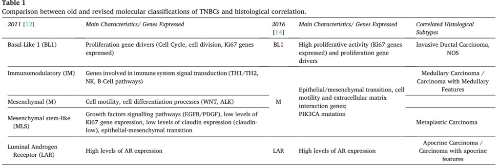
Original Molecular Classification of Triple Negative Breast Cancer (TNBC)

  • Following the description of the main intrinsic breast cancer molecular subtypes based on gene expression analysis:
    • Lehmann et al. demonstrated the complex and heterogeneous nature of TNBCs, and that their definition simply based on interpretation of histopathological features might be of limited efficacy in understanding prognostic behaviour and therapeutic implications
  • They described six subtypes of TNBC based on gene expression analysis of 21 breast cancer data sets:
    • Basal-like type 1 (BL1):
      • Characterized by high proliferative activity demonstrated by elevated Ki67 mRNA expression
    • Basal-like type2 (BL2):
      • Showing basal-myoepithelial phenotype
    • Immunomodulatory (IM) subtype:
      • That includes gene ontologies related to immune cell processes involved in immune signal transduction (such as TH1/TH2, NK and B-cell receptor pathways), in absence of significant correlation with stromal inflammatory cell infiltrate
    • Mesenchymal (M) subtype:
      • That displays genetic patterns responsible for cell motility and cell differentiation processes (i.e. Wnt pathway, ALK pathway)
    • Mesenchymal stem-like (MLS) subtype:
      • Shows genetic profiles associated with growth factors signalling pathways (i.e. EGFR, PDGF) and, particularly, low rates of proliferation genes and low expression of claudins’ family proteins (lately described as claudin-low cancer subtype)
    • LAR subtypes (luminal androgen receptor subtype):
      • Express high levels of androgen receptor hormones (AR) and correlates also with tumors showing apocrine differentiation on histologic examination

Pathology of Triple Negative Breast Cancer Part 3

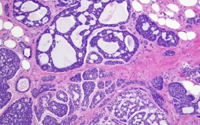
  • Adenoid cystic carcinoma:
    • 0.1% to 1% of all breast cancers
    • Low aggressive malignant potential
    • Myoepithelial differentiation
    • Exhibit tubular, trabecular, cribriform, and / or solid patterns
    • Cribriform is the classic pattern
    • Characterized by MYB-NFIB t(6;9)(q22-23;p23-24)
Adenoid Cystic Carcinoma of the Breast
Tumor Infiltrating Lymphocytes (TIL)
  • Tumor infiltrating lymphocytes (TIL):
    • Recommendations for assessing TILS in breast cancer:
      • Evaluated for the stromal component (% of stromal TIL)
      • Evaluated with the borders of the invasive tumor
      • Exclude TILs outside of the tumor border, around DCIS and normal lobules
      • Lymphocytes and plasma cells, exclude neutrophils
      • Full sections are preferred over biopsies:
        • Cores can be used in the pre therapeutic neoadjuvant setting
      • Average TILs in the tumor area (do not focus on hotspots)
      • The number of TILS correlate with complete pathologic response in the neoadjuvant setting
      • No formal recommendations for a clinically relevant TILS threshold(s) can be given at this stage
  • PD-L1 and Breast Cancer:
    • The PD-L1 on tumor cells, when combined with its PD-1 on immune cells:
      • Causes an inhibition of immune response mediated by CD8+ T cells
    • Breast tumor that have PD-L1 tend to have high number of TILs, and the majority are of the triple negative type
  • Tumors arising in BRCA 1 carriers:
    • BRCA 1 is involved in:
      • DNA repair
      • Cell cycle regulation
      • Transcriptional regulation
      • Chromatin remodeling
    • Loss of BRCA 1 leads to:
      • Deficiency in repair of DNA doble-strand breaks
    • 75% of all tumors developing in BRCA 1 germ line mutation carriers are TNBC:
      • High histologic grade
      • High proliferation rate
  • Residual cancer burden after neoadjuvant chemotherapy (NAC):
    • Parameters required to calculate residual cancer burden (RCB):
      • Submission of the entire area of the tumor bed
      • Tumor dimensions (at least in two dimensions)
      • Percentage invasive carcinoma in the tumor bed
      • Percentage of the in situ carcinoma in the tumor bed
      • The number of positive lymph nodes
      • The largest diameter of nodal metastasis
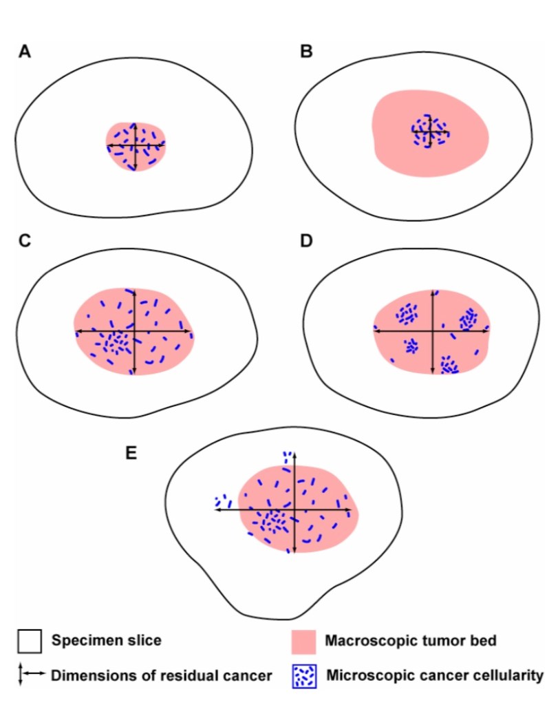
In these diagrams, the macroscopic tumor bed dimensions in examples A, C, D also define the final dimensions of the residual tumor bed after microscopic review. However, the macroscopic tumor bed dimensions in example B overestimate the extent of residual cancer, and so the dimensions of the residual tumor bed (d1 and d2) would be revised after microscopic evaluation of the extent of residual cancer in the corresponding slides from the gross tumor bed. In a different example (E), microscopic residual cancer extends beyond the confines of the macroscopic tumor bed. Again, the dimensions of the residual tumor bed (d1 and d2) would be revised after microscopic evaluation of the recognizable extent of residual cancer beyond the macroscopic tumor bed.
This approach accounts for differences in the concentration and distribution of residual cancer within a tumor bed. In the illustration above, the estimated % CA in example A would be high (in a small area), whereas the estimated % CA for examples C and D would be lower (in a larger area). In examples C and D, the estimated % CA would likely be similar, even though the distribution of cancer within the residual tumor bed is different in those two examples.
A practical way to estimate % CA in a slide is to encircle with ink dots the tumor bed on each slide from the grossly defined residual tumor bed (e.g., slides A1-A5 in the example above). Then use the microscope to estimate the cellularity in each microscopic field across the area of tumor bed. In each microscopic field, % CA can be estimated by comparing the proportion of residual tumor bed area containing cancer (invasive or in situ). Estimate an average of the readings for % CA in the cross-sectional area. The same can be done for in situ component (% CIS). Estimates are to the nearest 10%, but include 0%, 1%, and 5% for areas with low cellularity. The average cellularity within the tumor bed from each slide across the tumor bed can then be estimated (illustrated above).
  • It is recommended to repeat ER, PR, and HER2 on invasive TNBC after neoadjuvant therapy
  • Distant metastasis in patient with residual disease after NAC:
    • Factors associated with increased distant metastatic rate:
      • Positive pathologic LN status
      • Lymphovascular space invasion (LVSI)
      • Increasing clinical T and N stage
      • Multifocality
      • Extranodal extension

Pathology of Triple Negative Breast Cancer Part 2

  • Basal-like breast cancer:
    • Express genes of basal epithelium and demonstrate low expression of ER and HER2 related genes
    • Often (80%) ER negative, PR negative, and HER2 negative, EGFR positive, CK 5/6 positive
    • Most TNBC are basal-like and the reverse is true
  • Clinical presentation:
    • 6% to 27% of all breast cancers are basal-like
    • 1/5 of the cases are seen in younger patients:
      • 20% in patients less than 40 years
    • Often aggressive with poor prognosis
  • Pathology:
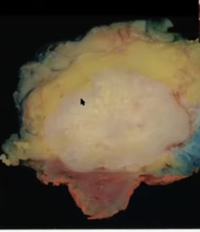
    • Macroscopic appearance:
      • Mostly well circumscribed masses
      • Geographic necrosis is common
  • Pathology:
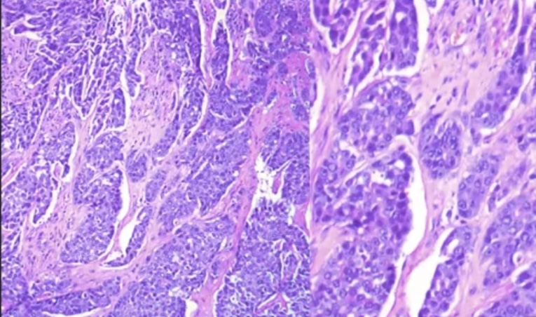
    • Invasive carcinoma NST with Medullary pattern
    • Solid architecture:
      • No tubule formation
    • High grade and high proliferation:
      • Ki-67 greater than 90%
    • High cell denisty and scant stroma
    • Pushing borders
    • Lymphocytic infiltrate at the tumor edge
    • Geographic or central necrosis
    • Syncytial arrangement, less cytoplasm, basaloid features, nuclei with coarse or vesicular chromatin and prominent nucleoli
Basal-like TNBC
Basal-Like TNBC
Immune Profile
  • Molecular Pathology in TNBC and Basal-Like:
    • High degree of genetic instability
    • Heterogeneity of gene copy number aberrations
    • 15% to 20% highly express genes of basal epithelium and demonstrate low expression of ER-related gene and HER2
    • TP53 (87%), PTEN mutation / loss (35%), RB1 mutation / loss (20%), Cyclin D1 amplification (58%), and CDK gain (25%)
    • P-cadherin, fatty acid-binding protein 7, c-kit, matrix metal lo-retina se 7, caveolin 1 and 2, metallothionein IX, TFG-beta receptor II
  • Histologic types of TNBC:
    • Metaplastic carcinomas
    • Adenoid cystic carcinoma
    • Secretory carcinoma
    • Triple negative tumors of luminal androgen receptor type (AR)
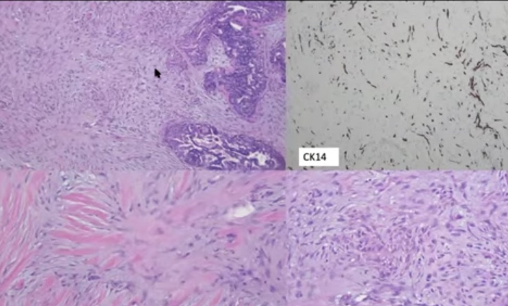
  • Metaplastic Carcinoma (MC):
    • A heterogenous group of invasive breast cancer
    • Components, including pindle cells, squamous cells , and matrix production:
      • Low-grade adenosquamous carcinoma
      • Fibromatosis-like metaplastic carcinoma
      • Spindle cell carcinoma
      • Squamous cell carcinoma
      • MC with heterozygous mesenchymal differentiation
      • Mixed metaplastic carcinoma
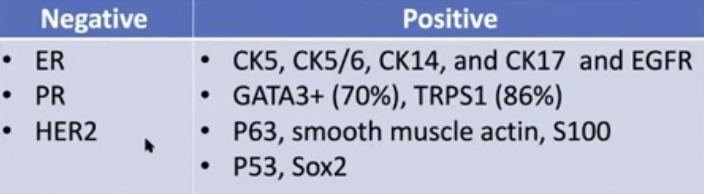
    • Immunohistochemical profile of MC
      • Positive:
        • AE1/AE3, 34BE12, CK5/6, CK14, p63, CK8/18, CK7, and CK19
      • Negative:
        • CD34, desmin, and SMMHC
TNBC – Metaplastic Carcinoma Spindle Cell Type
  • Secretory Carcinoma:
    • Rare low-grade type of breast cancer (< 0.1%) in adolescents
    • Partially circumscribed
    • Three histologic patterns:
      • Solid
      • Micro cystic (cysts simulating thyroid follicles)
      • Tubular
    • Neoplastic cells are uniform, round to polygonal, finely granular or vacuolated cytoplasm containing dense eosinophilia secretion
    • Characterized by fusion gene NTRK-ETV6 t(12;15)(p13;q25)
Secretory Carcinoma

Pathology of Triple Negative Breast Cancer Part 1

  • Definition:
    • Triple negative breast cancer (TNBC) is a subtype of breast tumor lacking hormone receptors expression and HER2 gene amplification:
      • Represents 24 % of newly diagnosed breast neoplasms
    • TNBC is usually characterized by poor prognosis and lack of wide choice therapeutic agents due to the absence of targetable hormone receptors and HER2 expression:
      • Therefore is considered a very interesting and challenging topic for breast cancer research
    • TNBC is a functional term that defines a wide spectrum of entities:
      • With different biology and clinical behavior, with marked genetic, transcriptional, histologic and clinical differences
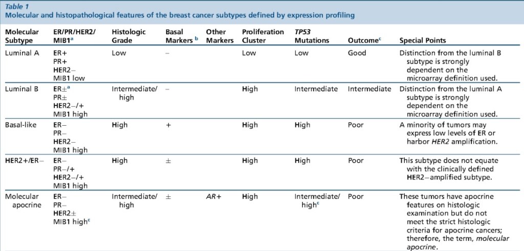
    • The definition of a new classification for breast cancer based on its gene expression pattern divided breast tumors into four “intrinsic subtypes”:
      • Luminal subtype:
        • Divided in Luminal A and Luminal B:
          • Characterized by estrogen receptor gene expression
      • The HER2 subtype:
        • Characterized by HER2 gene amplification
      • The so-called basal-like subtype:
        • A particular breast cancer showing positivity for basal and myoepithelial markers and lack of hormone receptors and HER2 gene amplification
      • “Normal breast-like” subtype:
        • With triple-negative phenotype but cellular derivation typical of normal breast epithelium
      • Notably, both the basal-like and normal breast-like subtypes were already recognized as triple negative
  • All breast cancers arise in the terminal duct lobular units (the functional unit of the breast) of the collecting duct
  • The histological and molecular characteristics:
    • Have important implications for therapy:
      • Several classifications on the basis of molecular and histological characteristics have been developed
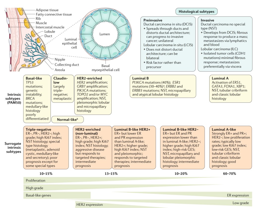
  • The histological subtypes described here (Figure) are the
    most frequent subtypes of breast cancer:
    • Ductal carcinoma (now referred to
      as ‘no special type’ (NST)
      ) and lobular carcinoma:
      • Are the invasive lesions
    • Their pre-invasive counterparts are:
      • Ductal carcinoma in situ and lobular carcinoma in situ (or lobular neoplasia), respectively
        • The intrinsic subtypes of Perou and Sorlie:
          • Are based on a 50-gene expression signature (PAM50)
        • The surrogate intrinsic subtypes are typically used clinically and are based on histology and
          immunohistochemistry expression of key proteins
          :
          • Estrogen receptor (ER), progesterone receptor (PR), human epidermal growth factor receptor 2
            (HER2) and the proliferation marker Ki-67
        • Tumors expressing ER and / or PR are termed ‘hormone receptor-positive’
        • Tumors not expressing ER, PR and HER2 are called ‘triple-negative’
  • The normal breast terminal duct lobular unit:
    • It is formed by two layers:
      • Inner or luminal layer (epithelial cell layer)
      • Outer or basal layer (myoepithelial cell layer)
Cancer can arise from both of this layers
  • Molecular subtypes that are triple negative:
    • Basal-like:
      • TP53 mutations
      • Genetic Instability
      • BRCA mutations
      • Medullary-like histology
      • Poorly differentiated
    • Claudin-low:
      • Largely triple negative
      • Metaplastic
    • Normal breast-like
    • Molecular apocrine
    • Interferon rich
We can classify TNBC according to the grade of the tumor. Representative micrographs of low-grade and high- grade variants of TNBC.
Histologic Types of TNBCs and their key genetic features/potential therapeutic targets.
  • TNBC can be classified into:
    • Low-grade and high-grade histologic types
  • Several histologic types of low-grade TNBC, including:
    • Salivary gland-like tumors of the breast and solid papillary carcinoma with reverse polarity:
      • Are underpinned by specific / pathognomonic genetic alterations
    • In contrast, acinic cell carcinoma and high-grade variants of TNBC have somatic genomic landscape similar to those of conventional TNBC
    • Low- grade variants of metaplastic breast carcinomas (MBCs) are unlikely to be underpinned by specific genetic alterations; however, the genetic analyses performed to date included only a few or single cases
    • Progression to high-grade TNBC has been described in most low-grade forms of TNBC:
      • However, it occurs at a different rate
    • Whereas fairly common in acinic cell carcinoma, it is a rare event in the salivary gland-like tumors of the breast and solid papillary carcinoma with reverse polarity
    • It should be noted that evidence for the presence of PRKD1 E710D mutations or PRKD1/2/3 rearrangements in polymorphous carcinoma of the breast remains to be documented
#Arrangoiz #BreastSurgeon #CancerSurgeon #SurgicalOncologist #Surgeon #Doctor #MountSinaiMedicalCenter #MSMC #Miami #Me

Ductal Carcinoma In Situ (DCIS) Radiation Oncology Perspective Part 3

  • Radiation techniques for DCIS – Hypofractionation
    • ASTRO evidence-based guideline from 2018:
      • Stage (including DCIS vs invasive breast cancer):
        • Statement KQ1G:
          • Hypofractionation whole breast irradiation (WBI) may be used as an alternative to conventional fractionation (CF) CF-WBE in patients with DCIS
            • Recommendation strength: conditional
            • Quality of evidence: Moderate
            • Consensus: 86%
      • Age, grade, and margins for DCIS:
        • Statement KQ2D:
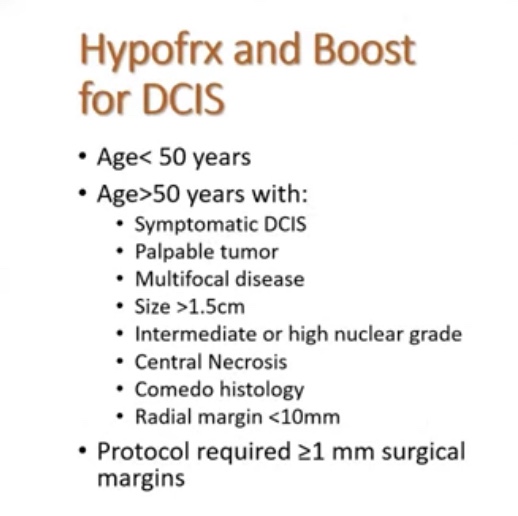
          • A tumor boost may be used for patients with DCIS who meet any of the following criteria:
            • Age =/< 50 years
            • High grade
            • Close (< 2 mm) or positive margins
          • Recommendation strength: conditional
          • Quality of evidence: Moderate
          • Consensus: 92%
  • This two statements from ASTRO rely of data from two randomized trials:
    • The DBCG Hypo Trial:
      • Entry criteria:
        • > 40 years of age
        • BCS for node-negative breast cancer
        • DCIS (13% of the cohort)
      • Primary endpoint:
        • Grade 2 to 3 breast induration assuming no inferiority regarding locoregional recurrence
      • Median follow-up of 7.26 years
The local control were the same between hypofractionation vs conventional fractionation.
Grade 2 to 3 induration rates were similar between hypofractionation vs conventional fractionation.
BIG 3-07 / TROG 07.01 Trial
  • BIG 3-07 / TROG 07.01:
    • Background:
      • Whole breast irradiation (WBI) after conservative surgery for ductal carcinoma in situ (DCIS) reduces local recurrence.
      • They investigated whether a tumor bed boost after WBI improved outcomes, and examined radiation dose fractionation sensitivity for non-low-risk DCIS.
    • Methods:
      • The study was an international, randomized, unmasked, phase 3 trial involving 136 participating centres of six clinical trials organisations in 11 countries (Australia, New Zealand, Singapore, Canada, the Netherlands, Belgium, France, Switzerland, Italy, Ireland, and the UK).
      • Eligible patients were women aged 18 years or older with unilateral, histologically proven, non-low-risk DCIS treated by breast-conserving surgery with at least 1 mm of clear radial resection margins.
      • They were assigned to one of four groups (1:1:1:1) of no tumour bed boost versus boost after conventional versus hypofractionated WBI, or randomly assigned to one of two groups (1:1) of no boost versus boost after each center prespecified conventional or hypofractionated WBI.
      • The conventional WBI used was 50 Gy in 25 fractions, and hypofractionated WBI was 42.5 Gy in 16 fractions. A boost dose of 16 Gy in eight fractions, if allocated, was delivered after WBI.
      • Patients and clinicians were not masked to treatment allocation. The primary endpoint was time to local recurrence.
    • Findings:
      • Between June 25, 2007, and June 30, 2014, 1608 patients were randomly assigned to have no boost (805 patients) or boost (803 patients).
      • Conventional WBI was given to 831 patients, and hypofractionated WBI was given to 777 patients.
      • Median follow-up was 6.6 years.
      • The 5-year free-from-local-recurrence rates were 92.7% (95% CI 90·6-94·4%) in the no-boost group and 97.1% (95·6-98·1%) in the boost group (hazard ratio 0·47; 0·31-0·72; p<0·001).
      • The boost group had higher rates of grade 2 or higher breast pain (10% [8-12%] vs 14% [12-17%], p=0·003) and induration (6% [5-8%] vs 14% [11-16%], p<0·001).
    • Interpretation:
      • In patients with resected non-low-risk DCIS, a tumor bed boost after WBI reduced local recurrence with an increase in grade 2 or greater toxicity.
      • The results provide the first randomised trial data to support the use of boost radiation after postoperative WBI in these patients to improve local control.
      • The international scale of the study supports the generalizability of the results.
  • Radiation Techniques for DCIS:
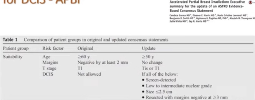
    • Accelerated partial breast irradiation:
ASTRO Evidence Based Consensus Statement
  • Summary:
    • Moderately hypo-fractionation WBI is a standard treatment
    • Consider boost for:
      • High grade DCIS
      • > 2 cm tumors
      • Positive or < 2 mm margins
      • Pre-menopausal patients
      • Patients less than 50 years of age
    • APBI in DCIS is safe and effective option in ASTRO “suitable”candidates
      • Not all techniques (just validated with external beam radiation)
    • Consider hormone therapy in aggressively minded patients or those wishing to decrease risk of contralateral breast cancer
    • Consider genomic assay assistance to aid in radiation decisions in select patients:
      • Postmenopausal patients with otherwise low-risk disease
#Arrangoiz #BreastSurgeon #CancerSurgeon #SurgicalOncologist #Doctor # Surgeon #MountSinaiMedicalCenter #MSMC #Miami #Mexico BreastCancer #BreastCancerAwareness

Ductal Carcinoma In Situ (DCIS) Radiation Oncology Perspective Part 2

  • The second generation trials in DCIS:
    • Started to think if there is a sub group of patients that we can omit radiation therapy
  • This trials looked into high risk factors for local regional recurrence (LRR) in DCIS:
    • Prognostic factors associated with increased locoregional recurrence:
      • Age less than 50 years
      • Size > than 2 cm
      • Grade 3
      • Positive of close < margins
    • Additional risk factors:
      • Symptomatic:
        • Palpable / bloody discharge
      • Comedo, solid types of DCIS
      • Black race
      • ER and / or PR negative
  • What is the optimal surgical margin in DCIS?
  • The use of a 2-mm margin as the standard for an adequate margin in DCIS treated with whole-breast irra- diation is associated with lower rates of IBTR and has the potential to decrease re-excision rates, improve cosmetic outcomes, and decrease health care costs:
    • Clinical judg- ment should be used in determining the need for further surgery in patients with negative margins narrower than 2 mm.
  • The second generation DCIS trials:
    • RTOG 9804:
      • They included:
        • Mammographically detected DCIS
        • Low to intermediate-grade DCIS
        • Less than 2.5 cm
        • Margins =/> than 3 mm
      • This study has two different perspectives:
        • Although the results are significant:
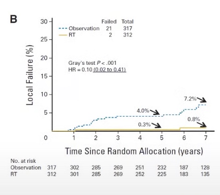
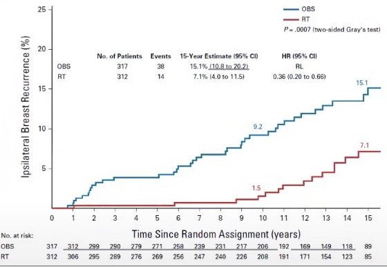
          • Local recurrence (LR) rates without RT are less than 10%
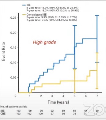
Longer follow-up of the RTOG 9804 trial: LR rates were as high as 15.1% in omitted radiation arm
  • The second generation DCIS trials:
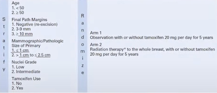
    • ECOG E5194:
      • From 1997 to 2022:
        • Has only one arm
      • Inclusion criteria:
        • Non-palpable DCIS
        • Cohort 1:
          • Low to intermediate grade DCIS < than 2 cm
        • Cohort 2:
          • High grade DCIS < than 1 cm
        • > 3 mm margins / no residual calcifications on postoperative mammogram
        • Lumpectomy alone:
          • NO radiation
        • Starting in the year 2000:
          • Patients could take tamoxifen
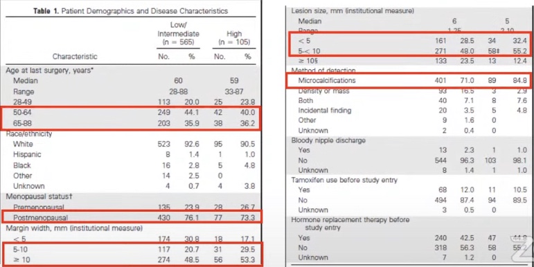
Like in the RTOG 9804 trial patients in the ECOG E5194 trial tended to be older, postmenopausal, have wider margins of resection, and had smaller tumors (detected on mammogram).
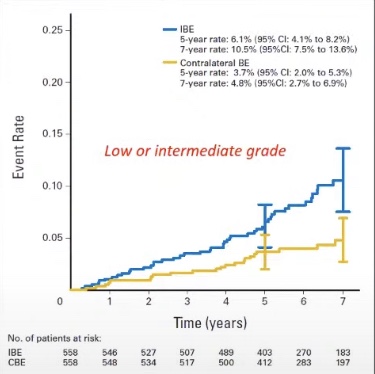
With long-term follow-up in the ECOG E5194 the LR rates in high risk DCIS were almost 25% and in lower risk DCIS was 14.4%, but local control rates were still high and these data should be taken into account in share decision making.
  • What can we use to help us determine risk of recurrence in DCIS?
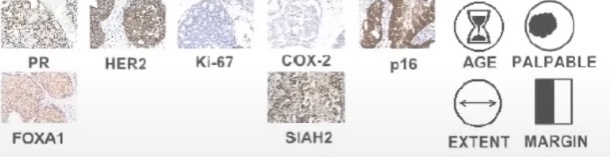
    • Genomic Assays – DCISion RT (Prelude DX):
      • Seven gene biological risk signature developed from three cohorts:
        • UCSF (n=324)
        • Uppsala Univeristy Hospital, Sweden (n=458)
        • University of Massachusetts (n=300)
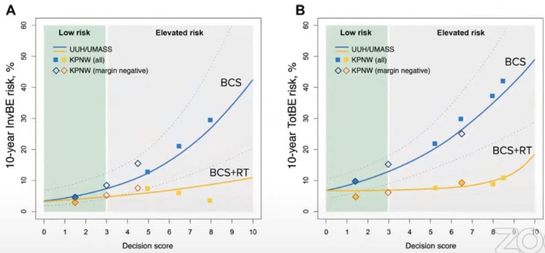
      • Decision score 0 to 10:
        • Low risk 0 to 3
        • High risk score > 3 to 10
      • This biological signature was validated in a retrospective cohort

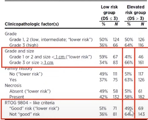
The patients that were defined as low risk by clinical and pathological factors were could actually be higher risk patients based on genetic analysis in 41% to 49% of the cases. 34% to 36% of high risk DCIS patients were found to be low risk by DCISion RT.
  • This tool is available but it has not be validated in a prospective clinical trial