Blog

Happy Thanksgiving

Unknown

Happy thanksgiving from Sociedad Quirúrgica S.C.


Happy thanksgiving de parte del equipo de Sociedad Quirúrgica S.C.

 

Examen Clínico de la Glándula Tiroides

  • La glándula tiroides está situada en la región anterior del cuello (en la unión de los dos tercios superiores con el tercio inferior):
    • Consta de dos lóbulos laterales unidos en su base por un istmo, que le confiere una forma de “U” o de mariposa; y tiene un peso aproximado de 20 a 30 g. 
    • El lóbulo derecho es ligeramente mayor que el izquierdo.
    • Cada lóbulo tiene unos 5 cm de largo y 2 cm de ancho.


  • El examen físico de la glándula tiroides se realiza mediante inspección, palpación, y auscultación. 
    • Normalmente, la glándula tiroides no se aprecia en la inspección y prácticamente, no se palpa, si es de tamaño normal, solo se logra su palpación con especial cuidado y con reglas precisas.
    • Si durante la inspección se detecta un aumento de volumen, se debe realizar también la auscultación de la glándula (presencia o ausencia de soplo en la glándula tiroides vascularizadas) y la medición del cuello.
      • La glándula está unida a la tráquea y se eleva cuando el individuo traga.
      • Para que la inspección y la palpación sean de mayor utilidad, usted debe explorarla también, mientras el sujeto traga; tenga preparado un vaso de agua, para que la persona trague sorbos, cuando se lo indique.
  • Es importante tener presente que la glándula tiroides puede estar aumentada de tamaño, aunque dentro de los límites normales, por distintas circunstancias: país, altitud, pubertad, embarazo, menstruación, etc:
    • Es decir, en los estados fisiológicos de alarma de cualquier orden, en la que puede aumentar de volumen y tornarse más activa fisiológicamente.


  • Inspección:

    • Observe la región anterior del cuello de frente y de perfil, de ser posible con una iluminación tangencial, que puede ayudar a detectar mejor, cambios sutiles en el contorno o la simetría. 
      • Normalmente solo puede verse el istmo glandular, sobre todo al tragar, con mayor frecuencia en mujeres jóvenes.
    • Primero, pida a la persona que mantenga la cabeza y el cuello en una posición normal y relajada. 
    • Observe si existe alguna desviación de la tráquea, así como las delimitaciones del cartílago tiroides y del cartílago cricoides, y fíjese si hay algún aumento de volumen. 
    • Después, pida que extienda ligeramente el cuello, inclinando la cabeza hacia atrás, y que trague un sorbo de agua. 
    • Observe en ese momento, el movimiento simétrico hacia arriba de la tráquea y los cartílagos laríngeos y, de existir algún aumento de volumen, si este también se desplaza.


  • Palpación:

    • En la palpación de la glándula tiroides hay que tener en cuenta básicamente, además de la forma y el tamaño, ya descritos, su consistencia y la sensibilidad. 
    • La glándula normalmente tiene una textura parecida a la de la goma, lo que le confiere una consistencia elástica a la palpación. 
    • No son normales las masas más duras, en forma de nódulos, o que puedan distinguirse de su textura habitual. 
    • La palpación no produce habitualmente dolor, aunque la persona puede experimentar ligera molestia. 
    • Una palpación tiroidea dolorosa es anormal, como se observa en algunas formas de tiroiditis. 
    • La palpación es mejor hacerla con el sujeto sentado, situándose el explorador, primero por detrás, y luego, por delante y por los lados.


    • Abordaje posterior:
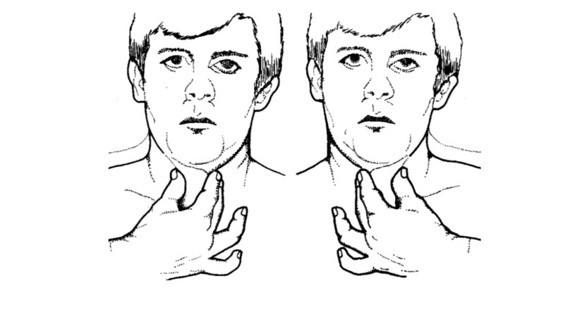
      • Párese detrás de la persona, que debe estar sentada con el cuello ligeramente flexionado, para relajar los músculos. 
      • Sé realiza entonces, la palpación del tiroides utilizando la técnica de Quervain, que consiste en rodear el cuello con ambas manos, con los pulgares descansando sobre la nuca y los cuatro dedos restantes hacia los lóbulos de cada lado.
        • Primero coloque ligeramente los pulpejos de sus dedos índice y del medio, por debajo del cartílago cricoides, para localizar y palpar el área del istmo. 
        • Repita la maniobra mientras la persona traga un sorbo de agua, lo que causa elevación del istmo y permite precisar aún más su textura, como de goma o elástica.
        • Después, pídale que incline ligeramente su cabeza hacia el lado izquierdo, para palpar el lóbulo derecho. 
        • Utilice los dedos en el lado opuesto para desplazar la glándula en dirección lateral, hacia el lado derecho, de manera que los dedos que palpan puedan sentir mejor el lóbulo. 
        • Pida a la persona que trague, mientras examina el lóbulo. 
        • Repita el procedimiento en el lado opuesto.


    • Abordaje anterior:
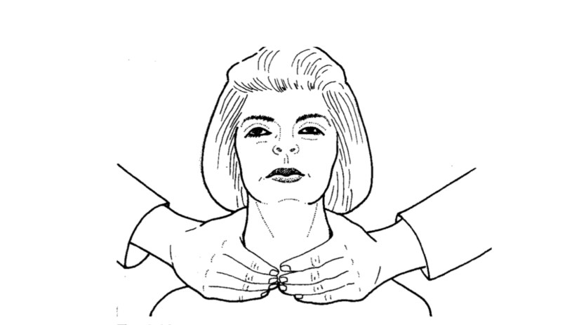
      • Párese frente a la persona, cuyo cuello debe estar relajado, pero ligeramente en extensión, para exponer mejor la glándula subyacente. 
      • Ahora las manos se colocan alrededor del cuello, pero con los pulgares en el plano anterior, que son los que palpan. 
      • Palpe los lóbulos tiroideos utilizando las dos técnicas descritas a continuación:
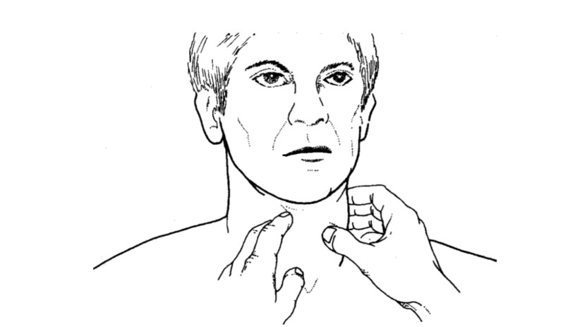
        • De frente al sujeto, el pulgar de cada mano palpa sucesivamente el lóbulo del lado opuesto, en busca de nódulos (maniobra de Crile).
        • Palpe también los lóbulos laterales, con una variante de la técnica anterior (maniobra de Lahey):
          • Se coloca el pulpejo de un dedo pulgar contra la cara lateral de la tráquea superior, empujando hacia el lado opuesto, con lo que el lóbulo del lado hacia el que se empuja, se exterioriza más hacia delante y puede ser más accesible al pulgar de la otra mano; esta maniobra se completa con la deglución, mientras se palpa.
  • Auscultación:

    • Recuerde la necesidad o no de auscultar el tiroides, especialmente cuando se sospecha hiperfunción de la glándula.

Presentation1Presentation2Presentation3

Rodrigo Arrangoiz MS, MD, FACS cirujano de tumores de cabeza y cuello / cirugia endocrina / cirugía oncológica es experto en el manejo de patología  de la glándula tiroides.

Su entrenamiento fue el siguiente:

• Cirugia general y gastrointestinal:
• Michigan State University:
• 2004 al 2010image-48• Cirugia oncológica / tumores de cabeza y cuello / cirugia endocrina:
• Fox Chase Cancer Center (Filadelfia):
• 2010 al 2012image-39• Maestria en ciencias (Clinical research for healthprofessionals):
• Drexel University (Filadelfia):
• 2010 al 2012image-50• Cirugia de tumores de cabeza y cuello / cirugiaendocrina
• IFHNOS / Memorial Sloan Kettering Cancer Center:
• 2014 al 2016image-51

https://www.cirugiatiroides.com

Goals of the treatment of cancer of the oral cavity

The goals of the treatment of cancer of the oral cavity are:

  • Cure of the cancer

  • Preservation or restoration of speech, mastication, swallowing, and external appearance

  • Minimization of the sequelae of treatment such as dental decay, osteonecrosis of the mandible, and trismus

  • Awareness of the risk of subsequent primary tumors and their management.

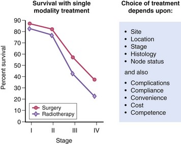
Surgery and radiotherapy can be used as a single modality or in combination for the treatment of cancer of the oral cavity.

B9780323055895000081_f008-041-9780323055895

Treatment Approaches:

  • In general, early-stage (stage I or II) head and neck tumors may be treated using a single modality (surgery or radiotherapy), whereas advanced disease (stage III or IV) frequently benefits from multimodality therapy.

    • The best therapeutic approach for the primary tumor depends on the anatomic site.

    • Most neck disease can be treated equally well with surgery or radiation, thus the modality chosen to treat the neck is based on which modality is selected for the primary.

    • When the primary tumor is treated with irradiation, the regional lymphatics “at-risk” are incorporated into the treatment fields.

    • Neck dissections should remain standardized (ie, complete anatomic dissections, as opposed to “berry picking” or random biopsy) in these settings to avoid incomplete surgery.

Philadelphia Illustration Dept./Elsevier
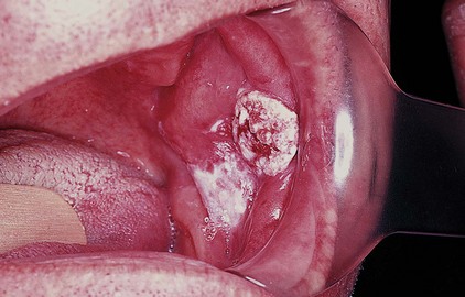
Buccal SCC

Rodrigo Arrangoiz MS, MD, FACS a head and neck surgeon and is a member of Sociedad Quirúrgica S.C at the America British Cowdray Medical Center.

He is first author on some publications on oral cavity cancer:

Training:

• General surgery:

• Michigan State University:

• 2004 al 2010

• Surgical Oncology / Head and Neck Surgery / Endocrine Surgery:

• Fox Chase Cancer Center (Filadelfia):

• 2010 al 2012

• Masters in Science (Clinical research for health professionals):

• Drexel University (Filadelfia):

• 2010 al 2012

• Surgical Oncology / Head and Neck Surgery / Endocrine Surgery:

• IFHNOS / Memorial Sloan Kettering Cancer Center:

• 2014 al 2016

Pathology of Oral Cavity Cancer

A. Squamous Cell Carcinoma:

– More than 90% of all head and neck cancers in the West are squamous cell carcinomas.

B. Histologic grade:

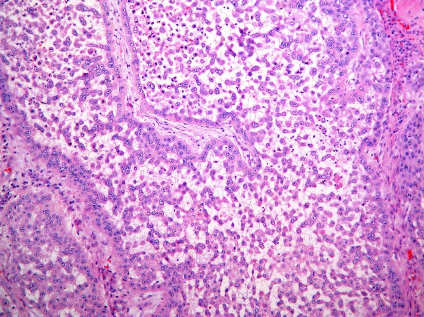
1. There are three histologic grades based on the amount of keratinization:
– A well-differentiated tumor is characterized by more than 75% keratinization.
– A moderately differentiated tumor, is characterized by 25% to 50% keratinization.
– A poorly differentiated tumor is characterized by less than 25% keratinization.

Unknown6713853577_41cf97059a_b

  • Histologic grade has not been a consistent predictor of clinical behavior.

C. Features that predict aggressive behavior include:

1. Perineural spread
2. Lymphatic invasion
3. Tumor spread beyond the lymph node capsule (Extranodal extension – ENE).

D. HPV-positive tumors tend to be non-keratinizing and poorly differentiated.


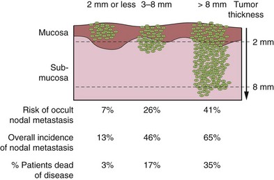
  • The most important histological feature of the primary tumor that affects selection of treatment and eventual prognosis is its depth of infiltration.

    • Thin and superficially invasive lesions have a lower risk of regional lymph node metastasis, are highly curable, and offer an excellent prognosis.

    • On the other hand, thicker lesions that deeply infiltrate the underlying soft tissues have a significantly increased incidence of regional lymph node metastasis and an adverse impact on prognosis.

  • The risk of lymph node metastasis and survival rates in relation to the thickness of the primary lesion for T1 and T2 squamous carcinomas of the oral tongue and floor of mouth are shown in the Figure.

B9780323055895000081_f008-039-9780323055895


  • Although it would be ideal to know the exact thickness of the lesion before surgical intervention, in many instances having that information before surgical excision of the primary tumor is clinically impractical.

  • In general, however, thickness of the lesion as appreciated by palpation is a reasonably good indicator of deeply invasive lesions versus superficial lesions to estimate the extent of soft tissue and/or bone resection for the primary lesion and to decide on the need for elective dissection of the regional lymph nodes at risk in a neck with negative clinical findings.

    • Pooled data from the literature on the thickness of the primary tumor for selection of elective treatment of the neck indicate that lesions thicker than 4 mm have a progressively increased risk of lymph node metastasis.

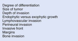
  • The presence of perineural invasion and the presence of lymphovascular tumor emboli are prognostic indicators for tumor control and survival.

  • Local recurrence also is affected by the pattern of tumor infiltration, with single cell infiltration more unfavorable than a broad, “pushing” border.

  • As one would expect, negative margins are another key factor in locoregional control.

    • If a tumor is present less than 5 mm from the resection margin, it should be stated in the pathology report because the presence of lesions (i.e., severe dysplasia/carcinoma in situ and invasive carcinoma) within this distance has been reported to be associated with a significant risk of local recurrence.

    • Histological features of the primary tumor that have an impact on therapeutic outcomes are shown in the figure.

B9780323055895000081_f008-040-9780323055895


  • Rodrigo Arrangoiz MS, MD, FACS a head and neck surgeon and is a member of Sociedad Quirúrgica S.C at the America British Cowdray Medical Center.

    He is first author on some publications on oral cavity cancer:

    Training:

    • General surgery:

    • Michigan State University:

    • 2004 al 2010

    • Surgical Oncology / Head and Neck Surgery / Endocrine Surgery:

    • Fox Chase Cancer Center (Filadelfia):

    • 2010 al 2012

    • Masters in Science (Clinical research for health professionals):

    • Drexel University (Filadelfia):

    • 2010 al 2012

    • Surgical Oncology / Head and Neck Surgery / Endocrine Surgery:

    • IFHNOS / Memorial Sloan Kettering Cancer Center:

    • 2014 al 2016

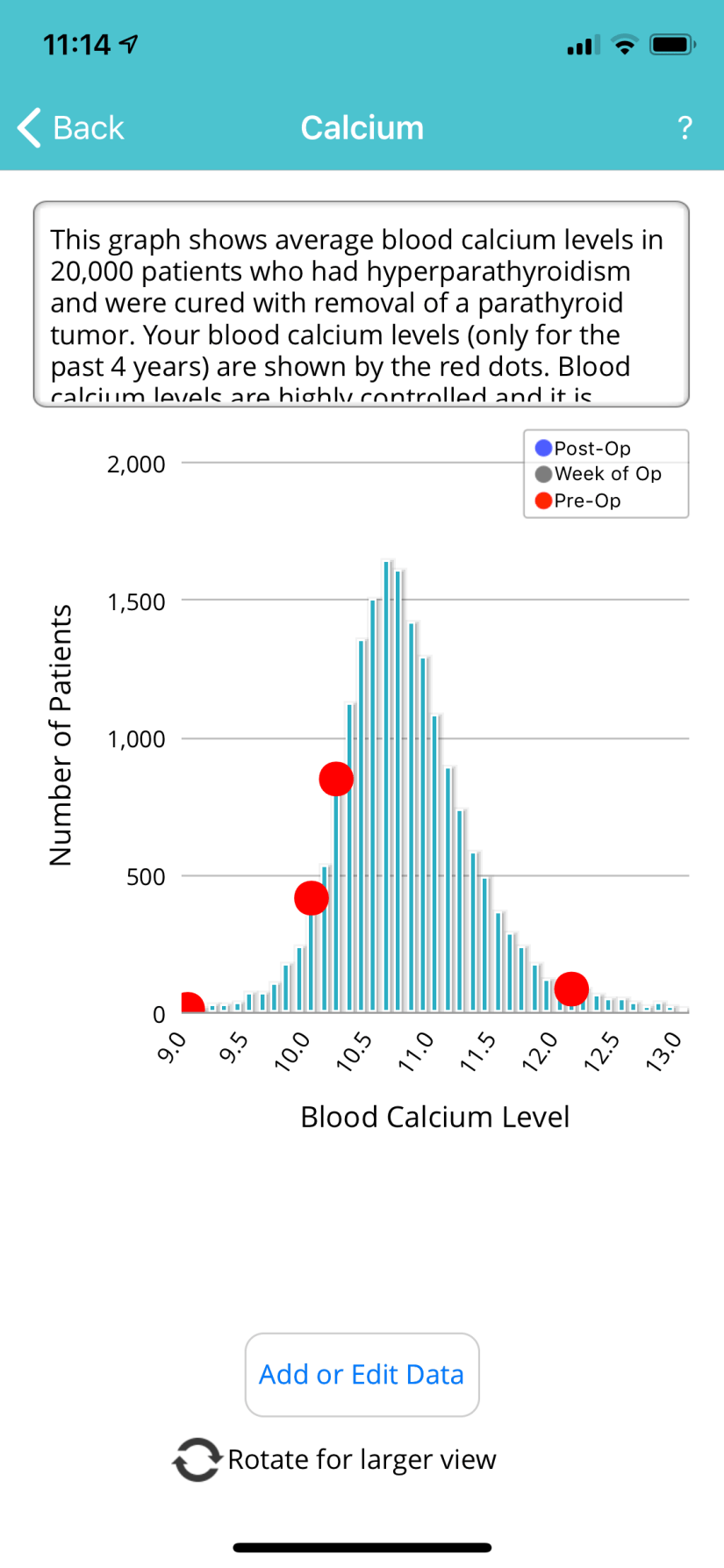
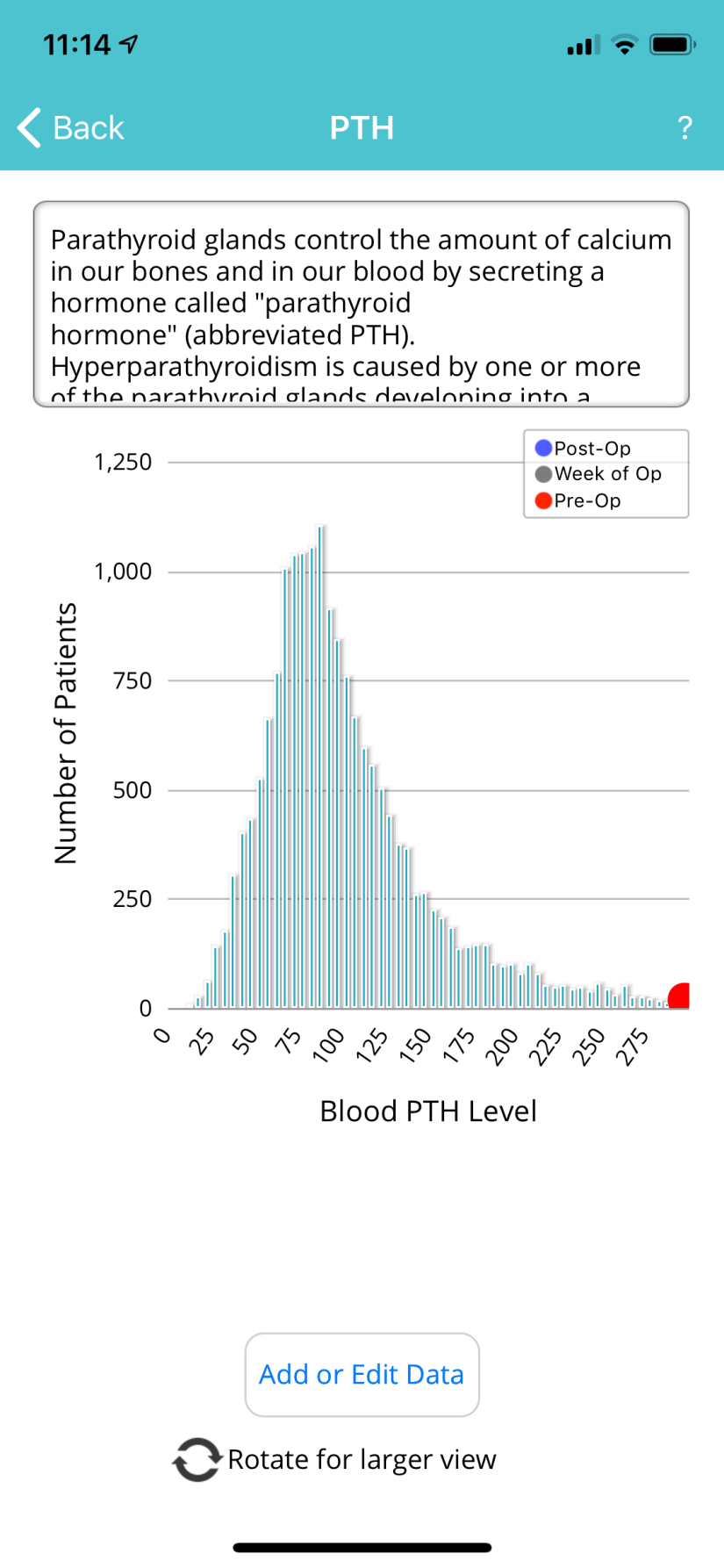
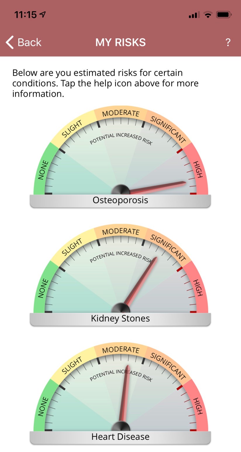
Hiperparatiroidismo Primario

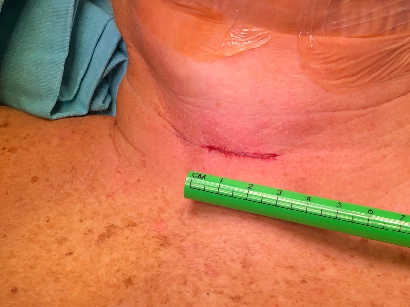
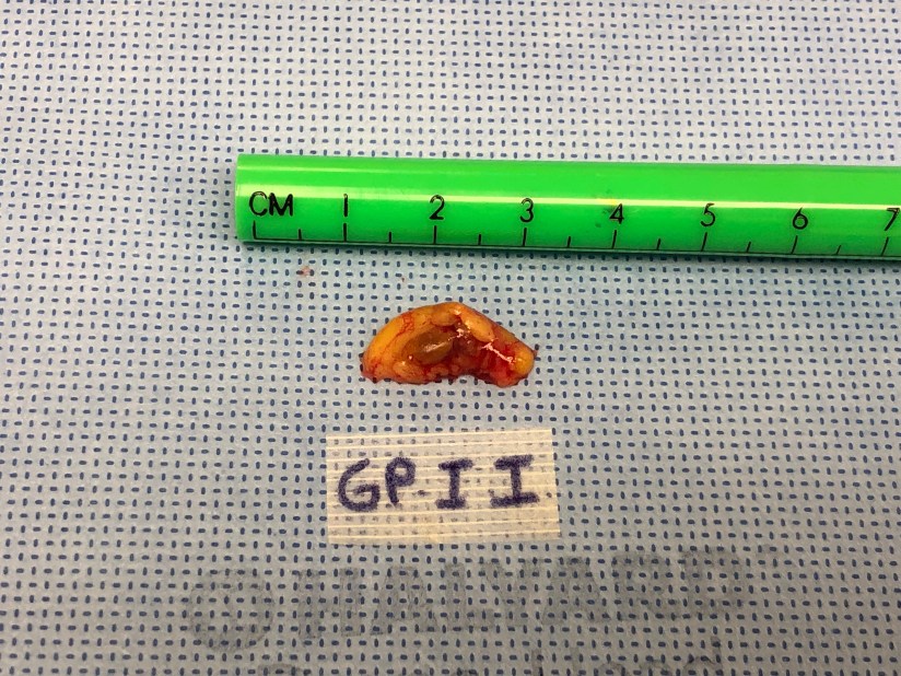
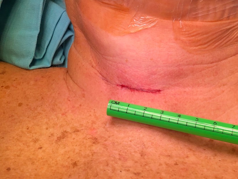
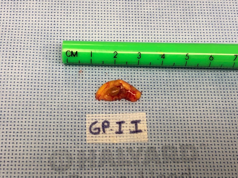
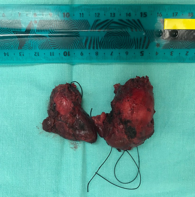
Paciente 69 años con hiperparatiroidismo primario operada por el Rodrigo Arrangoiz MS, MD, FACS miembro de Sociedad Quirúrgica SC

Paratiroidectomia radioguiada con exploración de las cuatro glándulas a través de una incisión de 2 cm de una adenoma paratiroideo

http://www.hiperparatiroidismo.info

Hiperparatiroidismo

Mujer 75 años con hiperparatiroidismo primario #arrangoiz #cirugiaendocrina #cirugiacabezaycuello http://www.hiperparatiroidismo.info

Carcinoma Papilar de Tiroides

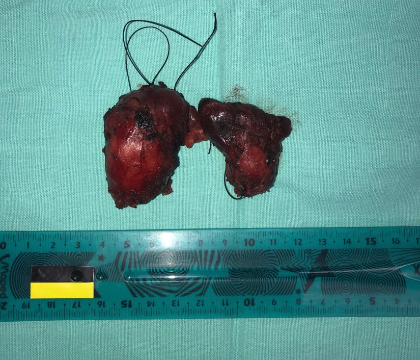
Mujer 32 años con carcinoma papilar de tiroides cT2, cN0, cM0 estadio I

http://www.cirugiatiroides.com

Cirugía de Tiroides

http://www.cirugiatiroides.com


Rodrigo Arrangoiz MS, MD, FACS cirujano de tumores de cabeza y cuello / cirugia endocrina / cirugía oncológica es experto en el manejo de patología  de la glándula tiroides.

Su entrenamiento fue el siguiente:

• Cirugia general y gastrointestinal:
• Michigan State University:
• 2004 al 2010

Cirugia oncológica / tumores de cabeza y cuello / cirugia endocrina:
• Fox Chase Cancer Center (Filadelfia):
• 2010 al 2012

• Maestria en ciencias (Clinical research for healthprofessionals):
• Drexel University (Filadelfia):
• 2010 al 2012

• Cirugia de tumores de cabeza y cuello / cirugiaendocrina
• IFHNOS / Memorial Sloan Kettering Cancer Center:
• 2014 al 2016
Staging of oral cavity cancer

The staging of primary tumors of the oral cavity as published by the American Joint Committee on Cancer/International Union Against Cancer is widely accepted.

  • The surface dimensions and local extent of the tumor are the most important parameters for primary tumor staging in the oral cavity.

  • One glaring deficiency of this staging system is the lack of the depth of infiltration of the tumor as a parameter for staging.

  • However, accurately assessing the tumor depth clinically is difficult:

    • Deeply infiltrating primary tumors have an increased risk of regional metastases and death due to disease compared with superficial tumors of the same T stage.

    • Thus a more aggressive therapeutic approach, including elective treatment of regional lymph nodes, should be considered for patients with early T-staged but deeply infiltrating tumors.

    • On the other hand, advanced T-staged but relatively superficial lesions have a better prognosis.

The depth of infiltration of the primary tumor as a parameter for staging has been added in the new 8th edition of the AJCC staging system:

Tx – Primary tumor cannot be assessed

Tis – Carcinoma in situ

T1 – Tumor < 2 cm, < 5 mm depth of invasión (DOI).
= DOI is depth of invasión and not tumor thickness.

T2 – Tumor < 2 cm, DOI > 5 mm and < 10 mm
or tumor > 2 cm but < 4 cm, and < 10 mm DOI

T3 – Tumor > 4 cm or any tumor > 10 mm DOI

T4 – Moderately advanced or very advanced local disease

T4a – Moderately advanced local disease

= (Lip) Tumor invades through cortical bone or involves the inferior alveolar nerve, floor of mouth, or skin of face (i.e., chin or nose)

= (Oral cavity) Tumor invades adjacent structures only (e.g., through cortical bone of the mandible or maxilla, or involves the maxillary sinus or skin of the face)

Note: Superficial erosión of bone/tooth socket (alone) by a gingival primary is not sufficient to classify a tumor as T4.

T4b – Very advanced local disease
Tumor invades masticator space, pterygoid plates, or skull base and/or encases the internal carotid artery

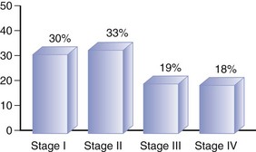
The stage distribution of patients with squamous cell carcinoma of the oral cavity is shown in the figure:

B9780323055895000081_f008-027-9780323055895

Rodrigo Arrangoiz MS, MD, FACS a head and neck surgeon and is a member of Sociedad Quirúrgica S.C at the America British Cowdray Medical Center.

He is first author on some publications on oral cavity cancer:


Training:

• General surgery:

• Michigan State University:

• 2004 al 2010

• Surgical Oncology / Head and Neck Surgery / Endocrine Surgery:

• Fox Chase Cancer Center (Filadelfia):

• 2010 al 2012

• Masters in Science (Clinical research for health professionals):

• Drexel University (Filadelfia):

• 2010 al 2012

• Surgical Oncology / Head and Neck Surgery / Endocrine Surgery:

• IFHNOS / Memorial Sloan Kettering Cancer Center:

• 2014 al 2016