Blog

Medullary Thyroid Carcinoma Article

Nueva publicación de Sociedad Quirúrgica S.C. sobre cáncer medular de tiroides

Rodrigo Arrangoiz MS, MD, FACS autor principal del artículo miembro de Sociedad Quirúrgica S.C. con especialidad en cirugía de tumores de cabeza y cuello / cirugía endocrina / cirugía oncológica

http://www.sociedadquirurgica.com

http://www.cirugiatiroides.com

http://www.hiperparatiroidismo.info
http://www.remedypublications.com/american-journal-of-otolaryngology-and-head-and-neck-surgery/articles/pdfs_folder/ajohns-v1-id1026.pdf

Tongue Anatomy II

  • Musculature of the Tongue:

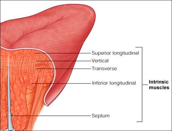
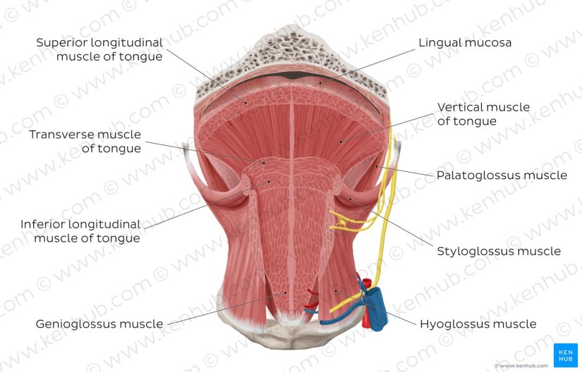
    • The intrinsic muscles (bilateral superior and inferior longitudinal, transverse, and vertical muscles) interdigitate and have no tissue spaces, which allows invasive cancers to spread easily.

ce507-fig04-intrinsic-tongue-muscles

H8rDTg2yQrkuDKTAMNqhg_the-muscles-of-the-tongue_english

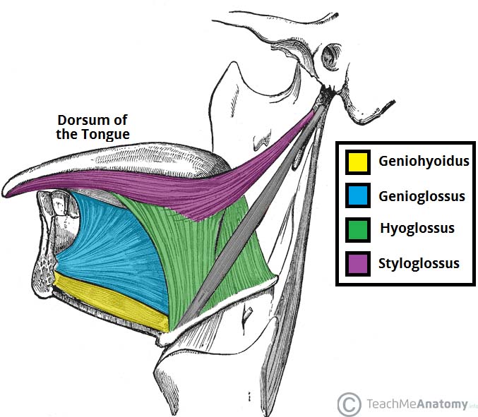
  • On the other hand, infiltration of the extrinsic muscles of the tongue (genioglossus, hyoglossus, styloglossus, and palatoglossus) is a feature of locally advanced cancer.

extrinsic

  • The arterial supply to the tongue and floor of the mouth is from the dorsal lingual, sublingual, and deep lingual branches of the lingual artery.

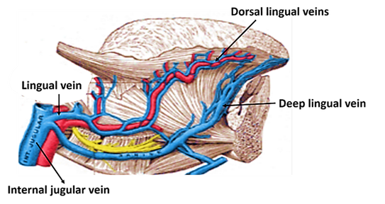
  • The venous drainage of the tongue is into the lingual veins, which drain into the facial and retromandibular veins, which join to form the common facial vein. 

    • Vasculature Tongue:

      • Similar to most of the head and neck region, the tongue derives its arterial blood supply from the external carotid artery.

      • The lingual artery branches off the external carotid artery deep to the stylohyoid muscle:

        • At first, it travels superomedially; after a short distance, it changes direction and moves anteroinferiorly.

        • The hypoglossal nerve (cranial nerve XII) crosses over it laterally before it enters the tongue deep in the hyoglossus muscle.

      • Within the tongue, the lingual artery gives rise to its 3 main branches:

        • The dorsal lingual artery:

          • The dorsal lingual artery supplies the base of the tongue

        • The deep lingual artery:

          • The deep lingual artery travels on the lower surface of the tongue to the tip.

        • The sublingual artery:

          • A branch to the sublingual gland and the floor of the mouth is known as the sublingual artery.

Blood-supply-of-tongue

  • The veins of the tongue parallel the lingual artery branches:

    • The deep lingual vein begins at the tip of the tongue and travels posteriorly to join the sublingual vein:

      • This veins drains into the dorsal lingual vein, which accompanies the lingual artery.

    • The dorsal lingual vein drains into the lingual veins, which drain into the facial and retromandibular veins, which join to form the common facial vein:

    • Directly or indirectly, this vein empties into the internal jugular vein.

veins-of-tongue

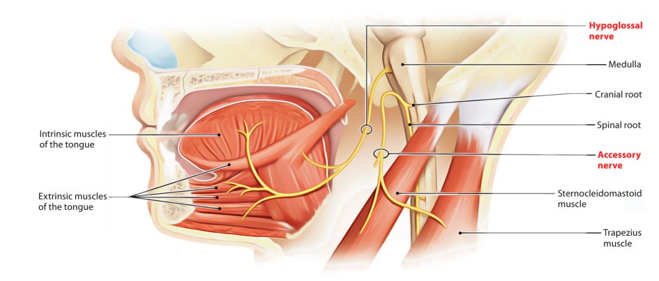
  • The hypoglossal nerve provides motor innervations to all muscles of the tongue except the palatoglossus, which is supplied by the pharyngeal plexus.

  • The lingual nerve is the sensory nerve to the anterior two thirds of the tongue, the floor of the mouth, and the lower gum, while taste sensation is carried along the chorda tympani branch of the facial nerve.

Hypoglossal-and-accessory-nerve-1

Rodrigo Arrangoiz MS, MD, FACS a head and neck surgeon and is amember of Sociedad Quirúrgica S.C at the America British Cowdray Medical Center.

He is first author on some publications on oral cavity cancer:

Training:

• General surgery:

• Michigan State University:

• 2004 al 2010

• Surgical Oncology / Head and Neck Surgery / Endocrine Surgery:

• Fox Chase Cancer Center (Filadelfia):

• 2010 al 2012

• Masters in Science (Clinical research for health professionals):

• Drexel University (Filadelfia):

• 2010 al 2012

• Surgical Oncology / Head and Neck Surgery / Endocrine Surgery:

• IFHNOS / Memorial Sloan Kettering Cancer Center:

• 2014 al 2016

The Tongue

  • Overview:

    • The tongue is a mass of muscle that is almost completely covered by a mucous membrane.

    • It occupies most of the oral cavity and oropharynx.

    • It is known for its role in taste, but it also assists with mastication (chewing), deglutition (swallowing), articulation (speech), and oral cleansing.

    • Five cranial nerves contribute to the complex innervation of this multifunctional organ.

    • The embryologic origins of the tongue first appear at 4 weeks’ gestation:

      • The body of the tongue forms from derivatives of the first branchial arch:

        • This gives rise to two lateral lingual swellings and one median swelling (known as the tuberculum impar):

          • The lateral lingual swellings slowly grow over the tuberculum impar and merge, forming the anterior two thirds of the tongue.

      • Parts of the second, third, and fourth branchial arches give rise to the base of the tongue.

    • Occipital somites give rise to myoblasts, which form the intrinsic tongue musculature.

Extrinsic-Muscles-of-the-Tongue

H8rDTg2yQrkuDKTAMNqhg_the-muscles-of-the-tongue_english

  • Gross Anatomy:

    • From anterior to posterior, the tongue has 3 surfaces:

      • Tip

      • Body

      • Base.

    • The tip is the highly mobile, pointed anterior portion of the tongue.

    • Posterior to the tip lies the body of the tongue, which has dorsal (superior) and ventral (inferior) surfaces.

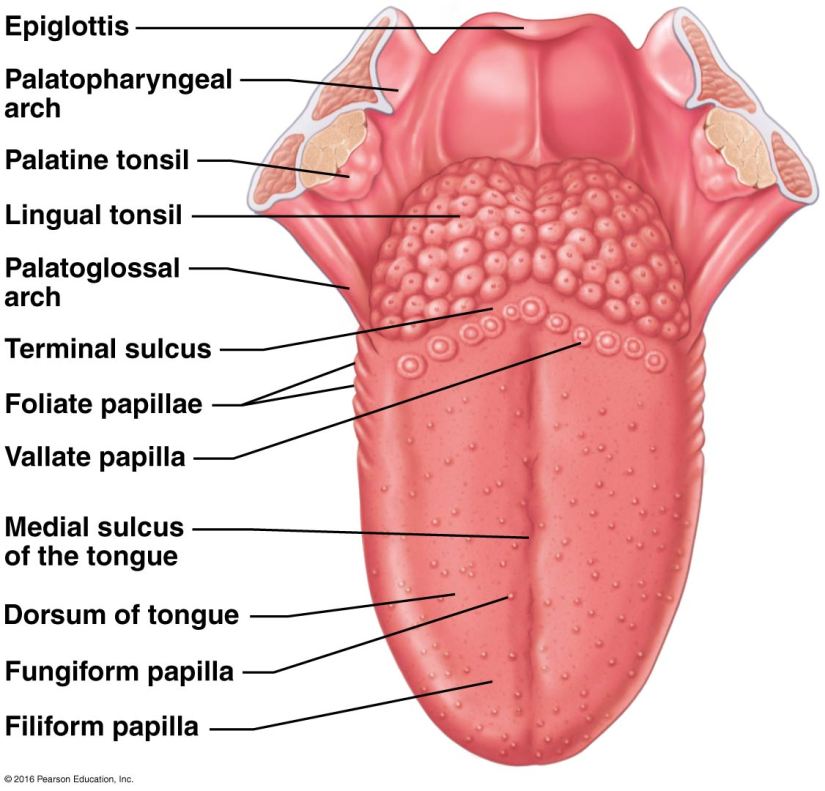
    • The median sulcus of the tongue separates the body into left and right halves.

    • The terminal sulcus, or groove, is a V-shaped furrow that separates the body from the base of the tongue:

      • At the tip of this sulcus is the foramen cecum, a remnant of the proximal thyroglossal duct.

    • The base of tongue contains the lingual tonsils, the inferiormost portion of Waldeyer’s ring.

  • Lingual papillae:

    • The surface of the body of the tongue derives its characteristic appearance from the presence of lingual papillae:

      • Which are projections of lamina propria covered with epithelium.

    • The four types of lingual papillae are as follows:

      • Vallate (circumvallate)

      • Foliate

      • Filiform

      • Fungiform

    • The vallate papillae (circumvallate) are flat, prominent papillae that are surrounded by troughs:

      • In humans, there are 8 to 12 vallate papillae, located directly anterior to the terminal sulcus.

        • The ducts of the lingual glands of von Ebner secrete lingual lipase into the surrounding troughs to begin the process of lipolysis.

    • The foliate papillae are small folds of mucosa (short vertical folds) located along the lateral surface of the tongue:

      • They are located on the sides at the back of the tongue, just in front of the palatoglossal arch of the fauces:

        • The foliate papillae appear as a series of red colored, leaf–like ridges of mucosa.

      • There are four or five vertical folds, and their size and shape is variable.

      • They are covered with epithelium, lack keratin and so are softer, and bear many taste buds:

        • Approximately 1000 taste buds.

      • They are usually bilaterally symmetrical.

      • Sometimes they appear small and inconspicuous, and at other times they are prominent.

      • Because their location is a high risk site for oral cancer, and their tendency to occasionally swell, they may be mistaken as tumors or inflammatory disease.

      • Serous glands drain into the folds and clean the taste buds.

      • Lingual tonsils are found immediately behind the foliate papillae and, when hyperplastic, cause a prominence of the papillae.

    • The filiform papillae:

      • Are the most numerous of the lingual papillae.

      • They are fine, small, cone-shaped papillae covering most of the dorsum of the tongue.

      • They cover most of the front two-thirds of the tongue’s surface.

      • They appear as very small, conical or cylindrical surface projections, and are arranged in rows which lie parallel to the sulcus terminalis:

        • At the tip of the tongue, these rows become more transverse.

      • They are responsible for giving the tongue its texture and are responsible for the sensation of touch.

      • Unlike the other kinds of papillae, filiform papillae do not contain taste buds.

    • The fungiform papillae are mushroom shaped (generally red in color) and are dispersed most densely along the tip and lateral surfaces of the tongue:

      • Humans have approximately 200 to 300 fungiform papillae.

    • Each vallate, foliate, and fungiform papilla contains taste buds (250, 1000, and 1600 taste buds, respectively):

      • Each taste bud is innervated by several nerve fibers.

      • In humans, all taste buds can perceive the five different taste qualities:

        • Salt

        • Sweet

        • Bitter

        • Acid

        • Umami.

file-5b48ef2de4636

  • Each taste bud consists of taste receptor, basal cell , and edge cells.

  • When a taste molecule binds to a taste receptor, the receptor cell depolarizes:

    • Causing an influx of Ca++, which results in the release of an unknown neurotransmitter.

    • Following depolarization, the afferent neural pathway depends on the location of the taste bud that was stimulated:

      • In the anterior two thirds of the tongue, the chorda tympani branch of the facial nerve (cranial nerve VII) is stimulated.

      • The lingual-tonsillar branch of the glossopharyngeal nerve (cranial nerve IX) relays taste information from the posterior third of the tongue.

  • Taste fibers from the anterior two thirds of the tongue first travel with the lingual nerve and then are relayed to the chorda tympani nerve:

    • This nerve enters the temporal bone from the infratemporal fossa, where it joins the facial nerve and travels to the geniculate ganglion, where its pseudounipolar cell bodies are located.

    • From the geniculate ganglion the taste fibers travel in the nervus intermedius to the nucleus of the solitary tract located in the medulla oblongata.

  • Similarly, taste fibers from the posterior one third of the tongue travel with the lingual-tonsillar nerve to the inferior glossopharyngeal ganglion and then to the nucleus of the solitary tract located in the medulla oblongata.

  • Second-order neurons then project taste fibers to the parabrachial nucleus of the pons.

  • The central tegmental tract carries taste sensation from the pons to the thalamus.

  • The pathway ends in the frontal operculum and insular cortex.

12605tn

Ess-13-01


Rodrigo Arrangoiz MS, MD, FACS a head and neck surgeon and is amember of Sociedad Quirúrgica S.C at the America British Cowdray Medical Center.

He is first author on some publications on oral cavity cancer:

Training:

• General surgery:

• Michigan State University:

• 2004 al 2010

• Surgical Oncology / Head and Neck Surgery / Endocrine Surgery:

• Fox Chase Cancer Center (Filadelfia):

• 2010 al 2012

• Masters in Science (Clinical research for health professionals):

• Drexel University (Filadelfia):

• 2010 al 2012

• Surgical Oncology / Head and Neck Surgery / Endocrine Surgery:

• IFHNOS / Memorial Sloan Kettering Cancer Center:

• 2014 al 2016

Clínica Multidisciplinaria de Patología de Mama

Rodrigo ARRANGOIZ MS, MD, FACS miembro del equipo de Sociedad Quirúrgica S.C. y uno de los cirujanos oncólogos de la clínica multidisciplinaria de patología de mama

#Arrangoiz #SurgicalOncology #CirugiaOncologica #BreastSurgery #CirugiadeMama


Cuando agendé su cita y le pregunten quien lo refirió: Contesten que vieron la publicidad en las redes sociales de Sociedad Quirúrgica S.C. y el Dr. Arrangoiz les dará un 50% de descuento de la consulta de primera vez.


Su entrenamiento fue el siguiente:

• Cirugia general y gastrointestinal:
• Michigan State University:
• 2004 al 2010image-48• Cirugia oncológica / tumores de cabeza y cuello / cirugia endocrina:
• Fox Chase Cancer Center (Filadelfia):
• 2010 al 2012image-39• Maestria en ciencias (Clinical research for healthprofessionals):
• Drexel University (Filadelfia):
• 2010 al 2012image-50• Cirugia de tumores de cabeza y cuello / cirugiaendocrina

• IFHNOS / Memorial Sloan Kettering Cancer Center:
• 2014 al 2016image-51

http://www.sociedadquirurgica.com

Carcinoma Papilar de Tiroides Variante Células en Estoperol (Hobnail)

  • Corona LE, Monserrat L, Lopez Bernal, Octavio Cesar, Torres Lara, Arrangoiz R. Carcinoma papilar de tiroides variante en estoperol: presentatción de un caso y sus caracteristicas en la BAAF.

    • XVIII Congreso Nacional de la FEDPATMEX, IX Congreso de la división Mexicana de la academia internacional de patologia. Noviembre 2018

CONSTANCIA CELULAS EN ESTOPEROL

Rodrigo Arrangoiz MS, MD, FACS cirujano de tumores de cabeza y cuello / cirugia endocrina / cirugía oncológica es experto en el manejo de patología  de la glándula tiroides.

Su entrenamiento fue el siguiente:

• Cirugia general y gastrointestinal:
• Michigan State University:
• 2004 al 2010image-48• Cirugia oncológica / tumores de cabeza y cuello / cirugia endocrina:
• Fox Chase Cancer Center (Filadelfia):
• 2010 al 2012image-39• Maestria en ciencias (Clinical research for healthprofessionals):
• Drexel University (Filadelfia):
• 2010 al 2012image-50• Cirugia de tumores de cabeza y cuello / cirugiaendocrina
• IFHNOS / Memorial Sloan Kettering Cancer Center:
• 2014 al 2016image-51

https://www.cirugiatiroides.com

Ameloblastoma

The ameloblastoma is a benign odontogenic tumor of epithelial origin that exhibits a locally aggressive behavior with a high level of recurrence, being believed theoretically to come from:

  • Dental lamina remains, the enamel organ in development, epithelial cover of odontogenic cysts or from the cells of the basal layer of the oral mucosa.

Especially larger, aggressive lesions require a more radical surgical approach resulting in large jaw defects.

Of all swellings of the oral cavity:

  • 9% are odontogenic tumors and within this group:

    • Ameloblastoma accounts for 1% of lesions

The WHO defines it as a locally invasive polymorphic neoplasia that often has a follicular o plexiform pattern in a fibrous storm:

  • Its behavior has been described as being benign but locally aggressive.

  • In 20% of all cases the tumor can be found in the upper jaw, predominantly in the canine or molar region.

  • Within the mandible (80% of the cases):

    • 70% are located in the molar region or the ascending ramus

    • 20% in the premolar region

    • 10% in the anterior part. 

  • Ameloblastomas occur with equal frequency in both genders.

  • The age range is usually between the first and the seventh decade of life with a mean in the fourth decade.

Clinically, ameloblastomas can be classified into 4 groups:

  • Uni cystic

  • Solid or multi cystic

  • Peripheral

  • Malignant

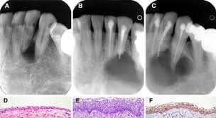
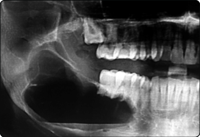
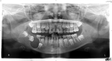
The unicystic ameloblastoma usually appears as a “cystic” lesion with either an intraluminal or an intramural proliferation of the cystic lining:

  • Radiographically, it may resemble a well-circumscribed slow-growing radiolucency.

1-s2.0-S1991790213000792-gr1

Unknown

Unknown-1

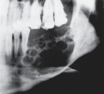
Multicystic ameloblastoma can infiltrate into the adjacent tissue and has the ability to recur and even metastasize:

  • Its prevalence is a slightly older age group than the unicystic ameloblastoma.

  • Radiographically, the appearance is generally unilocular or multilocular.

Fig-4-Solid-recurrent-multicystic-ameloblastoma-with-erosion-of-the-buccal-lingual079

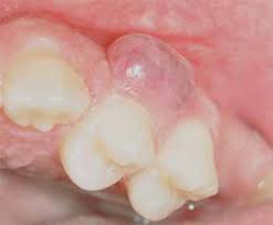
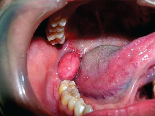
Peripheral ameloblastoma mostly appears in the alveolar mucosa.

  • It is a soft-tissue version of an ameloblastoma but can also involve the underlying bone.

UnknownNatlJMaxillofacSurg_2013_4_2_245_127662_f2

The malignant ameloblastoma is a rare entity.

  • It is defined as an ameloblastoma that has already metastasized but still maintains its classical microscopic features.

A histological classification subdivides ameloblastoma into:

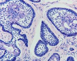
  • Follicular ameloblastoma

Unknown

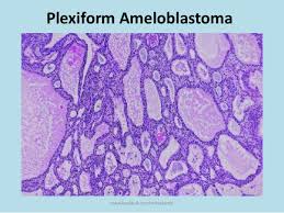
  • Plexiform ameloblastoma

Unknown-1

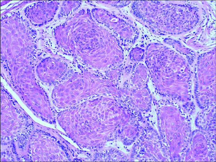
  • Acanthomatous ameloblastoma

acanthomatous-ameloblastoma-There-central-cells-show-more-mature-squamous

  • Granular ameloblastoma

JPharmBioallSci_2015_7_5_276_155955_u2

In most cases the tumor is asymptomatic, presenting as an incidental finding on orthopantomography.

  • The most common symptoms are facial swelling, pain, malocclusion, loosening of teeth, ill-fitting dentures, periodontal diseases or ulceration, oroantral fistulas and nasal airway obstruction.

Recurrence rates of ameloblastoma are reportedly as high as 15% to 25% after radical treatment and 75% to 90% after conservative treatment.

  • Therefore, wide resection of the jaw in accordance with the treatment of malignant tumors is usually recommended for ameloblastomas.

Rodrigo Arrangoiz MS, MD, FACS a head and neck surgeon and is amember of Sociedad Quirúrgica S.C at the America British Cowdray Medical Center.

He is first author on some publications on oral cavity cancer:

Training:

• General surgery:

• Michigan State University:

• 2004 al 2010

• Surgical Oncology / Head and Neck Surgery / Endocrine Surgery:

• Fox Chase Cancer Center (Filadelfia):

• 2010 al 2012

• Masters in Science (Clinical research for health professionals):

• Drexel University (Filadelfia):

• 2010 al 2012

• Surgical Oncology / Head and Neck Surgery / Endocrine Surgery:

• IFHNOS / Memorial Sloan Kettering Cancer Center:

• 2014 al 2016

Oral Submucous Fibrosis (OSF)

  • In 1952, Schwartz coined the term atrophica idiopathica mucosa oris to describe an oral fibrosing disease he discovered in 5 Indian women from Kenya.

    • Joshi subsequently coined the termed oral submucous fibrosis (OSF) for the condition in 1953.

  • Oral submucous fibrosis is a chronic debilitating disease of the oral cavity characterized by inflammation and progressive fibrosis of the submucosal tissues (lamina propria and deeper connective tissues):

    • Oral submucous fibrosis results in marked rigidity and an eventual inability to open the mouth.

    • The buccal mucosa is the most commonly involved site, but any part of the oral cavity can be involved, even the pharynx

  • Oral submucous fibrosis (OSF) is a premalignant condition caused by betel chewing:

    • It is very common in Southeast Asia but has started to spread to Europe and North America.

    • The condition is well recognized for its malignant potential and is particularly associated with areca nut chewing, the main component of betel quid.

    • Betel quid chewing is a habit practiced predominately in Southeast Asia and India that dates back for thousands of years.

      • It is similar to tobacco chewing in westernized societies.

    • The mixture of this quid, or chew, is a combination of the areca nut (fruit of the Areca catechu palm tree, erroneously termed betel nut) and betel leaf (from the Piper betel, a pepper shrub), tobacco, slaked lime (calcium hydroxide), and catechu (extract of the Acacia catechu tree).

      • Lime acts to keep the active ingredient in its freebase or alkaline form, enabling it to enter the bloodstream via sublingual absorption.

      • Arecoline, an alkaloid found in the areca nut, promotes salivation, stains saliva red, and is a stimulant:

        • Major constituents of betel quid are arecoline from betel nuts and copper, which are responsible for fibroblast dysfunction and fibrosis.

Unknown

Unknown-1

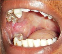
Oral-Submucous-Fibrosis-Picture

  •  OSF can lead to squamous cell carcinoma, a risk that is further increased by concomitant tobacco consumption.

  • OSF is a diagnosis based on clinical symptoms and confirmation by histopathology.

    • Hypovascularity leading to blanching of the oral mucosa, staining of teeth and gingiva, and trismus are major symptoms:

      • Symptoms of oral submucous fibrosis (OSF) include the following:

        • Progressive inability to open the mouth (trismus) due to oral fibrosis and scarring

          • Oral pain and a burning sensation upon consumption of spicy foodstuffs

            • Increased salivation

            • Change of gustatory sensation

            • Hearing loss due to stenosis of the eustachian tubes

            • Dryness of the mouth

            • Nasal tonality to the voice

            • Dysphagia to solids (if the esophagus is involved)

            • Impaired mouth movements (eg, eating, whistling, blowing, sucking)


  • Oral submucous fibrosis is clinically divided into three stages, and the physical findings vary according:
    • Stage 1:
      • Stomatitis includes erythematous mucosa, vesicles, mucosal ulcers, melanotic mucosal pigmentation, and mucosal petechia.


  • Stage 2:
    • Fibrosis occurs in ruptured vesicles and ulcers when they heal, which is the hallmark of this stage.
      • Early lesions demonstrate blanching of the oral mucosa.
      • Older lesions include vertical and circular palpable fibrous bands in the buccal mucosa and around the mouth opening or lips, resulting in a mottled, marblelike appearance of the mucosa because of the vertical, thick, fibrous bands running in a blanching mucosa.
      • Specific findings include the following:
        • Reduction of the mouth opening (trismus)
        • Stiff and small tongue
        • Blanched and leathery floor of the mouth 
        • Fibrotic and depigmented gingiva
        • Rubbery soft palate with decreased mobility
        • Blanched and atrophic tonsils
        • Shrunken budlike uvula
        • Sinking of the cheeks, not commensurate with age or nutritional status


  • Stage 3:
    • Leukoplakia is precancerous and is found in more than 25% of individuals with oral submucous fibrosis.
    • Speech and hearing deficits may occur because of involvement of the tongue and the eustachian tubes.

Rodrigo Arrangoiz MS, MD, FACS a head and neck surgeon and is amember of Sociedad Quirúrgica S.C at the America British Cowdray Medical Center.He is first author on some publications on oral cavity cancer:

Training:

• General surgery:

• Michigan State University:

• 2004 al 2010• Surgical Oncology / Head and Neck Surgery / Endocrine Surgery:

• Fox Chase Cancer Center (Filadelfia):

• 2010 al 2012• Masters in Science (Clinical research for health professionals):

• Drexel University (Filadelfia):

• 2010 al 2012
• Surgical Oncology / Head and Neck Surgery / Endocrine Surgery:

• IFHNOS / Memorial Sloan Kettering Cancer Center:

• 2014 al 2016

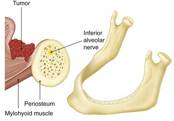
Pathway by which oral cancers invade the mandible

To determine the need and extent of mandible resection, it is essential to understand the pathway by which oral cancers invade the mandible.

  • Primary carcinomas of the lip, buccal mucosa, tongue, and floor of the mouth extend along the surface mucosa and the submucosal soft tissues to approach the attached labial, buccal, or lingual gingiva.

  • From this point, the tumor does not extend directly through intact periosteum and cortical bone toward the cancellous part of the mandible because the periosteum acts as a significant protective barrier.

  • Instead, the tumor advances from the attached gingiva toward the alveolus.

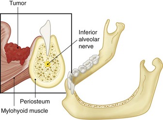
    • In patients with teeth, the tumor extends through the dental socket into the cancellous part of the bone and invades the mandible in that fashion (Figure).

Pathway by which oral cancers invade the mandible
Tumor invasion of the dentate mandible occurs through the dental socket to the cancellous bone and then to the alveolar canal.

  • In edentulous patients, the tumor extends up to the alveolar process and then infiltrates the dental pores in the alveolar ridge and extends to the cancellous part of the mandible (Figure)

Tumor invasion of the edentulous mandible occurs through the dental pores on the alveolar process to the cancellous bone and then to the alveolar canal.


  • Thus even in patients with early invasion of the mandible, a marginal mandibulectomy is feasible because the cortical part of the mandible inferior to the roots of the teeth remains uninvolved and can be safely spared.

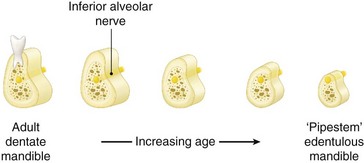
  • In edentulous patients, however, the feasibility of marginal mandibulectomy depends on the vertical height of the body of the mandible.

    • With aging, the alveolar process recedes and the mandibular canal comes closer to the surface of the alveolar process.

      • As shown in the Figure, the resorption of the alveolar process eventually leads to a “pipestem” mandible in elderly patients.

Vertical height and location of the alveolar canal in dentate and edentulous mandibles.

  • The ability to perform a satisfactory marginal mandibulectomy in such patients is almost impossible because the probability of iatrogenic fracture or postsurgical spontaneous fracture of the remaining portion of the mandible is very high.

  • Similarly, in patients who have received previous radiotherapy, a marginal mandibulectomy should be performed with extreme caution.

    • The probability of pathological fracture at the site of the marginal mandibulectomy in such patients is very high.

  • When the tumor extends to involve the cancellous part of the mandible, a segmental mandibulectomy must be performed.

  • A segmental mandibulectomy also may be required in patients with massive primary tumors with significant soft tissue disease in the proximity of the mandible. 

Rodrigo Arrangoiz MS, MD, FACS a head and neck surgeon and is amember of Sociedad Quirúrgica S.C at the America British Cowdray Medical Center.He is first author on some publications on oral cavity cancer:

Training:

• General surgery:

• Michigan State University:

• 2004 al 2010• Surgical Oncology / Head and Neck Surgery / Endocrine Surgery:

• Fox Chase Cancer Center (Filadelfia):

• 2010 al 2012• Masters in Science (Clinical research for health professionals):

• Drexel University (Filadelfia):

• 2010 al 2012
• Surgical Oncology / Head and Neck Surgery / Endocrine Surgery:

• IFHNOS / Memorial Sloan Kettering Cancer Center:

• 2014 al 2016