Blog

Management of Clinical N1 Disease After Neoadjuvant Chemotherapy in Patients with Breast Cancer

cpg170019fa

Following neoadjuvant chemotherapy for breast cancer:

  • Pathologic complete response (pCR) in the axillary nodes:
    • Is found in 40% of patients presenting with biopsy proven node-positive disease.
      • This observation led to several clinical trials including:
        • American College of Surgeons Oncology Group (ACOSOG Z1071)
        • SENTINA (Arm C)
        • SN FNAC
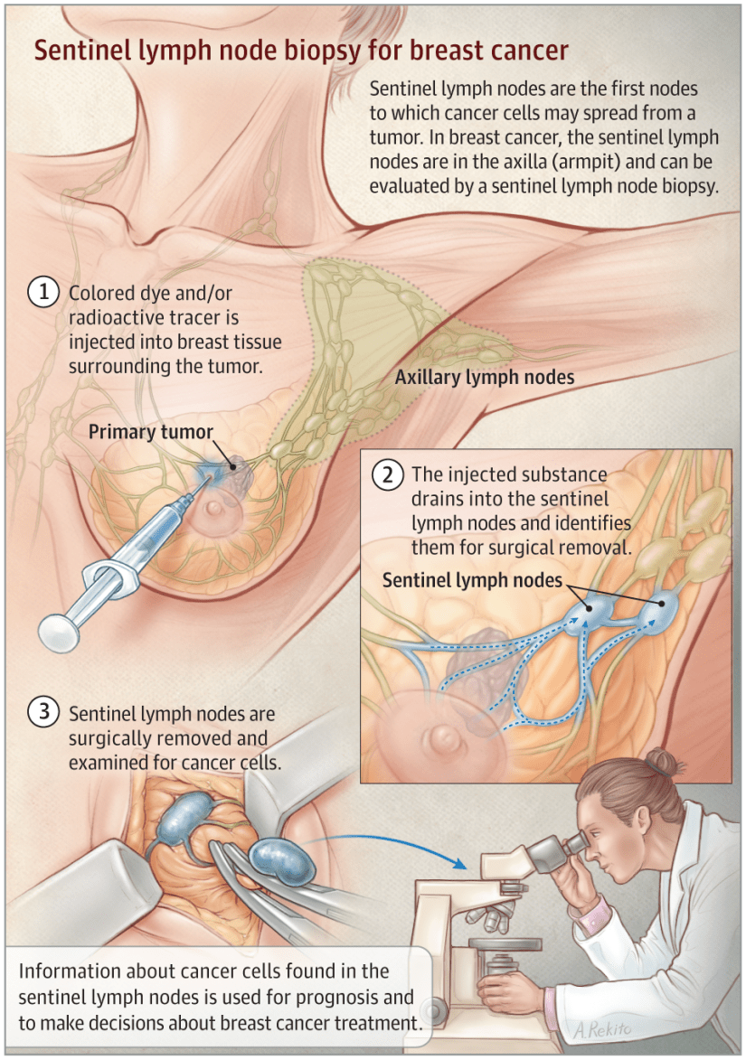
          • This trials evaluated the feasibility and accuracy of sentinel lymph node mapping (SLNM) and sentinel lymph node biopsy (SLNB) dissection as axillary staging after neoadjuvant chemotherapy in clinical N1 disease.
          • Although the false-negative rate of SLNB in this setting was above the predetermined threshold, several important factors associated with and impacting the false negative rate were described.
            • This included:
              • Use of dual agent mapping technique
              • Evaluation of at least 3 SLNs
              • Immunohistochemistry (IHC) evaluation in addition to H&E staining
              • Excision of the clipped (biopsy proven) node.
  • The ACOSOG Z1071 trial evaluated the false-negative rate (FNR) in patients with clinical N1 disease in whom at least 2 SLNs were identified:
    • This FNR was 12.6%.
    • The FNR was improved to 10.8%:
      • In patients in whom both radioisotope and blue dye were used.
    • In addition, the FNR in the Z1071 trial varied based on the number of SLNs identified:
      • For patients with a single SLN identified,:
        • The FNR was 31.5%
      • For those with 2 SLNs identified:
        • The FNR was 21.1%
      • For those with 3 or more SLNs identified:
        • The FNR was 9.1%
  • Data from the SENTINA trial also showed an improved FNR in patients in whom 3 or more SLNs were identified:
    • The trial was a 4-arm multicenter study.
    • Arm C was similar to the ACOSOG Z1071 trial in that it included patients who were clinically node-positive and converted to clinically node-negative (non-palpable) after chemotherapy:
      • The FNRs were 24.3% when a single SLN was identified
      • 18.5% when 2 SLNs were removed
      • Less than 10% when 3 or more SLNs were removed
    • Similar to the Z1071 trial:
      • The FNR was lower (8.6%) for patients who underwent mapping with radioisotope and blue dye.
  • The more recently reported SN FNAC study required the use of immunohistochemistry and considered SLN metastases of any size, including isolated tumor cells, to be positive:
    • Using this definition,:
      • The FNR was 8.4%.
    • If isolated tumor cells identified by immunohistochemistry had been considered negative:
      • The FNR would have increased to 13.3%.
  • The results of these findings led to refined technique termed targeted axillary dissection (TAD):
    • This procedure involves SLNB dissection and selective targeted excision of the clipped (biopsy proven) axillary node.
    • Response is documented during chemotherapy with ultrasound and clinical exam.
    • In selected patients planned for TAD, the clipped node is preoperatively localized with an I-125 radioactive seed or wire.
    • Intraoperatively, dual lymphatic mapping for SLN identification is employed and targeted excision of the clipped node is performed.
    • Excision is confirmed with specimen radiograph.
    • In approximately 23% of cases the clipped node will not be a sentinel node.
    • Compared with ALND:
      • The false-negative rate of this approach was:
        • 10.1% with sentinel node biopsy alone
        • 4.2% for excision of the clipped node alone
        • 2.0% for TAD.
    • In carefully selected patients TAD alone may offer an accurate approach to axillary staging and assessment of residual disease with limited morbidity.

Rodrigo Arrangoiz MS, MD, FACS a surgical oncologist and is a member of Sociedad Quirúrgica S.C at the America British Cowdray Medical Center in Mexico City:

  • He is an expert in the management of breast cancer.

    • If you have any questions about SLNM and SLNB after neoadjuvant chemotherapy in breast cancer please fill free to contact Dr. Arrangoiz.

Training:

• General surgery:

• Michigan State University:

• 2004 al 2010

• Surgical Oncology / Head and Neck Surgery / Endocrine Surgery:

• Fox Chase Cancer Center (Filadelfia):

• 2010 al 2012

• Masters in Science (Clinical research for health professionals):

• Drexel University (Filadelfia):

• 2010 al 2012

• Surgical Oncology / Head and Neck Surgery / Endocrine Surgery:

• IFHNOS / Memorial Sloan Kettering Cancer Center:

• 2014 al 2016

#Arrangoiz

#Surgeon

#Cirujano

#SurgicalOncologist

#CirujanoOncologo

#BreastSurgeon

#CirujanodeMama

#CancerSurgeon

#CirujanodeCancer

http://www.sociedadquirurigca.com
 

img_0832-2img_0833-2img_0834-2img_0835-2img_0836-2img_0837-2

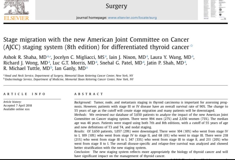
Thyroid Cancer

img_1389

#Arrangoiz

#ThyroidExpert

#ThyroidSurgeon

#CancerSurgeon

#HeadandNeckSurgeon

#SurgicalOncologist

#CirugiadeTumoresdeCabezayCuello

#CirugiaTiroides

#ExpertoenTiroides

#CirujanodeCancer

#CirujanoOncologo

http://www.sociedadquirurgica.com

http://www.cirugiatiroides.com

Acinic Cell Carcinoma (AcCC)

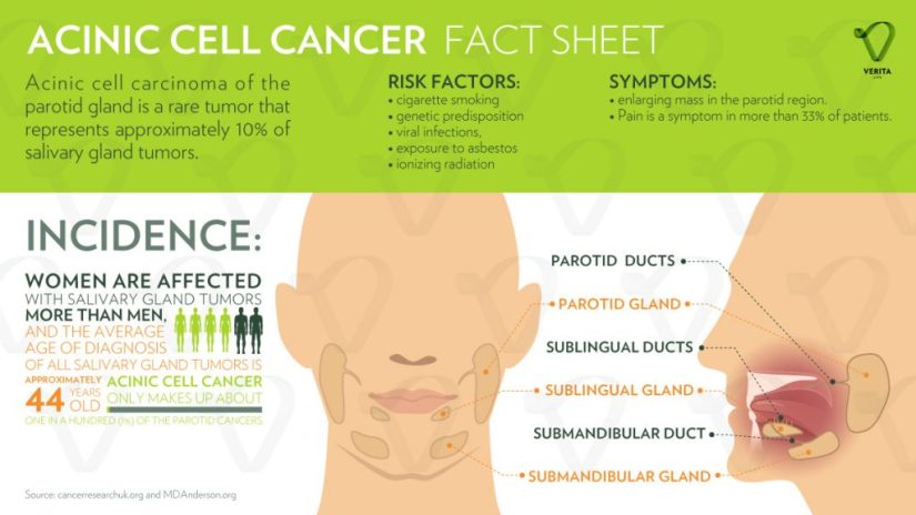
acinic-cell-cancer-infographic-1024x576.jpg

  • Acinic cell carcinoma (AcCC) is the second most common malignant tumor involving the parotid gland:

    • Representing 15% of malignant parotid gland neoplasms:

      • It accounts for approximately:

        • 5% to 17% of all salivary gland tumors

        • 10% to 17% of all malignant salivary gland tumors

  • The parotid gland is composed almost exclusively of serous type acini:

    • It is the most common site of AcCC:

      • 80% to 90% of cases

    • Other reported sites are:

      • The palate:

        • Up to 15% of the cases

      • Submandibular gland:

        • 4% of the cases

      • Sublingual gland:

        • 1% of the cases

  • AcCC occurs most often in:

    • The fifth decade of life

    • In women more often than in men (ratio 3:2)

  • It is generally a low-grade carcinoma with indolent behavior. 

  • Recurrences are usually delayed, sometimes after decades:

    • In a series reported by Ellis et al:

      • 12% of tumors recurred

      • 8% metastasized:

        • Regional lymph nodes, bone, lung, and brain:

          • The regional lymph nodes were the most common site of metastases:

            • Spafford et al. reported cervical lymphadenopathy to be present in 10% to 15% of cases of AcCC and Spiro et al. in a series of 67 cases with AcCC treated before 1968, identified five cases (7.5%) with cervical metastases at the time of initial treatment.

  • It is interesting that this tumor is the most common bilateral malignant salivary gland neoplasm:

    • Although its bilateral presentation is not nearly as common as the bilateral presentations of benign tumors:

      • Warthin’s tumor and pleomorphic adenoma

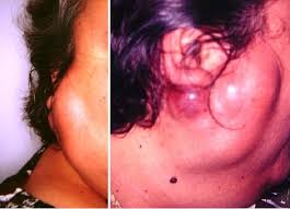
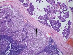
  • AcCC are typically encased in a fibrous capsule:

    • Grossly resembling round circumscribed nodules with a tan surface:

      • The cut surface is solid but may show cystic degeneration and hemorrhage

images

  • Histologically, there are five cell types:

    • Serous acinar cells (explaining the predilection for the parotid gland)

    • Cells with clear cytoplasm

    • Intercalated ductal cell

    • Nonspecific glandular cell

    • Vacuolated cell

i1543-2165-124-12-1835-f01.jpgUnknownUnknown-1

  • The microscopic recognition of AcCC also requires a strong appreciation for its varied growth pattern:

    • There are four histologic growth patterns:

      • Solid

      • Microcystic

      • Papillary

      • Follicular

    • Caution must be taken not to misread:

      • The solid pattern as normal parotid parenchyma

      • The papillary-cystic pattern as cystic mucoepidermoid carcinoma

      • The follicular pattern as metastatic thyroid carcinoma

    • Serous acinar differentiation is developed most fully in the acinic cell.

    • These cells have dark round nuclei and granular purplish cytoplasm.

  • The diagnosis of AcCC may be difficult to establish, especially when some other cell type dominates the histopathology picture:

    • Some examples of this are the predominance of clear cells might cause confusion with mucoepidermoid carcinoma, clear cell adenocarcinoma, and metastatic renal cell carcinoma:

      • In these circumstances, the diagnostic acinic cells can be identified using a periodic acid-Schiff(PAS) reagent (their cytoplasmic secretory granules are PAS positive and diastase resistant).

  • Overall survival has been crudely estimated to be about 84%:

    • Survival at 5 years has been reported between 76%  to 90%, but fell to 56% at 20 years:

      • Emphasizing the need for long-term follow-up

Rodrigo Arrangoiz MS, MD, FACS a head and neck surgeon / surgical oncologist and is a member of Sociedad Quirúrgica S.C at the America British CowdrayMedical Center in Mexico City:

  • He is an expert in the management of salivary gland neoplasms:

    • If you have any questions about salivary gland neoplasms  please fill free to ask Dr. Arrangoiz

Training:

• General surgery:

• Michigan State University:

• 2004 al 2010

• Surgical Oncology / Head and Neck Surgery / Endocrine Surgery:

• Fox Chase Cancer Center (Filadelfia):

• 2010 al 2012

• Masters in Science (Clinical research for health professionals):

• Drexel University (Filadelfia):

• 2010 al 2012

• Surgical Oncology / Head and Neck Surgery / Endocrine Surgery:

• IFHNOS / Memorial Sloan Kettering Cancer Center:

• 2014 al 2016

#Arrangoiz

#HeadandNeckSurgeon

#SurgicalOncologist

#Surgeon

#CancerSurgeon

#CirujanodeCabezayCuello

#CirujanoOncologo

http://www.sociedadquirurgica.com

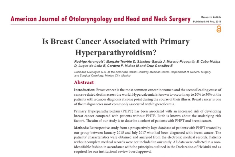
Is Breast Cáncer Associated with a primary Hyperparathyroidism?

Artículo publicado por el equipo de Sociedad Quirúrgica SC

Autor principal Rodrigo ARRANGOIZ MS, MD, FACS

remedypublications.com/american-journal-of-otolaryngology-and-head-and-neck-surgery/articles/pdfs_folder/ajohns-v2-id1033.pdf

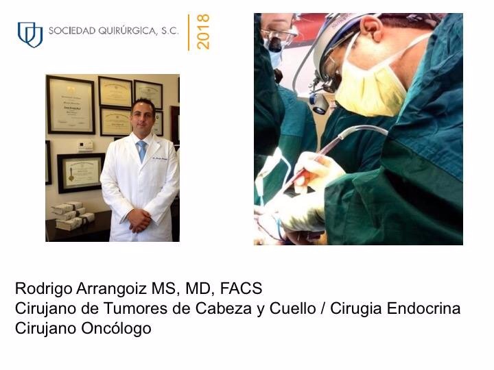
TOPDOCTORS Award a Rodrigo Arrangoiz MS, MD, FACS

Rodrigo Arrangoiz MS, MD, FACS miembro de Sociedad Quirúrgica S.C. fue reconocido por #TOPDOCTORS como médico de excelencia en el año 2018:


Presentation1


#Arrangoiz

#ThyroidExpert

#ThyroidSurgeon

#CancerSurgeon

#HeadandNeckSurgeon

#SurgicalOncologist

#CirugiadeTumoresdeCabezayCuello

#CirugiaTiroides

#ExpertoenTiroides

#CirujanodeCancer

#CirujanoOncologo


Hobnail Variant of Papillary Thyroid Carcinoma

👉Trabajo de Sociedad Quirurgica SC, investigador principal Rodrigo Arrangoiz MS, MD, FACS fue presentado en el Miami Thyroid Oncology Symposium Febrero 15 al 16 del 2019.

👉Hobnail Variant of Papillary Thyroid Carcinoma

👉El presentador fue Raúl Moran MD residente de cirugía del hospital ABC.

https://docs.google.com/presentation/d/1j5ZWoGK251CMV44CkzbokQtNc-weNmCVS7WocsDiZDE/edit?usp=sharing


20d943b1-6f20-49c3-9754-2f3756051c9312fc1993-1232-40ac-91a0-f394c7a8de2e2131cd57-aa86-4187-801d-bb4d2ab2d6ef5b081828-c940-488d-bd45-21f33bd5f84f

Rodrigo Arrangoiz MS, MD, FACS cirujano de tumores de cabeza y cuello / cirugía endocrina / cirugía oncológica miembro de Sociedad Quirúrgica S.C. experto en el manejo del cáncer de tiroides:

  • Cumple con los requisitos determinados por el Dr. Ashok Saha para realizar cirugía de tiroides de manera efectiva y segura:

Rodrigo Arrangoiz MS, MD, FACS es miembro de la American Thyroid Association:

2019 membership certificate arrangoiz, rodrigo

Entrenamiento:

  • Cirugia general y gastrointestinal:

• Michigan State University:

• 2004 al 2010image-48

• Cirugia oncológica / tumores de cabeza y cuello / cirugia endocrina:

• Fox Chase Cancer Center (Filadelfia):

• 2010 al 2012

image-39

• Maestria en ciencias (Clinical research for healthprofessionals):

• Drexel University (Filadelfia):

• 2010 al 2012

image-50

• Cirugia de tumores de cabeza y cuello / cirugiaendocrina

• IFHNOS / Memorial Sloan Kettering Cancer Center:

• 2014 al 2016

image-51

http://www.sociedadquirurgica.com

http://www.hiperparatiroidismo.info

http://www.cirugiatiroides.com


#Arrangoiz

#ThyroidExpert

#ThyroidSurgeon

#CancerSurgeon

#HeadandNeckSurgeon

#SurgicalOncologist

#CirugiadeTumoresdeCabezayCuello

#CirugiaTiroides

#ExpertoenTiroides

#CirujanodeCancer

#CirujanoOncologo 


Pathway by Which Oral Cancers Invade the Mandible

To determine the need and extent of mandible resection, it is essential to understand the pathway by which oral cancers invade the mandible .

  • Primary carcinomas of the lip, buccal mucosa, tongue, and floor of the mouth extend along the surface mucosa and the submucosal soft tissues to approach the attached labial, buccal, or lingual gingiva.

  • From this point, the tumor does not extend directly through intact periosteum and cortical bone toward the cancellous part of the mandible because the periosteum acts as a significant protective barrier .

  • Instead, the tumor advances from the attached gingiva toward the alveolus.

    • In patients with teeth, the tumor extends through the dental socket into the cancellous part of the bone and invades the mandible in that fashion (Figure).

Pathway by which oral cancers invade the mandible
Tumor invasion of the dentate mandible occurs through the dental socket to the cancellous bone and then to the alveolar canal.

In edentulous patients, the tumor extends up to the alveolar process and then infiltrates the dental pores in the alveolar ridge and extends to the cancellous part of the mandible (Figure)

Tumor invasion of the edentulous mandible occurs through the dental pores on the alveolar process to the cancellous bone and then to the alveolar canal.

  • Thus even in patients with early invasion of the mandible, a marginal mandibulectomy is feasible because the cortical part of the mandible inferior to the roots of the teeth remains uninvolved and can be safely spared.

  • In edentulous patients, however, the feasibility of marginal mandibulectomy depends on the vertical height of the body of the mandible .

    • With aging, the alveolar process recedes and the mandibular canal comes closer to the surface of the alveolar process.

      • As shown in the Figure, the resorption of the alveolar process eventually leads to a “pipestem” mandible in elderly patients.

Vertical height and location of the alveolar canal in dentate and edentulous mandibles.

The ability to perform a satisfactory marginal mandibulectomy in such patients is almost impossible because the probability of iatrogenic fracture or postsurgical spontaneous fracture of the remaining portion of the mandible is very high.

  • Similarly, in patients who have received previous radiotherapy, a marginal mandibulectomy should be performed with extreme caution.

    • The probability of pathological fracture at the site of the marginal mandibulectomy in such patients is very high.

When the tumor extends to involve the cancellous part of the mandible, a segmental mandibulectomy must be performed.

  • A segmental mandibulectomy also may be required in patients with massive primary tumors with significant soft tissue disease in the proximity of the mandible. 

20d943b1-6f20-49c3-9754-2f3756051c93

Rodrigo Arrangoiz MS, MD, FACS a head and neck surgeon and a member of Sociedad Quirúrgica S.C at the America British Cowdray Medical Center.

He is first author on some publications on oral cavity cancer:

Training:

• General surgery:

• Michigan State University:

• 2004 al 2010

image-38

• Surgical Oncology / Head and Neck Surgery / Endocrine Surgery:

• Fox Chase Cancer Center (Filadelfia):

• 2010 al 2012

image-49

• Masters in Science (Clinical research for health professionals):

• Drexel University (Filadelfia):

• 2010 al 2012

image-50

• Surgical Oncology / Head and Neck Surgery / Endocrine Surgery:

• IFHNOS / Memorial Sloan Kettering Cancer Center:

• 2014 al 2016

image-51

Parotidectomia Realizada por Rodrigo Arrangoiz MS, MD, FACS


Rodrigo Arrangoiz MS, MD, FACS a head and neck surgeon / surgical oncologist and is a member of Sociedad Quirúrgica S.C at the America British CowdrayMedical Center in Mexico City:

  • He is an expert in the management of salivary gland neoplasms:

    • If you have any questions about salivary gland neoplasms  please fill free to ask Dr. Arrangoiz

Training:

• General surgery:

• Michigan State University:

• 2004 al 2010

• Surgical Oncology / Head and Neck Surgery / Endocrine Surgery:

• Fox Chase Cancer Center (Filadelfia):

• 2010 al 2012

• Masters in Science (Clinical research for health professionals):

• Drexel University (Filadelfia):

• 2010 al 2012

• Surgical Oncology / Head and Neck Surgery / Endocrine Surgery:

• IFHNOS / Memorial Sloan Kettering Cancer Center:

• 2014 al 2016

#Arrangoiz

#HeadandNeckSurgeon

#SurgicalOncologist

#Surgeon

#CancerSurgeon

#CirujanodeCabezayCuello

#CirujanoOncologo

http://www.sociedadquirurgica.com

Facial Nerve Anatomy for Surgeons

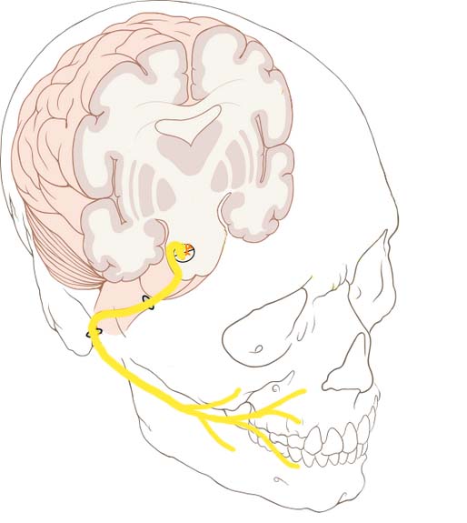
Overview-of-Anatomical-Course-of-the-Facial-Nerve.jpg

  • The facial nerve, (CN VII), is the seventh paired cranial nerve. 
    • The facial nerve is associated with the derivatives of the second pharyngeal arch.
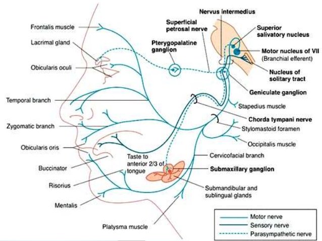
    • Motor: 
      • Innervates the muscles of facial expression, the posterior belly of the digastric, the stylohyoid and the stapedius muscles.
    • Sensory:
      • A small area around the concha of the auricle.
    • Special Sensory:
      • Provides special taste sensation to the anterior 2/3 of the tongue.
    • Parasympathetic:
      • Supplies many of the glands of the head and neck, including:
        • Submandibular and sublingual salivary glands.
        • Nasal, palatine and pharyngeal mucous glands.
        • Lacrimal glands.

images

  • Anatomical Course:
    • The course of the facial nerve is very complex:
      • There are many branches, which transmit a combination of sensory, motor and parasympathetic fibres.
    • Anatomically, the course of the facial nerve can be divided into two parts:
      • Intracranial:
        • The course of the facial nerve through the cranial cavity, and the cranium itself.
      • Extracranial:
        • The course of the facial nerve outside the cranium, through the face and neck.

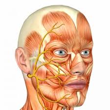
Schematic-of-the-Course-and-Branches-of-the-Facial-Nerve.jpg

  • Intracranial portion:
    • The nerve arises in the pons, an area of the brainstem. 
    • It begins as two roots:
      • A large motor root 
      • Small sensory root:
        • The part of the facial nerve that arises from the sensory root is sometimes known as the intermediate nerve).
    • The two roots travel through the internal acoustic meatus:
      • A 1 cm long opening in the petrous part of the temporal bone. 
        • Here, they are in very close proximity to the inner ear.
    • Still within the temporal bone:
      • The roots leave the internal acoustic meatus, and enter into the facial canal:
        • The facial canal is a ‘Z’ shaped structure.
        • Within the facial canal, three important events occur:
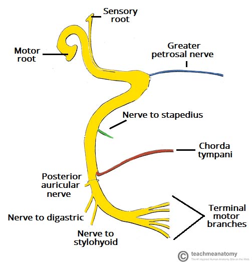
          • Firstly the two roots fuse to form the facial nerve.-
          • Next, the nerve forms the geniculate ganglion:
            • A ganglion is a collection of nerve cell bodies
          • Lastly, the nerve gives rise to:
            • Greater petrosal nerve:
              • Parasympathetic fibers:
                • To mucous glands of the head and neck and lacrimal gland.
            • Nerve to stapedius muscle:
              • Motor fibres to stapedius muscle of the middle ear.
            • Chorda tympani:
              • Special sensory fibers to the anterior 2/3 tongue
              • Parasympathetic fibers to the submandibular and sublingual glands.
    • The facial nerve then exits the facial canal (and the cranium) via the stylomastoid foramen (in a lateral position):
      • This is an exit located just posterior to the styloid process of the temporal bone.
  • Extracranial portion of the facial nerve:
    • After exiting the skull:
      • The facial nerve turns superiorly to run just anterior to the outer ear.
    • The first extracranial branch to arise is the posterior auricular nerve:
      • It provides motor innervation to the some of the muscles around the ear.
    • Immediately distal to this, motor branches are sent to the posterior belly of the digastric muscle and to the stylohyoid muscle.
    • The main trunk of the nerve:
      • Now termed the motor root of the facial nerve:
        • Continues anteriorly and inferiorly into the parotid gland:
          • The facial nerve does not contribute towards the innervation of the parotid gland:
            • Which is innervated by the glossopharyngeal nerve).
        • Within the parotid gland, the nerve terminates by splitting into five branches:
          • Temporal branch
          • Zygomatic branch
          • Buccal branch
          • Marginal mandibular branch
          • Cervical branch
        • These branches are responsible for innervating the muscles of facial expression.
  • Motor Functions:
    • Branches of the facial nerve are responsible for innervating many of the muscles of the head and neck.
    • All these muscles are derivatives of the second pharyngeal arch.
    • The first motor branch arises within the facial canal:
      • The nerve to stapedius muscle:
        • The nerve passes through the pyramidal eminence to supply the stapedius muscle in the middle ear.
      • Between the stylomastoid foramen, and the parotid gland, three more motor branches are given off:
        • Posterior auricular nerve:
          • Ascends in front of the mastoid process
          • Innervates the intrinsic and extrinsic muscles of the outer ear.
          • It also supplies the occipital part of the occipitofrontalis muscle.
        • Nerve to the posterior belly of the digastric muscle:
          • Innervates the posterior belly of the digastric muscle (a suprahyoid muscle of the neck):
            • It is responsible for raising the hyoid bone.
        • Nerve to the stylohyoid muscle:
          • Innervates the stylohyoid muscle (a suprahyoid muscle of the neck):
            • It is responsible for raising the hyoid bone.
      • Within the parotid gland, the facial nerve terminates by bifurcating into five motor branches:
        • These innervate the muscles of facial expression:
          • Temporal branch:
            • Innervates the frontalis, orbicularis oculi and corrugator supercili
          • Zygomatic branch:
            • Innervates the orbicularis oculi.
          • Buccal branch:
            • Innervates the orbicularis oris, buccinator and zygomaticus muscles.
          • Marginal Mandibular branch:;
            • Innervates the mentalis muscle.
            • Innervates the depressor anguli oris and the depressor labii inferioris
          • Cervical branch:;
            • Innervates the platysma.

1478347175_the-facial-nerve.jpg

  • Special sensory functions:
    • The chorda tympani branch of the facial nerve is responsible for innervating the anterior 2/3 of the tongue with the special sense of taste:

      • The nerve arises in the facial canal
      • Travels across the bones of the middle ear
      • Exiting via the petrotympanic fissure, and entering the infratemporal fossa:
        • Here, the chorda tympani ‘hitchhikes’ with the lingual nerve:

          • The parasympathetic fibres of the chorda tympani stay with the lingual nerve:

            • But the main body of the nerve leaves to innervate the anterior 2/3 of the tongue.

  • Parasympathetic functions:

    • The parasympathetic fibres of the facial nerve are carried by the greater petrosal and chorda tympani branches:

      • Greater Petrosal Nerve:

        • The greater petrosal nerve arises immediately distal to the geniculate ganglion within the facial canal:

          • It then moves in anteromedial direction:

            • Exiting the temporal bone into the middle cranial fossa.

              • From here, its travels across (but not through) the foramen lacerum:

                • Combining with the deep petrosal nerve to form the nerve of the pterygoid canal:

                  • The nerve of pterygoid canal then passes through the pterygoid canal (Vidian canal) to enter the pterygopalatine fossa, and synapses with the pterygopalatine ganglion.

                    • Branches from this ganglion then go on to provide parasympathetic innervation to the mucous glands of the oral cavity, nose and pharynx, and the lacrimal gland.

  • Chorda Tympani:
    • The chorda tympani also carries some parasympathetic fibres:
      • These combine with the lingual nerve (a branch of the trigeminal nerve) in the infratemporal fossa and form the submandibular ganglion:
        • Branches from this ganglion travel to the submandibular and sublingual salivary glands.

20d943b1-6f20-49c3-9754-2f3756051c93.jpg

Rodrigo Arrangoiz MS, MD, FACS a head and neck surgeon / surgical oncologist and is a member of Sociedad Quirúrgica S.C at the America British Cowdray Medical Center in Mexico City:

  • He is an expert in the management of head and neck cancers.


Training:

• General surgery:

• Michigan State University:

• 2004 al 2010

• Surgical Oncology / Head and Neck Surgery / Endocrine Surgery:

• Fox Chase Cancer Center (Filadelfia):

• 2010 al 2012

• Masters in Science (Clinical research for health professionals):

• Drexel University (Filadelfia):

• 2010 al 2012

• Surgical Oncology / Head and Neck Surgery / Endocrine Surgery:

• IFHNOS / Memorial Sloan Kettering Cancer Center:

• 2014 al 2016

#Arrangoiz

#Surgeon

#Cirujano

#SurgicalOncologist

#CirujanoOncologo

#BreastSurgeon

#CirujanodeMama

#CancerSurgeon

#CirujanodeCancer

#HeadandNeckSurgeon

#CirujanodeCabezayCuello

http://www.sociedadquirurigca.com
¿Estás Cansado, Podrías Tener Hiperparatiroidismo Primario

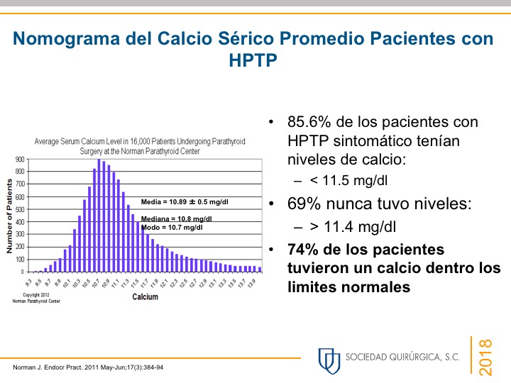
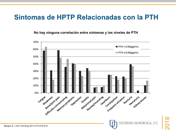
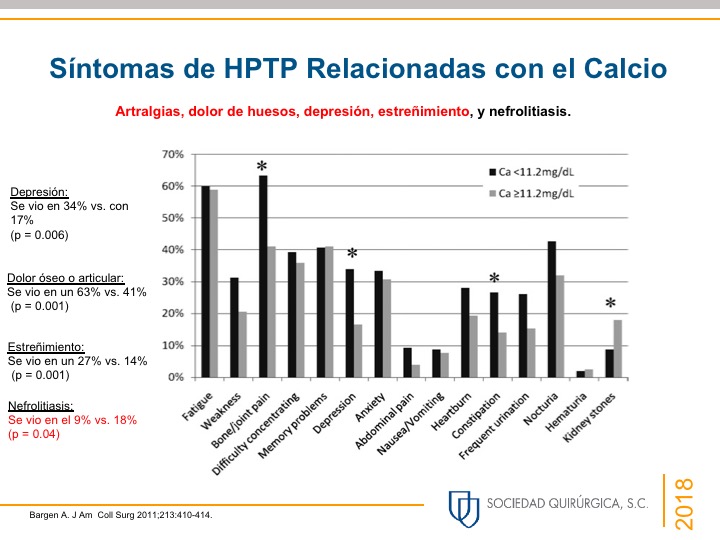
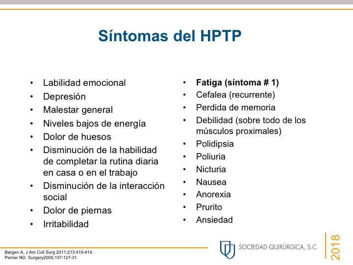
Slide2Slide2Slide2Slide1Slide2Slide1

20d943b1-6f20-49c3-9754-2f3756051c93

Rodrigo Arrangoiz MS, MD, FACS cirujano de tumores de cabeza y cuello / cirugía endocrina / cirugía oncológica miembro de Sociedad Quirúrgica S.C. experto en el manejo del hiperparatiroidismo:

  • Rodrigo Arrangoiz MS, MD, FACS es miembro de la American Thyroid Association:

2019 membership certificate arrangoiz, rodrigo

Publicaciones sobre el hiperparatiroidismo del miembro de Sociedad Quirúrgica Rodrigo Arrangoiz MS, MD, FACS experto en cirugía endocrina:


Rodrigo Arrangoiz MS, MD, FACS cirujano de tumores de cabeza y cuello / cirugía endocrina / cirugía pionero en México de la paratiroidectomia radioguiada mínimamente invasiva:


Entrenamiento:

  • Cirugia general y gastrointestinal:

• Michigan State University: 2004- 2010


image-48

• Cirugia oncológica / tumores de cabeza y cuello / cirugia endocrina:

• Fox Chase Cancer Center (Filadelfia):

• 2010 al 2012

image-39

• Maestria en ciencias (Clinical research for healthprofessionals):

• Drexel University (Filadelfia):

• 2010 al 2012

image-50

• Cirugia de tumores de cabeza y cuello / cirugiaendocrina

• IFHNOS / Memorial Sloan Kettering Cancer Center:

• 2014 al 2016

image-51

http://www.sociedadquirurgica.com
http://www.hiperparatiroidismo.info

http://www.cirugiatiroides.com
#Arrangoiz

#CirugiaEndocrina

#EndocrineSurgery

#HeadandNeckSurgeon

#CirujanodeTumoresdeCabezayCuello