Blog

Isolated Mediastinal Goiter (Substernal Goiter Type III)

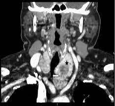
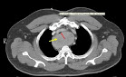
  • Although rare, thyroid glands within the mediastinum may exist without connection to the normal cervical orthotopic gland:
    • Such purely isolated mediastinal goiters represent only 0.2% to 3% of all goiters requiring surgical treatment
  • Such lesions are important to recognize because unlike all other types of substernal goiters:
    • Blood supply of the isolated mediastinal goiter may be through purely mediastinal arteries (including the aorta, subclavian, internal mammary, thyrocervical trunk, and innominate) and veins:
      • This is extremely important in planning their surgical resection
  • This entity is best termed isolated mediastinal goiter:
    • Other terms have been used, including aberrant mediastinal and ectopic mediastinal goiter
  • Three explanations exist for isolated mediastinal goiter:
    • Embryologic fragmentation of the thyroid anlagen with hyperdescent, likely associated with cardiac and great vessel descent, may explain some cases of isolated mediastinal goiter
    • Alternatively, isolated mediastinal goiter may form as an exophytic nodule, through progressive attenuation of the nodule-thyroid stalk
    • Finally, the isolated mediastinal goiter may form as a parasitic nodule representing a thyroid tissue fragment implant in the upper mediastinum from past goiter surgery:
      • I have seen such implants also within the peri-thyroid area and posterior to the upper cervical segment of the carotid artery

#Arrangoiz #ThyroidSurgeon #ThyroidExpert #HeadandNeckSurgeon #CancerSurgeon #HeadandNeckSurgeon #MultinodularGoiter #SubsternalGoiter #CASO #CenterforAdvancedSurgicalOncology

Substernal Goiter: Type II Posterior Mediastinal Goiters

  • Most surgical and radiographic series suggest that substernal goiters affect:
    • The anterior mediastinum in approximately 85% of patients:
      • Extension into the anterior mediastinum brings the thyroid:
        • Anterior to the subclavian and innominate vessels and anterior to the RLN
      • The relationship of the anterior mediastinal goiter to the RLN is as in the normal cervical gland:
        • That is, that the nerve is deep
    • The posterior mediastinum in approximately 15% of patients:
      • When substernal goiter expands to the posterior mediastinum:
        • It excavates the region posterior to the trachea, pushing the trachea anteriorly and splaying the great vessels anteriorly:
          • The thyroid then comes to rest in a space posterior to the innominate vein, carotid sheath contents, innominate and subclavian arteries, RLN, and inferior thyroid artery
      • Of importance, the relationship of the thyroid gland and the RLN is reversed as compared with the normal cervical orthotopic gland-RLN relationship:
        • The RLN is ventral to the inferior component of the thyroid and, if not recognized early on, can be stretched or cut by even the most meticulous thyroid surgeon
        • The nerve can also be entrapped between components of the posterior mediastinal goiter; even in these circumstances, a portion of the goiter will be deep to the RLN:
          • Such posterior mediastinal goiters can come to rest in a space bounded inferiorly by the azygous vein, posteriorly by the vertebral column, laterally by the first rib, medially by the trachea and esophagus, and anteriorly by the carotid sheath, subclavian and innominate vessels, superior vena cava, and phrenic and recurrent laryngeal nerves
  • Posterior mediastinal goiter (type IIA):
    • Can occur ipsilateral to the cervical thyroid gland of origin or may come to rest through retrotracheal extension in the contralateral thorax (substernal goiter type IIB)
  • Extension to the right thorax is more commonly seen as a result of:
    • Aortic arch and associated branch vessels obstructing the left posterior mediastinal descent pathway
  • Contralateral thoracic extension in the posterior mediastinum may occur either:
    • Behind the trachea and esophagus (IIB1) or between trachea and esophagus (IIB2)
  • Axial CT scanning and barium swallow help to determine this pattern
  • Generally the right chest caudal extension:
    • Is limited at the level of the azygous arch

#Arrangoiz #HeadandNeckSurgeon #ThyroidSurgeon #ThyroidExpert #MultinodularGoiter #SubsternalGoiter #CASO #CenterforAdvancedSurgicalOncology

Hipercalcemia Hipocalciurica Familiar

👉 Una de las causas de hiperparatiroidismo primario persistente puede ser la Hipercalcemia hipocalciúrica familiar (FHH) no reconocida causada por una mutación del gen CaSR, lo que lleva a niveles bajos de calcio en la orina de 24 horas.

👉Más información: https://www.ncbi.nlm.nih.gov/pubmed/30449544

👉CheckYourCalcium (realízate un calcio total en sangre).

👉Para más información: http://www.hiperparatiroidismo.info

# CheckYourCalcium #Arrangoiz #ParathyroidExpert #ParathyroidSurgeon #Hiperparatiroidism #Hipercalcemia #CheckYourCalcium #HeadandNeckSurgeon #SociedadQuirurgica

Parathyroidectomy

👉Operative cure rates of 95% to 98% with complication rates of 1% to 2% are possible when parathyroidectomy is performed by experienced surgeons.” – Dr. Rodrigo Arrangoiz

👉Para más información: http://www.hiperparatiroidismo.info

#Arrangoiz #ParathyroidExpert #ParathyroidSurgeon #Hiperparatiroidism #Hipercalcemia #HeadandNeckSurgeon #SociedadQuirurgica #Hiperparatiroidismo #ExpertoenParatiroides #CirujanodeParatiroides

Definition of Goiter

  • Both greatest diameter and goiter weight have been used to define thyroid enlargement:
    • In studies, methods for determining goiter size range from:
      • Physical examination measured in centimeters, to physical examination estimated in grams, to surgical specimen measured in centimeters or grams
      • Preoperative imaging diameters may also be used
  • The definition of goiter varies substantially among reports:
    • McHenry 80 g as the threshold value
    • Russell 100 g as the threshold value
    • Clark 200 g as the threshold value
  • Studies investigating radioiodine treatment for multinodular goiter:
    • Often define significant goiter as:
      • Greater than 100 g
  • Hegedus, Nygaard, and Hansen found that goiter surgical specimens:
    • Averaged:
      • 30 g for unilateral resection
      • 64 g for bilateral resection
  • Katlic, Grillo, and Wang reported that:
    • The average weight of substernal goiter was:
      • 104 g (range 25 to 357 g)
      • Greatest diameter averaging 9 cm (range 5 to 19 cm)
  • In a series of more than 200 cervical and substernal goiters treated at Massachusetts Eye and Ear Infirmary and Massachusetts General Hospital:
    • The mean weight was 143 g
    • The mean goiter size was 10.5 cm
  • The World Health Organization (WHO) 1960 grading system for clinical assessment of goiter defines:
    • Stage 0 as no enlargement
    • Stages 1 to 3 describe progressive goiter enlargement:
      • Stage 1A:
        • Includes patients with palpable abnormalities
      • Stage 1B:
        • Includes patients with palpable and visual abnormalities with the neck in extension
      • Stage 2:
        • Is defined as a goiter that is visible with the neck in neutral position
      • Stage 3:
        • As a goiter that is able to be visualized at a considerable distance
  • The WHO 1994 goiter classification system is more streamlined:
    • Grade 0:
      • Is defined as no palpable or visual abnormality
    • Grade 1:
      • Is defined as a palpable thyroid mass that is not visualized with the neck in neutral position
    • Grade 2:
      • As a visually apparent mass with the neck in neutral position
  • Substernal GoiterSynonyms:
    • Substernal goiter and its subtypes have been termed:
      • Retrosternal, subclavicular, intrathoracic, mediastinal, aberrant, wandering, and spring goiter, as well as goiter mobile and goiter plongeant
  • Numerous definitions and classification schemes have been proposed for substernal goiter:
    • Lahey and Swinton defined substernal goiter as:
      • A “gland in which the greatest diameter of the intrathoracic component by x-ray was well below the upper aperture of the thoracic inlet
    • Crile, in 1939, simply defined substernal goiter as:
      • A lesion extending to the aortic arch
    • Lindskog and Goldenberg in 1957 defined substernal goiter as:
      • A goiter whose lower border radiographically reaches the transverse process of the fourth thoracic vertebra or lower
    • Katlic, Grillo, and Wang described substernal goiter as:
      • When greater than 50% of the goiter is present substernally
    • Sanders et al. defined substernal goiter as:
      • That which requires mediastinal exploration and dissection for removal
  • Substernal classification schemes:
    • Higgins based his classification scheme on the percentage of goiter in the neck versus the percentage of goiter in the chest with:
      • Greater than 50% in the neck being described as:
        • Substernal
      • Greater than 50% in the chest as:
        • Partially intrathoracic
      • Greater than 80% in the chest as:
        • Completely intrathoracic
    • Cho, Cohen, and Som offered a grading system relating grade to percentage of goiter within the chest:
      • Grade I is defined as 0% to 25% of the goiter within the chest
      • Grade II as 26% to 50% of the goiter within the chest
      • Grade III as 51% to 75% of the goiter within the chest,
      • Grade IV as greater than 75% of the goiter within the chest
    • Shahian offered an interesting and detailed classification scheme:
      • Type I substernal goiter is associated with the anterior mediastinal extension:
        • Type IA involves “isolated” anterior mediastinal disease
        • Type IB involves “extensive” substernal involvement
      • Type II involves posterior mediastinal involvement:
        • Type IIA being isolated posterior mediastinal goiter
        • Type IIB posterior mediastinal goiter with ipsilateral extension relative to the thyroid lobe of origin
        • Type IIC contralateral extension relative to the thyroid lobe of origin:
          • C1 being retrotracheal
          • C2 being retroesophageal course
  • A classification system for substernal goiters is most useful when it takes into account the features of substernal goiters that must be appreciated to extract them safely:
    • Substernal goiter simply as those goiters that are associated with substernal extension such that the thoracic component requires mediastinal dissection to facilitate extraction
    • All substernal goiters require axial computed tomographic (CT) scanning to differentiate between the various subtypes
    • Such differentiation provides tremendously useful surgical information

#Arrangoiz #ThyroidSurgeon #ThyroidExpert #HeadandNeckSurgeon #CancerSurgeon #EndocrineSurgery #MultinodularGoiter #SubsternalGoiter #CASO #CenterforAdvancedSurgicalOncology

Parathyroid Hormone Receptor

👉Parathyroid hormone (PTH) and PTH-related peptide (PTHrP) both bind and activate one receptor, the G protein-coupled PTH/PTHrP receptor, yet they fulfill very different biological functions – Dr. Rodrigo Arrangoiz

👉PTH is primarily produced in the parathyroid chief cells and is the major regulator of serum calcium.

👉When the level of blood calcium falls, PTH is released from the parathyroid glands and then functions to increase the level of blood calcium.

👉When the level of blood calcium rises, PTH secretion is suppressed.

👉This classic endocrine feedback loop stabilizes the level of blood calcium within a narrow range.

👉PTHrP, by contrast, is expressed in most tissues of the human body and acts locally as a paracrine/autocrine factor.

👉During fetal development and during lactation, PTHrP also acts as a hormone regulating calcium homeostasis.

👉Para más información: http://www.hiperparatiroidismo.info

#Arrangoiz #ParathyroidExpert #ParathyroidSurgeon #Hiperparatiroidism #Hipercalcemia #HeadandNeckSurgeon #SociedadQuirurgica #Hiperparatiroidismo #ExpertoenParatiroides #CirujanodeParatiroides

Preoperative Vitamin D Deficiency Is a Risk Factor for Postsurgical Hypoparathyroidism

  • Clin Thyroidol 2021;33:137–139.
  • Background
    • Hypoparathyroidism is a rare condition characterized by:
      • Absent or inappropriately low parathyroid hormone (PTH):
        • Resulting in hypocalcemia and hyperphosphatemia
    • The most common cause of hypoparathyroidism is:
      • Surgery-associated accidental removal, damage, or devascularization of the para‐ thyroid glands:
        • Accounting for up to 7.6% of thyroid surgeries
    • This cause of hypoparathyroidism can be divided into:
      • Transient:
        • Which resolves within 6 months after anterior neck surgery (75% of cases)
      • Permanent (chronic):
        • Which persists for ≥ 6 months after the surgery (25% of cases)
    • Several risk factors have been identified, including:
      • Total thyroidectomy
      • Substernal goiter localization
      • Lymph node dissection
      • Inexperienced surgeons
      • Previous neck surgery
      • Malabsorptive states
    • This systematic review and meta-analysis aimed to synthesize the best available evidence between the association of vitamin D deficiency and the risk of transient or permanent postsurgical hypoparathyroidism in patients who undergo thyroidectomy
  • Methods
    • This was a systematic review with meta-analysis limited to observational studies that follow the:
      • MOOSE (Meta-analyses Of Observational Studies in Epidemiology) guidelines
    • The inclusion criteria were:
      • Studies investigating the association of preoperative serum vitamin D (25-OHD) levels with the risk of postoperative hypoparathyroidism in patients who underwent thyroidectomy (whether partial or total)
    • A comprehensive literature search was conducted in MEDLINE (PubMed), Cochrane (CENTRAL), and Scopus to include all studies up to October 31, 2020
    • Additionally, gray literature was searched using the most relevant websites, and references in all selected studies were manually searched to identify additional eligible trials
    • Language of publication was restricted to English
    • Two investigators independently completed the primary search and extracted the data
    • For any discrepancies, a third researcher resolved the differences
    • The Newcastle–Ottawa scale was used for assessing the studies’ quality
    • Sensitivity and subgroup analysis were used to investigate confounding factors such as preoperative use of vitamin D and / or calcium supplements, thyroid pathology, type of surgery and surgeon’s volume, quality of studies, and study design
  • Results
    • All the studies included in this systematic review were published between 2009 and 2020
    • The qualitative analysis included 56 studies and the quantitative analysis 39 (22 prospective and 17 retro‐ spective)
    • A total of 755,585 participants (607,077 women) were analyzed
    • The sample size for each study ranged from 30 to 620,744 patients
    • Post-thyroidectomy patients with vitamin D deficiency (serum 25-OHD levels ≤ 20 ng/ml) or insufficiency (levels between 21 and 30 ng/ml) showed:
      • A higher risk of developing transient hypoparathyroidism:
        • As compared with patients with preoperative vitamin D sufficiency (serum 25-OHD levels > 30 ng/ml):
          • With high relative heterogeneity among studies (RR, 1.92; 95% CI, 1.50–2.45; I2, 85%)
        • These results remained statistically significant regardless of whether patients had mild (serum 25-OHD levels between 11 and 20 ng/ml [RR, 1.46; 95% CI, 1.10–1.94]) or severe (levels ≤ 10 ng/ml [RR, 1.98; 95% CI, 1.42–2.76]) vitamin D deficiency
    • Regarding permanent hypoparathyroidism:
      • There was an increased risk only in those with severe vitamin D deficiency (RR, 2.45; 95% CI, 1.30–4.63)
    • No differences were found in the subgroup analyses according to the type of study design or quality assessment
  • Conclusions
    • In this systematic review and meta-analysis, patients who underwent partial or total thyroidectomy and who had preoperative mild vitamin D deficiency or insufficiency were at increased risk for the development of transient postsurgical hypoparathyroidism
    • Furthermore, those with severe vitamin D deficiency are at increased risk for permanent hypoparathyroidism

#Arrangoiz #ParathyroidSurgeon #ThyroidSurgeon #ParathyroidExpert #ThyroidExpert #EndocrineSurgery #HeadandNeckSurgeon #PostThyroidectomyHypoparathyroidism #CASO #CenterforAdvancedSurgicalOncology

What Do Hürthle Cells Mean in Thyroid Nodule Aspirates?

  • Clin Thyroidol 2021;33:83–86.
  • Background
    • Hürthle cells (also called “oncocytes”):
      • Are follicular thyroid cells with increased mitochondrial content:
        • Characterized by:
          • Polygonal shape
          • Abundant granular eosinophilic cytoplasm
          • Prominent nucleoli
    • Hürthle cell change:
      • Is thought to represent metaplastic change:
        • In response to cellular stress and microenvironmental factors
    • Hürthle cells are found in both benign and malignant thyroid conditions, yet they can present diagnostic challenges and confusion for both cytopathologists and clinicians
    • Cytopathologists have attempted to use the proportion of Hürthle cells, if any are found, to help classify the malignancy risk in fine-needle aspiration (FNA) samples:
      • A Hürthle-cell neoplasm is a subtype of follicular neoplasm in which the FNA consists exclusively or nearly exclusively of Hürthle cells; if surgery is performed, this difference may correspond to either a:
        • Benign Hürthle-cell adenoma or a malignant Hürthle-cell carcinoma
    • But what about all other thyroid FNAs containing fewer Hürthle cells?
      • The current study aimed to determine the risk of malignancy (ROM) across all Bethesda categories in FNAs containing Hürthle cells, subclassified according to degree of Hürthle-cell change (mild, moderate, or predominant)
      • Ultimately, this information could help inform clinicians about how to interpret cytology reports that show the presence of Hürthle cells
  • Methods
    • This retrospective analysis studied all thyroid nodules that had undergone FNA with cytology reports that included:
      • The words Hürthle cell or oncocytes
      • Used the Bethesda System for Reporting Thyroid Cytopathology (BSRTC or “Bethesda,” or analogous/ Bethesda-inferred) categorization
      • Had undergone surgical resection at the Massachusetts General Hospital between 2000 and 2013
  • Results
    • The proportion of the 300 study FNA samples in each Bethesda category (I to VI) were:
      • 4.7, 37.7, 11, 41.6, 4, and 1%, respectively
    • Of the subset of 203 samples stratified according to Hürthle-cell content:
      • 29% had mild
      • 6% moderate
      • 65% predominant Hürthle cells
    • The ROM for each Bethesda category (I to VI) based on final histopathologic diagnosis was:
      • 7%, 7%, 15%, 21%, 50%, and 100%
    • The ROM for each Hürthle-cell category based on final histopathologic diagnosis was:
      • Mild – 3%
      • Moderate – 15%
      • Predominant – 21.4%.
    • The ROM in the study cohort was calculated for the combination of Bethesda score and Hürthle-cell category and compared to the multiinstitutional control group (that was not stratified by Hürthle-cell content)
    • The ROM for the study group was found to be equal or lower for each Bethesda category, except for one:
      • Hürthle cell-predominant, Bethesda II (benign) FNAs:
        • Which had a higher rate of malignancy (27.3%) than the Bethesda II control group:
          • 9.3%, not stratified for Hürthle-cell content
    • From the entire study cohort of nodules, 16% were malignant on surgical pathology, but only half of these were Hürthle-cell carcinomas; the second half were other cancers (mostly papillary, but also follicular and medullary carcinomas)
  • Conclusions
    • In this study, the presence of Hürthle cells did not generally increase the ROM for any given Bethesda category:
      • The one exception of increased ROM was the finding of Hürthle-cell predominance (>75%) from Bethesda II (benign) FNAs
  • If cytopathologists find the interpretation of Hürthle cells within thyroid FNA samples to be challenging, no wonder clinicians are even more confounded:
    • The alias oncocyte (which conjures oncology) doesn’t help
  • The connotation of poor prognosis of malignant Hürthle cell carcinomas is at least partly attribut- able to their lower avidity for radioactive iodine compared to other differentiated thyroid cancers
  • Although Hürthle-cell carcinomas represent only 3% to 5% of all thyroid malignancies:
    • Hürthle cells are found in both nonneoplastic conditions (Hashimoto’s thyroiditis being the most common) and neoplasms (which include benign and malignant follicular and Hürthle-cell tumors and even papillary and medullary carcinomas)
  • The current study aims to determine how Hürthle-cell presence in FNA samples relates to both Bethesda classification and the risk of malignancy
  • The study began with FNA samples whose reports mentioned Hürthle cells:
    • So the ROM rates are understandably different from those predicted by the 2017 Bethesda system (both excluding and including noninvasive follicular thyroid neoplasm with papillary-like nuclear features [NIFTP] as “malignant”) as well as from the multicenter control group, neither of which selected FNAs for Hürthle cells
    • The study further integrated Hürthle-cell content (mild, moderate, or predominant) with Bethesda classification, to reveal even more stratified ROM rates
    • The net results were that Hürthle cells did not increase ROM beyond that of the underlying Bethesda category, except in one group: Bethesda II with predominance of Hürthle cells
  • This study and its extrapolation have limitations:
    • There is no mention of whether the cytopathologist was blinded to final results, nor that the study institution (and perhaps even some of the same samples) were part of the multiinstitutional control cohort
    • The diagnosis and reporting of Hürthle cells is variable among cytopathologists, and Hürthle cells may especially be underreported in otherwis unequivocally benign FNAs
    • On the other hand, only highly experienced cytopathologists would likely label Hürthle-cell–predominant (>75%) FNAs as benign (Bethesda II) as was the case in 11 patients in this cohort, three of whom proved to have malignant tumors according to surgical pathology (for a rate of 27.3%)
    • The study time frame largely preceded the development of the Bethesda system (2009) its revision (2017), although the authors describe their reporting system as “analogous” to the BSRTC prior to 2009
    • Yet they excluded 77 cases that lacked Bethesda assignments, though these cases are relevant
    • Surgical pathology specimens were not reviewed, so some may have been called “malignant” that would now be called “NIFTP” or “nonmalignant
    • The cohort was biased toward patients selected for surgery, and there may have been worrisome clinical or sonographic features that contributed to the decision to operate
    • Finally, although there were “Hürthle-cell–predominant” samples across all Bethesda categories, the majority were in the Bethesda IV group, and the majority of these (81.3%) were benign according to surgical pathology
    • If the Hürthle-cell–predominant benign (Bethesda II) FNAs had been called “Bethesda IV follicular” (Hürthle cell) neoplasms / suspicious for follicular neoplasms, as would likely occur in the community at large, the overall ROM calculations would have been altered but thus more similar to those of the Bethesda system alone
  • In summary, the presence and proportion of Hürthle cells in FNA samples does not help the clinician interpret ROM beyond that predicted by the Bethesda system itself
  • The mere mention of Hürthle cells does not increase malignancy risk from that predicted by Bethesda category
  • The quest for understanding the significance of Hürthle cells continues

#Arrangoiz #ThyroidSurgeon #ThyroidExpert #SurgicalOncologist #CancerSurgeon #ThyroidCancer #HeadandNeckSurgeon #CASO #CenterforAdvancedSurgicalOncology

Lingering Questions about Active Surveillance for Papillary Thyroid Microcarcinomas

  • Clin Thyroidol 2021;33:128–130.
  • Background
    • Autopsy studies have shown that up to one third of adults who die of other causes:
      • Have latent or unsuspected papillary thyroid microcarcinomas (PTMC):
        • That is, papillary thyroid cancers (PTCs) of sizes ≤ 1 cm and frequently quite minute
    • For PTMCs detected during life (by ultrasound and / or fine-needle aspiration [FNA]):
      • The 2015 American Thyroid Association guidelines for the management of thyroid nodules and cancer recommend:
        • Thyroid lobectomy as a definitive treatment:
          • Provided there is no evidence of extrathyroidal extension (ETE), lymph node (LN) or distant metastases, or history of high risk
    • However, the guidelines also support the non‐ surgical alternative of active surveillance (AS) in such settings
    • Unease with AS arises from the fact that a small minority of PTMCs are associated with advanced features such as:
      • LN or distant metastases, lymphovascular invasion (LVI), or ETE:
        • Features that may be difficult to detect prior to surgical extirpation and histopathologic evaluation
    • The aim of this study was to investigate the incidence of and interrelation between such advanced pathologic features in a large database of PTMC cases, which make up about 30% of all diagnosed PTCs
  • Methods
    • This retrospective cohort study used the National Cancer Database (NCDB; 2010 to 2014) and analyzed adult patients with a primary diagnosis of PTMC who had undergone thyroid surgery
    • Independent factors assessed were age, gender, race, Deyo–Charlson comorbidity score, type of thyroidectomy, LN dissection (or not), radioactive iodine therapy (or not), and hospital PTMC volume
    • The association between each of these independent factors and the risk of advanced pathologic features was tested, and a multivariable logistic-regression model incorporated the factors that showed a significant association
    • Further, the association of each factor with overall survival was assessed using log-rank and Kaplan–Meier tests.
  • Results
    • The study cohort consisted of 30,180 patients, of whom 5628 (18.65%) had at least one advanced pathologic feature (LN metastasis, ETE, LVI, or distant metastasis)
    • The median follow-up was 38.93 months
    • The 5-year overall survival (OS) was 98.45%, which was similar to the OS of patients without advanced pathologic features (98.40%)
    • Most patients (82%) had no comorbidities
    • The majority of patients (82.64%) underwent total thyroidectomy, half (52.22%) underwent concomitant neck dissection, and a quarter (25.42%) received adjuvant radioactive iodine therapy.
    • Patients with advanced pathologic features:
      • Were more likely to be young (less than 55 years old)
      • Male
      • White
      • Treated in high-PTMC-volume hospitals (P<0.05 or stronger)
    • With regard to the association of pathologic features with OS:
      • Both central and lateral LN metastases, as well as gross ETE and distant metastases:
        • Were associated with decreased OS,
        • Whereas microscopic ETE and LVI were not
    • The presence of central or lateral LN metastasis or gross ETE:
      • Was also associated with distant metastasis
  • Conclusions
    • Since AS does not yield the opportunity for histopathologic evaluation of PTMC and related LNs:
      • Advanced pathologic features may go undetected and untreated in some patients (18.65% of patients in this cohort)
    • The authors propose that for PTMC, thyroid lobectomy offers both therapeutic and diag‐ nostic advantages for such patients and likely results in improved survival
  • Large cancers grow from smaller ones, and even knowing that one has a small (or “micro”) cancer is different from having a microscopic cancer found in one’s thyroid after death
  • The current study recommends diagnostic and therapeutic thyroid lobectomy for seemingly isolated PTMCs:
    • Owing to the limited ability to detect features, through clinical and radiographic examination, that have been associated with worse prognosis
  • Even more limited is the ability to predict future behavior of small cancers:
    • Are they truly indolent tumors?
    • Or are they potentially aggressive cancers that were just caught early?
    • In this NCDB cohort of over 30,000 patients with PTMC who underwent surgery:
      • So-called advanced features were identified in 18.65% of patients:
        • 8% had central LN metastases
        • 4% had lateral LN metastases
        • 6.7% had ETE
        • 4% had LVI
        • 0.4% had distant metastases
    • Yet the high (98.5%) 5-year survival rate was the same for patients with no versus those with any advanced pathologic features (although median follow-up in this study was just over 3 years)
    • The NCDB does not provide data on recurrence (or persistence) rate
  • Preoperative findings that led to surgery for these patients is unknown, but these findings likely contributed to selection bias
  • The time frame of the study (2010 to 2014) overlapped with the 7th edition of the AJCC staging system and 2009 ATA guidelines for the management of thyroid nodules and cancer, which promoted total thyroidectomy (rather than lobectomy) and suggested prophylactic central neck dissection for patients with PTC
  • Newer (2015) ATA guidelines and the 2017 8th edition AJCC staging system have helped to deescalate therapy through evidence-based deemphasis of certain features as “aggressive”
  • Time will tell how much further we have to go to achieve deescalation and deferred intervention (AS) or whether the pendulum will swing back, as suggested here
  • We remain hampered by the lack of biologic predictors, such as genetic markers, that portend bad behavior, independent of tumor size at the time of detection
  • This limitation is further aggravated by operator dependence and variable quality of neck ultrasonography
  • Perhaps the most impactful observation was that younger patients were more likely to harbor advanced pathologic features:
    • A finding that parallels the observation that under AS, PTMC is most likely to progress in younger patients
  • One caveat is that while refraining from FNA biopsy of suspicious but subcentimeter thyroid nodules:
    • Remember that a small percentage of such nodules may harbor medullary, rather than papillary carcinoma
    • A serum calcitonin test in such patients might go far in detecting and preventing progression of a truly more aggressive cancer
    • The current ATA guidelines for the management of medullary thyroid cancers advise that routine measurement of serum calcitonin concentrations for patients with thyroid nodules should be decided on an individual basis

#Arrangoiz #CancerSurgeon #ThyroidSurgeon #ThyroidExpert #EndocrineSurgery #HeadandNeckSurgeon #CASO #CenterforAdvancedSurgicalOncologist

Routine Parathyroid Autotransplantation?

👉Although most authors still favour the approach of selective (‘ready’) parathyroid autotransplantation, routine autotransplantation, a philosophical approach based on the understanding that while parathyroid autotransplantation is very effective, the viability of in situ vascularized parathyroids remains unpredictable with late ischaemia always a possibility.

👉The routine autotransplantation of at least one parathyroid gland during every total thyroidectomy, while unnecessary in most cases, provides insurance in cases where late ischaemia of the remaining glands actually occurs.

👉The parathyroid gland chosen for routine autotransplantation is either one determined to be non-viable on the basis of anatomical location or a failed knife-test, or else the least viable appearing of the remaining in situ glands.

👉Routine autotransplantion will be associated with an increase in the rate of temporary hypocalcaemia in the short term but has, in theory, the potential to reduce permanent hypoparathyroidism to zero.

#Arrangoiz

#ThyroidSurgeon

#ParathyroidSurgeon

#ThyroidExpert

#ParathyroidExpert

#HeadandNeckSurgeon

#Teacher