- Both greatest diameter and goiter weight have been used to define thyroid enlargement:
- In studies, methods for determining goiter size range from:
- Physical examination measured in centimeters, to physical examination estimated in grams, to surgical specimen measured in centimeters or grams
- Preoperative imaging diameters may also be used
- In studies, methods for determining goiter size range from:
- The definition of goiter varies substantially among reports:
- McHenry 80 g as the threshold value
- Russell 100 g as the threshold value
- Clark 200 g as the threshold value
- Studies investigating radioiodine treatment for multinodular goiter:
- Often define significant goiter as:
- Greater than 100 g
- Often define significant goiter as:
- Hegedus, Nygaard, and Hansen found that goiter surgical specimens:
- Averaged:
- 30 g for unilateral resection
- 64 g for bilateral resection
- Averaged:
- Katlic, Grillo, and Wang reported that:
- The average weight of substernal goiter was:
- 104 g (range 25 to 357 g)
- Greatest diameter averaging 9 cm (range 5 to 19 cm)
- The average weight of substernal goiter was:
- In a series of more than 200 cervical and substernal goiters treated at Massachusetts Eye and Ear Infirmary and Massachusetts General Hospital:
- The mean weight was 143 g
- The mean goiter size was 10.5 cm
- The World Health Organization (WHO) 1960 grading system for clinical assessment of goiter defines:
- Stage 0 as no enlargement
- Stages 1 to 3 describe progressive goiter enlargement:
- Stage 1A:
- Includes patients with palpable abnormalities
- Stage 1B:
- Includes patients with palpable and visual abnormalities with the neck in extension
- Stage 2:
- Is defined as a goiter that is visible with the neck in neutral position
- Stage 3:
- As a goiter that is able to be visualized at a considerable distance
- Stage 1A:
- The WHO 1994 goiter classification system is more streamlined:
- Grade 0:
- Is defined as no palpable or visual abnormality
- Grade 1:
- Is defined as a palpable thyroid mass that is not visualized with the neck in neutral position
- Grade 2:
- As a visually apparent mass with the neck in neutral position
- Grade 0:
- Substernal GoiterSynonyms:
- Substernal goiter and its subtypes have been termed:
- Retrosternal, subclavicular, intrathoracic, mediastinal, aberrant, wandering, and spring goiter, as well as goiter mobile and goiter plongeant
- Substernal goiter and its subtypes have been termed:
- Numerous definitions and classification schemes have been proposed for substernal goiter:
- Lahey and Swinton defined substernal goiter as:
- A “gland in which the greatest diameter of the intrathoracic component by x-ray was well below the upper aperture of the thoracic inlet”
- Crile, in 1939, simply defined substernal goiter as:
- A lesion extending to the aortic arch
- Lindskog and Goldenberg in 1957 defined substernal goiter as:
- A goiter whose lower border radiographically reaches the transverse process of the fourth thoracic vertebra or lower
- Katlic, Grillo, and Wang described substernal goiter as:
- When greater than 50% of the goiter is present substernally
- Sanders et al. defined substernal goiter as:
- That which requires mediastinal exploration and dissection for removal
- Lahey and Swinton defined substernal goiter as:
- Substernal classification schemes:
- Higgins based his classification scheme on the percentage of goiter in the neck versus the percentage of goiter in the chest with:
- Greater than 50% in the neck being described as:
- Substernal
- Greater than 50% in the chest as:
- Partially intrathoracic
- Greater than 80% in the chest as:
- Completely intrathoracic
- Greater than 50% in the neck being described as:
- Cho, Cohen, and Som offered a grading system relating grade to percentage of goiter within the chest:
- Grade I is defined as 0% to 25% of the goiter within the chest
- Grade II as 26% to 50% of the goiter within the chest
- Grade III as 51% to 75% of the goiter within the chest,
- Grade IV as greater than 75% of the goiter within the chest
- Shahian offered an interesting and detailed classification scheme:
- Type I substernal goiter is associated with the anterior mediastinal extension:
- Type IA involves “isolated” anterior mediastinal disease
- Type IB involves “extensive” substernal involvement
- Type II involves posterior mediastinal involvement:
- Type IIA being isolated posterior mediastinal goiter
- Type IIB posterior mediastinal goiter with ipsilateral extension relative to the thyroid lobe of origin
- Type IIC contralateral extension relative to the thyroid lobe of origin:
- C1 being retrotracheal
- C2 being retroesophageal course
- Type I substernal goiter is associated with the anterior mediastinal extension:
- Higgins based his classification scheme on the percentage of goiter in the neck versus the percentage of goiter in the chest with:
- A classification system for substernal goiters is most useful when it takes into account the features of substernal goiters that must be appreciated to extract them safely:
- Substernal goiter simply as those goiters that are associated with substernal extension such that the thoracic component requires mediastinal dissection to facilitate extraction
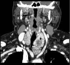
- All substernal goiters require axial computed tomographic (CT) scanning to differentiate between the various subtypes
- Such differentiation provides tremendously useful surgical information
#Arrangoiz #ThyroidSurgeon #ThyroidExpert #HeadandNeckSurgeon #CancerSurgeon #EndocrineSurgery #MultinodularGoiter #SubsternalGoiter #CASO #CenterforAdvancedSurgicalOncology
