Significant increase in incidence after mammography screening:
10-fold to 17-fold increase from 1970’s to 2004:
1 in 1300 mammograms
Represents 20% to 25% of all screen-detected breast neoplasias diagnosed annually
Based on autopsy data:
The prevalence of undetected DCIS may exceed 20% in women over 40:
Underscoring the concern for overdiagnosis:
Modeling studies estimate up to 65% of DCIS may be overdiagnosed
DCIS by it self:
Is not a risk to life
DCIS may progress to invasion and compromise survival:
If left untreated:
1 in 6 DCIS patients:
Progress to invasive breast cancer (IBC):
70% estimated to remain indolent
At this time we do not have any robust biomarkers:
That can quantify the risk of progression to IBC or
Help us separate indolent disease:
From the potentially dangerous lesions
Risk of overtreament:
The increase incidence of DCIS in mammographically detected cases:
Has not lead to a decrease in the incidence of IBC or reduction of IBC morality
Risk factors for progression / recurrence of DCIS:
The risk factors for IBC recurrence may be different from the risks factors for DCIS recurrence?
Risk of IBC recurrence:
African American race
Premenopausal status
Detection by palpation
Involved margins
High histologic grade
High p16 expression
Risk of IBC or DCIS recurrence:
DCIS size
Histology type
Comedo necrosis
Grade (High)
Young age
Close margins or positive margins
Patients with DCIS that recur with an IBC:
Some patients with DCIS may develop:
Progression of there disease:
30% (1 out of 6)
Some will have a de novo invasive breast cancer
Some might have a missed invasive cancer?
Patient that have a DCIS recurrence:
Might be a true in situ recurrence
De novo DCIS
Residual disease?
Studies are describing observations of events like:
Synchronous IBC
Subsequent ipsilateral or contralateral DCIS or IBC (often a mixture)
We have limited data on DCIS progression with paired molecular profiles
The most consistent biological feature of DCIS:
Heterogeneity:
In clinical presentation
Morphology
Protein expression:
Including receptor status
Gene expression
Genetic alterations
Epigenetic alterations
The heterogeneity is:
Between patients – within the lesion – and within cells in a single duct
Morphological features that help us predict progression are:
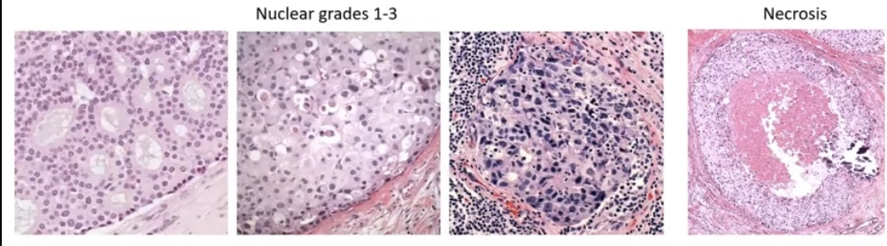
Histologic grading:
Combing the nuclear grade 1 to 3 and necrosis into a three tier system (the good, the bad, and the ugly):
Low / intermediate / high grade
Grade 1 to 3
Van Nuys Group 1 to 3
DIN 1 to 3
We all know that there is regression towards the mean and substantial interobserver variation
DCIS is classified by two complementary systems:
Nuclear grade (the primary classification):
Low (grade I), intermediate (grade II), and high (grade III):
Based on nuclear size, pleomorphism, chromatin pattern, nucleoli, mitotic activity, and cell polarization
Nuclear grade is the most reproducible and clinically relevant parameter
Architectural pattern (secondary / noncore):
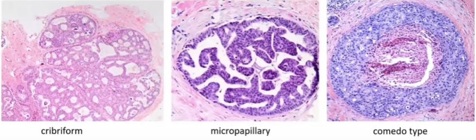
Solid, cribriform, micropapillary, papillary, and comedo:
Multiple patterns frequently coexist within a single lesion, limiting reproducibility
Morphologic Features Suggestive of Progression
Unclear prognostic value of the 3-tier system:
We suspect that:
Low to intermediate grade = low risk of progression
High grade system = high risk of progression or shorter time to progression
Maxwell, A.J. Eur.J.Surg.Oncol.,2018, Ryser, MD. J.Natl Cancer Inst., 2019:
Risk of ipsilateral recurrence (DCIS / IBC) at 10 years:
High grade 17.6% (95% CI=12.1-25.2%)
Non high grade 12.2 (95% CI=8.6-17.1%):
Including grade 2
There is overlap in the confidence intervals
Low grade DCIS are the lesions that might have:
Discontinuous growth (multifocal gaps up to 1 cm) and skip lesions that might lead to a:
Greater likelihood of residual disease and recurrence?
DCIS has heterogeneity of grade within the same lesion
Histologic subtype as a prognostic factor:
Subtype:
Cribriform:
Is more often a grade 1 lesion
Comedo type:
Is more often a grade 3 lesion
Usually histology subtype correlates with grade but:
There is often a mixture of growth patterns:
Compromising the use for prognostication
Can we use histology as a prognostic feature?
Tumor micro environment:
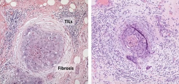
Could potentially be the most important morphologic feature suggestive of progression especially:
Circumferential periductal fibrosis and associated tumor infiltrating lymphocytes (TIL):
Indicating host reaction to the tumor cells
Tumor micro environment includes:
Myoepithelial cell layer
Tumor infiltrating lymphocytes (TIL)
Adipocytes
Fibroblasts
Matrix
The border around the myoepithelial layer might have prognostic value
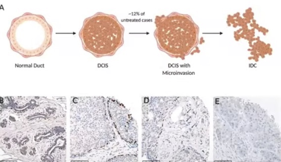
The myoepithelial cell layer acts as a gatekeeper:
Has tumor suppressive functions
The largest gene expression change from normal tissue to DCIS:
Occurs in the myoepithelial cell layer
DCIS is associated myoepithelial cell loss:
That leads the decrease tumor suppressor functions
The myoepithelial cell layer is lost in IBC
Myoepithelial cell layer acts as a GATEKEEPER (Tumor Suppressive Functions)
Disruption of the myoepithelial defense:
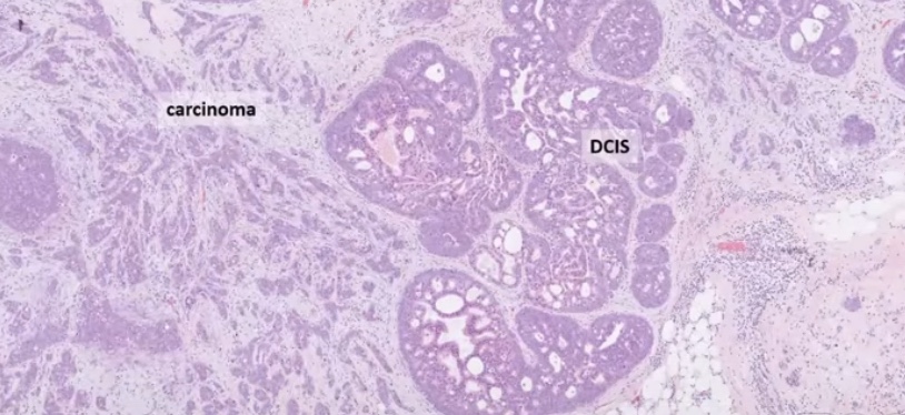
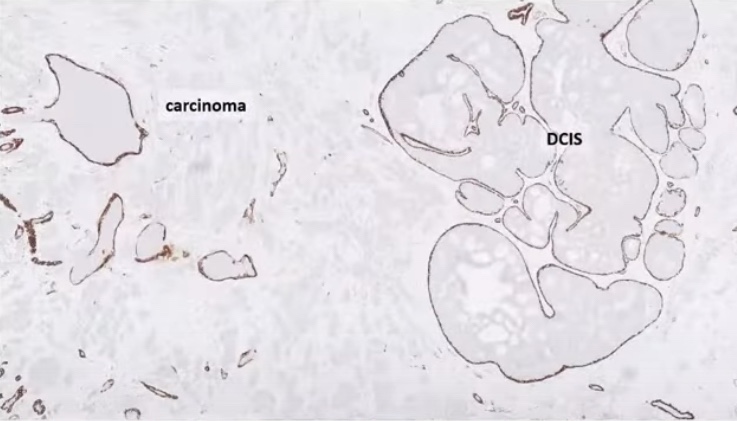
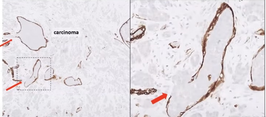
IBC with DCISBrown color – myoepithelium Arrows point to the disrupted myoepithelial cell layer (micro-invasion)
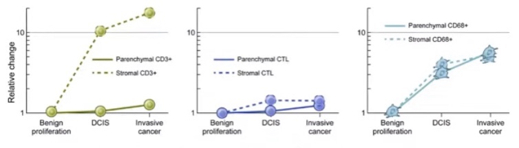
Conflicting data on prognostic value of TIL:
Some studies have reported no prognostic value of stromal TIL for subsequent recurrences:
Does the spatial location of the immune cells matter?
The TIL in direct contact with the DCIS might be more important that the TIL that are further away
Other studies have shown a correlation between higher levels of TIL and increased risk of subsequent IBC and a shorter (ipsilateral) recurrence-free survival
Avoids all the disadvantages of a lateral mandibulotomy and the sequelae of a midline mandibulotomy
A paramedian mandibulotomy offers significant advantages:
Wide exposure to the surgical field
Preservation of the geniohyoid and genioglossus muscles:
Leading to preservation of:
The hyomandibular complex
The only muscle requiring division is the mylohyoid muscle:
Which leads to minimal swallowing difficulties
A paramedian mandibulotomy does not cause denervation or devascularization of the skin of the chin or the teeth and mandible
Fixation at the mandibulotomy site is easy
The site of the mandibulotomy does not fall within the lateral portal of radiation therapy if the patient needs postoperative radiotherapy:
Thus at present a paramedian mandibulotomy remains an optimal surgical approach for access to posteriorly located larger lesions of the oral cavity and tumors of the oropharynx and parapharyngeal space
The 2025 ATA guidelines (Recommendation 11) formally endorse active surveillance (AS) as an acceptable management option for cT1aN0M0 papillary thyroid cancers (≤1 cm), graded as a conditional recommendation with low certainty evidence.
For recurrent / persistent nodal disease, the guidelines state that stable disease can continue monitoring, while bulky and / or invasive recurrent disease is generally best treated with surgery.
Recommendation 14 specifies that surgical resection is indicated when there is tumor growth ≥ 3 mm, new biopsy-proven lymph node metastases, distant metastases, extrathyroidal extension, or patient preference.
The NCCN Thyroid Carcinoma Guidelines (v1.2026) similarly note that AS is supported by low-quality evidence for papillary thyroid microcarcinoma ≤ 1 cm, with limited data for cancers >1 cm.
Quantitative Data on Progression During Active Surveillance
A 2026 systematic review and meta-analysis (10 studies, n = 841) provides the most comprehensive pooled data. The overall pooled progression rate during AS was 23% (95% CI, 12–34%), rising to 32% in biopsy-confirmed cases. Critically, after adjustment for follow-up loss, the estimated progression rate increased to 35% to 70%, suggesting that reported rates may substantially underestimate true progression. Subgroup analyses showed higher progression in patients with elevated baseline thyroglobulin (log-transformed Tg: 1.02 in progressors vs. −0.07 in stable, p < 0.05) and in those with advanced stage (≥ 5% Stage IV patients: 28% vs. 14%, p < 0.05).
Individual cohort studies provide additional granularity:
Jerkovich et al. (2022): 50 patients with cytology-confirmed metastatic lymph nodes (mean size 10.1 mm) under AS; 24% had lymph node growth at median 29 months. The only predictor of growth was a Tg rise ≥0.5 ng/mL (OR 16.2, p = 0.020). Median progression-free survival was 6.6 years. Among the 7 patients who underwent surgery after progression, none had structural incomplete response at follow-up.
Walter et al. (2023): 40 patients with metastatic cervical lymph nodes ≤2 cm; 35% progressed (17.5% enlargement ≥3 mm, 17.5% new metastasis) at median 27.5 months. Of 14 who progressed, 6 remained on AS with 5/6 showing no further progression.
Tufano et al. (2015): Lateral neck lymph nodes with suspicious ultrasonographic features demonstrated a low potential for structural progression—over a median of 3.5 years, only 9% increased by more than 5 mm.
Size Thresholds: Confirmed
The ATA-endorsed size thresholds for FNA biopsy consideration (and by extension, the boundary for observation) are ≥8 mm in the central compartment and ≥10 mm in the lateral compartment. The NCCN guidelines do not specify exact size cutoffs for AS of recurrent nodes but recommend “disease monitoring for non-progressive disease that is stable and distant from critical structures”.
Thyroglobulin Doubling Time as a Prognostic Marker
A Tg doubling time (Tg-DT) <1 year portends poor prognosis, while Tg-DT >2 years signifies good prognosis. The AHNS consensus statement recommends cross-sectional imaging at 4 to 6 month intervals when Tg-DT is <1 year, with longer intervals for longer doubling times.
A study by Albano et al. demonstrated that a TgDT threshold of ≤2.5 years predicted positive FDG-PET/CT results with 93% sensitivity and 87% specificity (AUC = 0.911), outperforming absolute Tg level alone. Ito et al. (2025) showed that combining unstimulated Tg ≥3 ng/mL with a Tg doubling rate ≥0.33/year identified the highest-risk group for both lymph node and distant recurrence.
FDG-PET Avidity: Strong Prognostic Data
FDG-PET avidity is a well-established marker of aggressive biology. Robbins et al. demonstrated that a positive FDG-PET/CT predicted a 7-fold increased risk of mortality compared with negative FDG findings. FDG uptake reflects tumor dedifferentiation and is a major negative predictive factor for response to RAI treatment. An SUVmax >5.0 has been associated with very high rates of disease progression even in the presence of mixed iodine-avid disease, and SUVmax >10 has been linked to shorter locoregional disease-free survival and anaplastic transformation. The 2025 ATA guidelines recommend FDG-PET/CT as both a diagnostic and prognostic tool in high-risk patients, particularly those with aggressive histologies or negative RAI imaging.
BRAF/TERT Mutations: Quantified Risk
The co-occurrence of BRAF V600E and TERT promoter mutations is the most powerful molecular predictor of aggressive behavior. A meta-analysis showed this combination was associated with reduced disease-specific survival (RR 15.09, 95% CI 7.75–29.37), far exceeding BRAF V600E alone (RR 5.34). Song et al. (2016) reported that co-existing BRAF + TERT mutations increased recurrence risk (HR 4.64 for BRAF/TERT; HR 5.36 for RAS/TERT) and mortality (HR 15.13 for BRAF/TERT; HR 14.75 for RAS/TERT). Ryu et al. (2026) demonstrated that TERT-mutant tumors show continuous recurrence over time without the expected decline seen in TERT-wildtype cases, and are associated with significantly higher mortality. Xing et al. (2025) showed that the BRAF/TERT genetic duet worsened survival across all AJCC stages.
Reoperative Surgery Outcomes
The 2025 ATA guidelines report that reoperative surgery results in 27% to 63% excellent responses, 5.7% to 13.3% biochemically incomplete responses, 10% to 44% structurally incomplete responses, and 8.6% to 30% indeterminate responses. Independent risk factors for incomplete response after reoperation include age >45 years, aggressive histology, and lymph node ratio >0.6 at initial resection.
The Central Tension: Is Progression Evidence That Surveillance Is Failing?
The assertion that “progression itself is evidence that surveillance is failing” requires nuance. The data suggest that not all progression is equal:
Slow, limited nodal growth in low-risk patients can often be managed with continued observation or delayed intervention without compromising outcomes. In the Jerkovich cohort, all 7 patients who underwent surgery after progression on AS achieved structural complete response. In the Walter cohort, 5 of 6 patients who remained on AS despite progression showed no further growth.
However, the 2026 meta-analysis raises a cautionary note: true progression rates may be 2–3 times higher than reported due to follow-up loss, and AS “should be considered with caution”. Trimboli and Piccardo (2026) argue that until stronger prospective data are available, AS in DTC should be applied within a well-defined multidisciplinary framework with transparent communication about uncertainties.
The evidence therefore supports a nuanced position: AS is appropriate and evidence-based for carefully selected patients with stable, low-volume recurrence, but documented progression—particularly when accompanied by rising Tg kinetics, FDG avidity, aggressive molecular profile, or threatened critical structures—should prompt serious consideration of intervention. The key is that AS is not a passive strategy but an active decision-making framework requiring serial reassessment.
Are available for resection of primary tumors of the oral cavity
The choice of a particularapproach will depend on factors such as:
The size and site of the primary tumor:
Anterior versus posterior
As well as its depth of invasion
Proximity to the mandible or maxilla
Factorssuch as dentition, size of the oral aperture, trismus, and the size and mobility of the tongue:
Also influence selection of the surgical approach
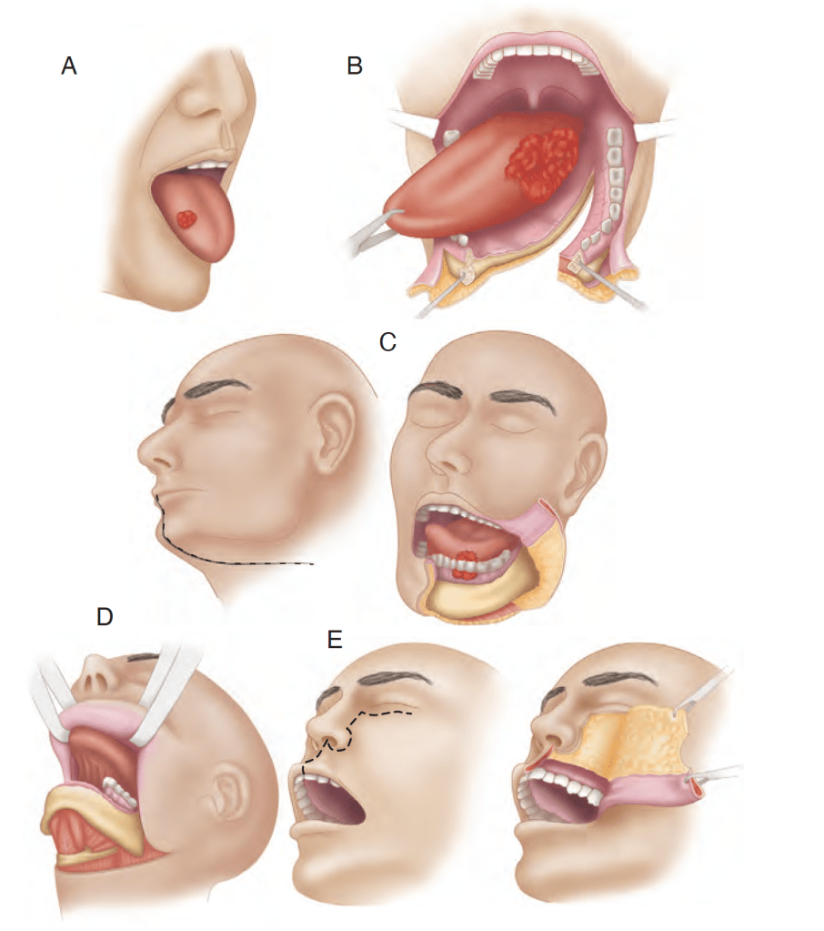
The various surgical approaches such as:
Peroral, mandibulotomy, lower cheek flap approach, visor flap approach, and upper cheek flap approach are shown in Figure
The transoral approach has wider applications with technologic advances using:
Lasers (transoral laser microsurgery [TLM]) and robotics (transoral robotic surgery [TORS])
Various surgical approaches. A, Peroral. B, Mandibulotomy. C, Lower cheek flap. D, Visor flap. E, Upper cheek flap.
When the peroral approach does not offer adequate exposure:
The visor flap or cheek flap approaches (upper or lower) become necessary
The visor flap approach:
Provides sufficient exposure for anteriorly located lesions:
But is not satisfactory for tumors located in the posterior oral cavity
The benefit of this approach is that it avoids a lower lip–splitting incision:
But produces permanent numbness of the chin:
Because the mental nerves need to be transected for adequate mobilization of the flap
It also may cause sagging of the lower lip and drooling:
Because of a loss of support and sensation:
Thus its utility is limited
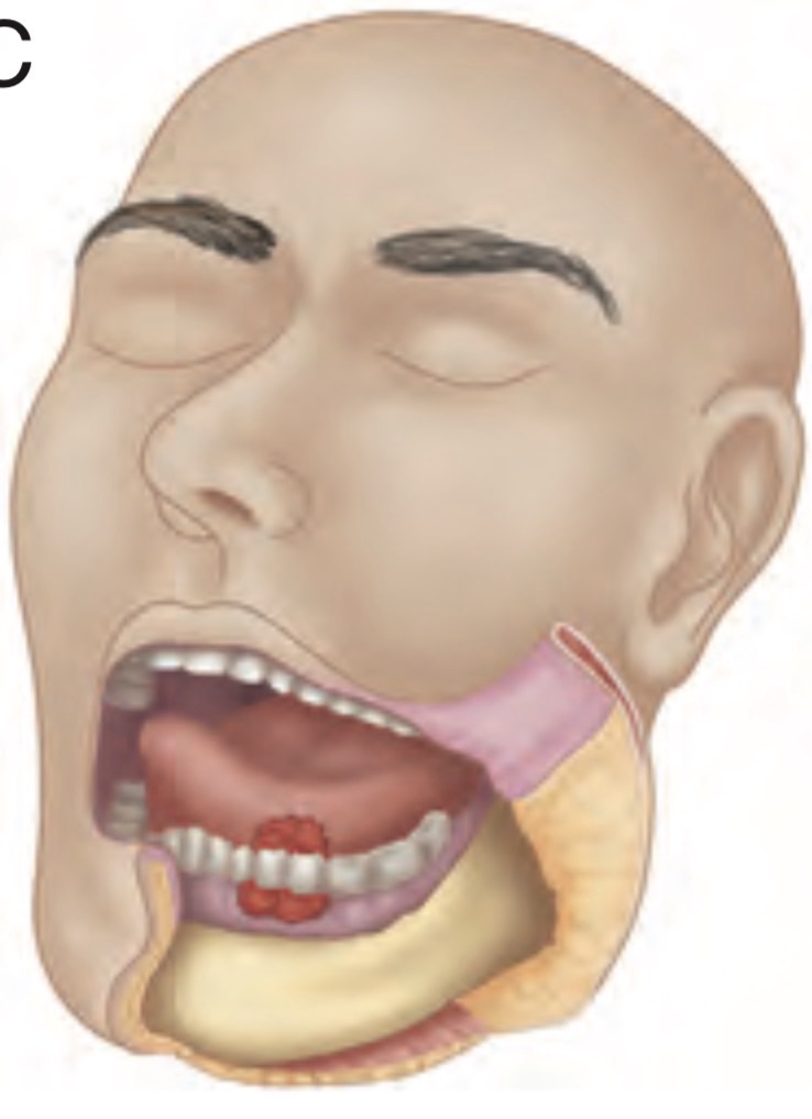
The lower cheek flap approach:
Requires a midline lip–splitting incision that is continued laterally into the neck for exposureand neck dissection:
This approach provides excellent exposure for nearly all tumors of the oral cavity:
Except those of the upper gum and hard palate
Mandible resection (marginal or segmental) and reconstruction:
Require the lower cheek flap approach in most instances
The lower cheek flap approach:
Is required for marginal or segmental mandibulectomy of tumors adjacent to the body of the mandible
The upper cheek flap approach (the Weber-Ferguson incision and its modifications):
Is required for resection of larger tumors of the hard palate and upper alveolus:
Particularly if they are posteriorly located
Access to larger tumors of the tongue, particularly those closer to the base of the tonguewhere the mandible is not involved:
Requires wider exposure for resection:
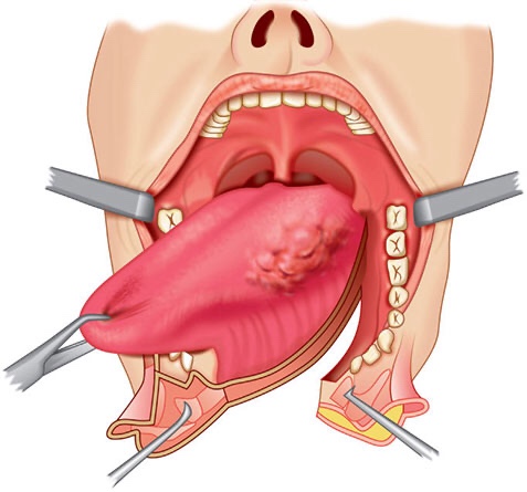
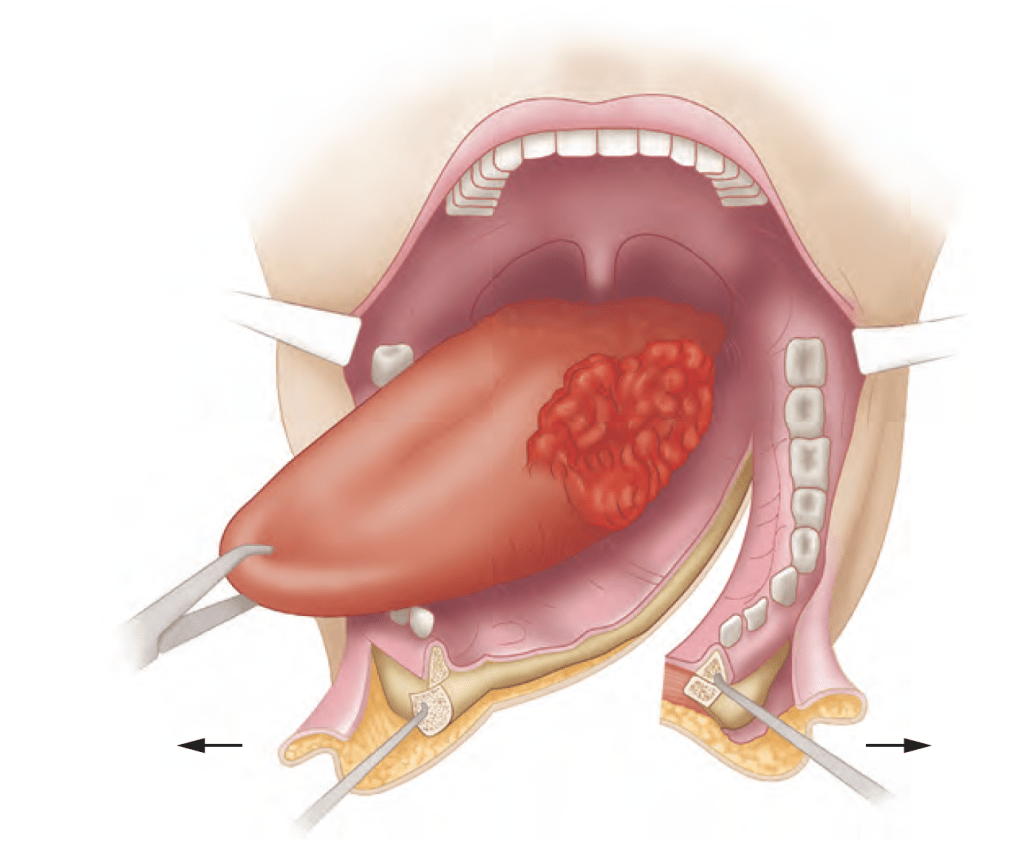
A mandibulotomy or mandibular osteotomy is an excellent mandible sparing surgical approach:
Designed to gain access to the oral cavity or oropharynx for resection of primary tumors otherwise not accessible through the open mouth or by the lower cheek flap approach (Figure)
A mandibulotomy is an excellent mandible-sparing surgical approach to gain access to bulky tumors of the oral cavity or oropharynx.
To determine the need and extent of mandible resection:
It is essential to understand the pathwayby which oral cancers invade the mandible
Primary carcinomas of the lip, buccal mucosa, tongue, and floor of the mouth:
Extend along the surface mucosa and the submucosal soft tissues:
To approach the attached labial, buccal, or lingual gingiva
From this point:
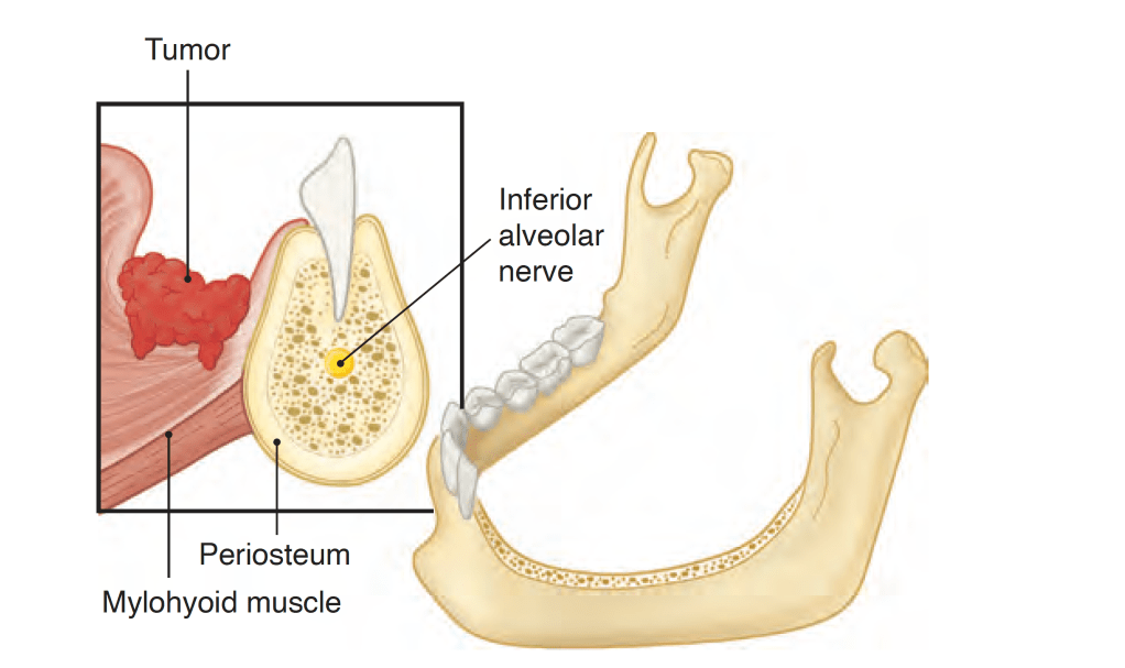
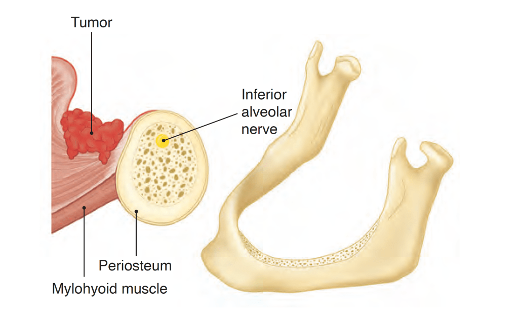
The tumor does not extend directly through intact periosteum and cortical bone toward the cancellous part of the mandible:
Because the periosteum acts as a significant protective barrier
Instead:
The tumor advances from the attached gingiva toward the alveolus:
In patients with teeth:
The tumor extends through the dental socket into the cancellous part of the bone and invades the mandible in that fashion (Figure)
Tumor invasion of the dentate mandible occurs through the dental socket to the cancellous bone and then to the alveolar canal
In edentulous patients:
The tumor extends up to the alveolar process and then infiltrates the dental pores in the alveolar ridge and extends to the cancellous part of the mandible (Figure)
Tumor invasion of the edentulous mandible occurs through the dental pores on the alveolar process to the cancellous bone and then to the alveolar canal.
Thus even in patients with early invasion of the mandible:
A marginal mandibulectomy is feasible because the cortical part of the mandible inferior to the roots of the teeth:
Remains uninvolved and can be safely spared
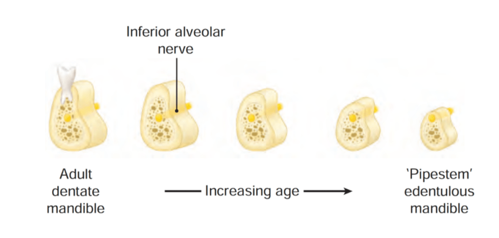
In edentulous patients, however, the feasibility of marginal mandibulectomy depends on the vertical height of the body of the mandible:
With aging, the alveolar process recedes and the mandibular canal comes closer to the surface of the alveolar process:
As shown in the Figure, the resorption of the alveolar process eventually leads to a “pipestem” mandible in elderly patients
Vertical height and location of the alveolar canal in dentate and edentulous mandibles.
The ability to perform a satisfactory marginal mandibulectomy in such patients is almost impossible:
Because the probability of iatrogenic fracture or postsurgical spontaneous fracture of the remaining portion of the mandible is very high:
Similarly, in patients who have received previous radiotherapy:
A marginal mandibulectomy should be performed with extreme caution:
The probability of pathological fracture at the site of the marginal mandibulectomy in such patients is very high
When the tumor extends to involve the cancellous part of the mandible:
A segmental mandibulectomy must be performed
A segmental mandibulectomy also may be required in patients with massive primary tumors with significant soft tissue disease in the proximity of the mandible
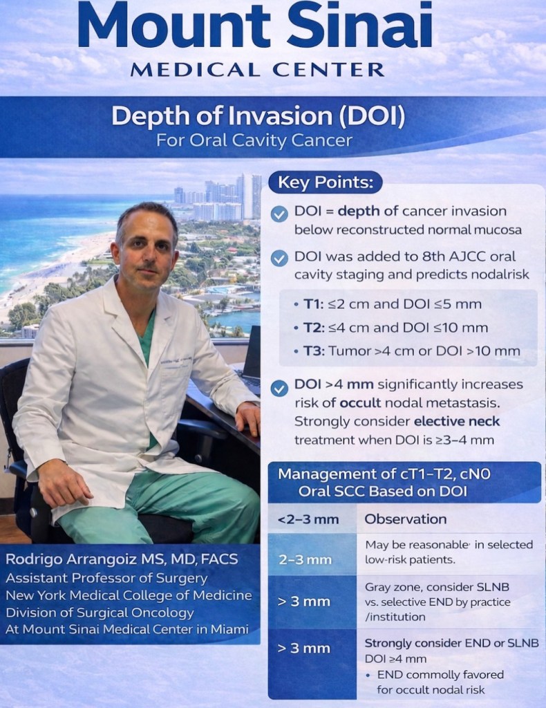
Rodrigo Arrangoiz MS, MD, FACS, FSSO is an Assistant Professor at New York Medical College School of Medicine Division of Surgical Oncology at Mount Sinai Medical Center
He is first author on some publications on oral cavity cancer:
Oral Tongue Cancer: Literature Review and Current Management
The oral cavity represents the entrance to the upper aerodigestive tract:
Which begins at the lips and ends at the anterior surface of the faucial arch
It is lined by:
Squamous epithelium:
With interspersed minor salivary glands
The oral cavity also contains the:
Dentoalveolar structures:
With the upper and lower dentition
The oral cavity is continuously exposed to inhaled and ingested carcinogens:
Thus it is the most common site for the origin of malignant epithelial neoplasms in the head and neck region
Known carcinogens for oral cavity carcinoma include:
Those present in tobacco, alcohol, and betel nuts
The association of human papilloma virus with oral cancer:
Is not as well established as in oropharyngeal cancers
Primary tumors of the oral cavity may arise from the:
Surface epithelium (most common)
Minor salivary glands
Submucosal soft tissues
Lesions of dentoalveolar origin:
Representing a unique group of neoplasms and cysts
The various anatomic sites within the oral cavity as described by the American Joint Committee on Cancer (AJCC) and International Union Against Cancer (UICC) staging system are shown below:
Anatomic sites within the oral cavity
More than 90% of malignant tumors in the oral cavity are:
Squamous cell carcinomas:
The remainder are:
Minor salivary gland carcinomas and other rare tumors
Most patients with cancer in the oral cavity are men:
Although the incidence of tongue cancer in women in the United States has progressively increased over the past several decades
In the Western world:
The tongue (> 50% of cases) and floor of the mouth:
Are the most common sites of origin for primary squamous cell carcinomas in the oral cavity:
However, the retromolar trigone and buccal mucosa:
Are the most frequently encountered primary sites in areas of the world where chewing of tobacco and / or betel nuts is common
The site distribution of various primary cancers in the oral cavity in the United States is shown bowls:
The site distribution of primary cancers in the oral cavity (MSKCC data 1985 to 2015; all patients re-staged according to AJCC 8th edition criteria).
– The goals of treatment of cancer of the oral cavity are:
Cure of the cancer
Preservation or restoration of:
Speech, mastication, swallowing, and external appearance
Minimization of the sequelae of treatment such as:
Dental decay, osteonecrosis of the mandible, and trismus
Awareness of the risk of subsequent primary tumors and their management
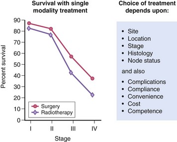
– Surgery and radiotherapy can be used as a single modality or in combination for the treatment of cancer of the oral cavity
– Treatment Approaches:
In general, early-stage (stage I or II) head and neck tumors:
May be treated using a single modality (surgery or radiotherapy):
Whereas advanced disease (stage III or IV) frequently benefits from multimodality therapy
The best therapeutic approach for the primary tumor depends on the anatomic site
Most neck disease can be treated equally well with surgery or radiation:
Thus the modality chosen to treat the neck is based on which modality is selected for the primary
When the primary tumor is treated with irradiation:
The regional lymphatics “at-risk” are incorporated into the treatment fields
Neck dissections should remain standardized (ie, complete anatomic dissections, as opposed to “berry picking” or random biopsy) in these settings to avoid incomplete surgery
Rodrigo ArrangoizSinai Medical Center in Miami, Florida:
He is first author on some publications on oral cavity cancer:
Oral Tongue Cancer: Literature Review and Current Management