Is defined as persistent or newly identified locoregional or distant metastases on imaging:
Usually in conjunction with elevated thyroglobulin (Tg) and / or anti-thyroglobulin antibody (TgAb) levels
It is one of four response-to-therapy categories in the ATA dynamic risk stratification system:
Alongside excellent, indeterminate, and biochemical incomplete responses
Criteria by Treatment Context:
The definition of SIR is consistent regardless of surgical extent:
Structural evidence of disease confirmed by suspicious imaging or biopsy-proven local or distant metastatic disease
The TSH goal for patients with SIR:
Is maintained below the normal reference range
Prevalence and Outcomes:
SIR occurs in approximately 2.8% to 10% of DTC patients after initial therapy
In a cohort of 501 patients treated with total thyroidectomy and RAI:
2.8% had SIR at initial assessment:
By last follow-up, 10.2% had structurally incomplete responses
Key outcome data include:
Patients with an initial SIR:
Had an 83.9% rate of continued structural disease over long-term follow-up (mean 10.3 years)
All patients categorized as SIR after total thyroidectomy without RAI:
Experienced continued presence of disease
SIR requires multidisciplinary management tailored to disease status (regional vs. distant metastases, iodine-avid vs. non-iodine-avid disease)
Management:
Management of SIR depends on:
Disease location, RAI avidity, resectability, and rate of progression
Locoregional disease:
Surgery is preferred for resectable locoregional recurrence:
With formal compartmental dissection for previously undissected basins
RAI therapy:
If radioiodine imaging is positive
Active surveillance:
Is appropriate for non-progressive disease that is stable and distant from critical structures:
Small-volume lymph node recurrences often show little progression over years
Local therapies (ethanol ablation, RFA, cryoablation):
May be considered for limited-burden nodal disease
RAI-refractory or progressive disease:
Somatic molecular testing for actionable mutations (BRAF, RET, NTRK, ALK fusions; dMMR/MSI/TMB) is recommended for advanced, progressive, or threatening disease
Systemic therapy for progressive and/or symptomatic disease:
Lenvatinib (preferred; PFS 18.3 vs. 3.6 months, ORR 65%)
Sorafenib (PFS 10.8 vs. 5.8 months)
Cabozantinib after prior VEGFR TKI (PFS 11.0 vs. 1.9 months)
Targeted therapies:
Dabrafenib / trametinib (BRAF V600E)
Selpercatinib / pralsetinib (RET fusion)
Larotrectinib / entrectinib (NTRK fusion)
Pembrolizumab (MSI-H/dMMR or TMB-H)
Disease monitoring is often appropriate for asymptomatic patients with indolent, non-progressive disease and no brain metastases:
TKI therapy may not be appropriate for stable or slowly progressive disease
TSH suppression should be maintained with TSH
References:
2025 American Thyroid Association Management Guidelines for Adult Patients With Differentiated Thyroid Cancer. Ringel MD, Sosa JA, Baloch Z, et al. Thyroid : Official Journal of the American Thyroid Association. 2025;35(8):841-985. doi:10.1177/10507256251363120.
SNMMI Procedure Standard/Eanm Practice Guideline for Nuclear Medicine Evaluation and Therapy of Differentiated Thyroid Cancer: Abbreviated Version. Avram AM, Giovanella L, Greenspan B, et al. Journal of Nuclear Medicine : Official Publication, Society of Nuclear Medicine. 2022;63(6):15N-35N.
Thyroid Carcinoma. National Comprehensive Cancer Network. Updated 2025-03-27. Thyroid Cancer. Chen DW, Lang BHH, McLeod DSA, Newbold K, Haymart MR. Lancet (London, England). 2023;401(10387):1531-1544. doi:10.1016/S0140-6736(23)00020-X.
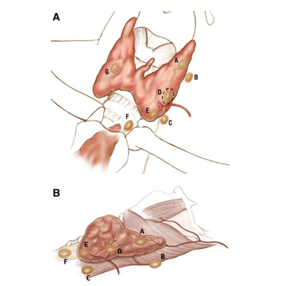
Type A glands are in the accepted, expected location of a normal parathyroid gland
Type B:
Behind the thyroid parenchyma
Type B glands are exophytic to the thyroid parenchyma and lie in the tracheoesophageal groove:
This category includes adenomas in:
Retroesophageal, retropharyngeal, high lateral pharyngeal, and carotid sheath locations
A ‘‘B+’’ subcategory can be used to document the location of adenomas above the level of the hyoid bone:
The ‘‘+’’ is meant to reflect cranial elevation
Type C:
Caudal to the thyroid parenchyma:
In the tracheoesophageal groove
A type C gland is more inferior than a type B gland on lateral images:
Located inferior to the inferior pole of the thyroid:
Closer to the clavicle
Type D:
Directly over the recurrent laryngeal nerve:
At the level of the inferior thyroid vessels
The dissection may be difficult:
Because a type D gland is dangerously close to the recurrent laryngeal nerve
Type E:
Located in the external aspect of the inferior pole of the thyroid
A type E gland is in a location that is:
More superficial in an anterior–posterior plane than the recurrent laryngeal nerve:
It is the easiest to resect
Type F:
‘‘Fallen’’ into the thyrothymic ligament:
Below the inferior pole of the thyroid in a pretracheal plane
A type F gland is frequently referred to as an ectopic gland:
Its resection usually involves:
Transcervical delivery of the thyrothymic ligament or superior portion of the thymus
Type G:
A gauge, true intrathyroidal gland location
Schematic representation of the nomenclature system for localization of parathyroid adenomas. Anterior view (a); right lateral view (b) of the superior thyroid pole is oriented to the left. The dotted circle depicts the region where the recurrent laryngeal nerve is most at risk
This nomenclature system has been designed that takes into account the pathologic position of the parathyroid glands (Figure):
Superior and inferior glands:
Are defined by the location of the gland’s pedicle and its relationship to the RLN:
Superior parathyroid glands:
Anatomically have a vascular pedicle superior and lateral to the RLN (type A through D glands)
Inferior parathyroid glands:
Anatomically have a vascular pedicle inferior and medial to the RLN (type D through F glands)
Type G glands:
Represent intrathyroidal parathyroid lesions
This information not only helps radiologists communicate potential parathyroid lesions of interest to surgeons:
But also helps surgeons direct their dissection in relation to the RLN
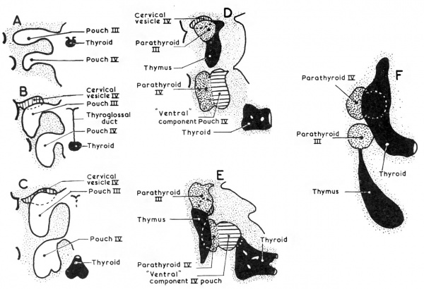
Develop as epithelial thickenings of the dorsal endoderm of the:
Third and fourth branchial pouches
The superior parathyroid glands:
Are derived from the fourth branchial pouch:
Which also gives rise to the ultimobranchial bodies:
The ventral aspect of these pouches is believed to fuse with the rudimentary fifth branchial pouches:
To from the ultimobranchial bodies
The superior parathyroid glands follow the migration of the ultimobranchial bodies:
Which descend a relative limited path toward the lateral thyroid region:
Ultimately giving rise to the parafollicular cells of the thyroid
The superior parathyroid glands separate from the ultimobranchial bodies:
As the median and lateral thyroid anlages fuse and incorporate the ultimobranchial bodies:
This separation event determines the final anatomic position of the superior parathyroid glands relative to the thyroid (Type A gland)
The inferior parathyroid glands:
Are derived from the third branchial pouch (dorsal aspect):
Along with the thymus (derived from the ventral aspect of the third branchial pouch)
The parathyroid glands:
Remain intimately connected with their respective branchial pouch derivatives
The normal anatomic location of the superior parathyroid glands:
Is more constant than the inferior parathyroid glands:
With 80% of the superior glands being found near the posterior aspect of the thyroid gland:
At the junction of the upper and middle portion of the thyroid lobes:
At the level of the cricoid cartilage:
Each gland with its own capsule of connective tissue:
Type A gland
Roughly one percent (1%) of the superior parathyroid glands:
May be found in the paraesophageal or retroesophageal space, retrolaryngeal space, high lateral pharyngeal, and carotid shealth locations:
Type B glands:
Behind the thyroid parenchyma:
Type B glands are exophytic to the thyroid parenchyma:
Lie in the tracheoesophageal groove
Type C glands:
Caudal to the thyroid parenchyma:
In the tracheoesophageal groove
A type C gland is more inferior than a type B gland on lateral images and located inferior to the inferior pole of the thyroid (closer to the clavicle)
Enlarged superior parathyroid glands:
May descend in the tracheoesophageal groove and come to lie below the inferior parathyroid glands (Type C gland)
Truly ectopic superior parathyroid glands:
Are extremely rare:
But may be localized to the middle or posterior mediastinum or in the aortopulmonary window
During intrauterine development:
The thymus and the inferior parathyroid glands:
Migrate caudally in the neck
The most common location for the inferior parathyroid glands:
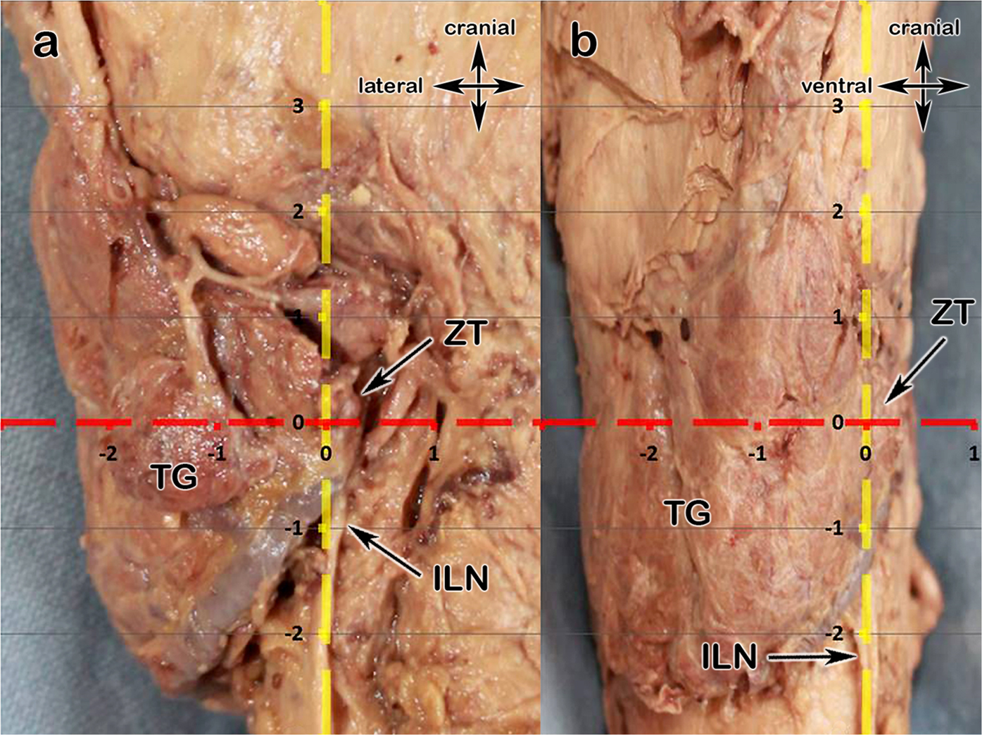
Is within 1 cm from a point centered where the inferior thyroid artery and the recurrent laryngeal nerve (RLN) cross
In roughly 50% of the cases:
The inferior parathyroid gland is located at the level of the inferior thyroid lobe:
Anterior of the posterolateral surface:
Type E:
Located in the external aspect of the inferior pole of the thyroid
A type E gland is in a location that is more superficial in an anterior-posterior plane than the recurrent laryngeal nerve
It is the easiest to resect
Approximately 15% to 50% of the inferior glands:
Are found in the thyrothymic ligament or the thymus
The inferior parathyroid gland is typically situated within a pocket of thymic derived fatty tissue:
But may be closely adherent to the thyroid capsule
The position of the inferior parathyroid glands:
However, tends to be more variable due to their longer migratory route:
Undescended inferior glands may be found near the:
Skull base, angle of the mandible, or above the superior parathyroid glands:
Along with an undescended thymus
The frequency of intrathyroidal glands:
Is approximately 2%
Superior parathyroid glands (4th branchial pouch):
Short migration
More constant location
Posterior to RLN, near cricothyroid joint
Inferior glands (3rd branchial pouch):
Long migration with thymus
Highly variable
Can be anywhere from angle of mandible → mediastinum
The classification is built on this predictable vs variable descent pattern
The Perrier Classification System
The authors propose categorizing adenomas based on their relationship to key anatomic landmarks, especially:
Thyroid gland
Recurrent laryngeal nerve (RLN)
Thymus
Carotid sheath
📍 Four Main Categories
Type A — Orthotopic (Normal Position)
Located in expected anatomical position
Adjacent to thyroid gland
Most common
👉 Clinical relevance:
Ideal for minimally invasive parathyroidectomy (MIP)
High concordance with sestamibi + ultrasound
Type B — Ectopic but Cervical
Includes:
Retroesophageal
Carotid sheath
Intrathyroidal
High cervical (undescended)
👉 Key point: Still in the neck but outside usual location
👉 Surgical implication:
May require focused but modified approach
Intrathyroidal → partial thyroid resection
Type C — Thymic / Thyrothymic
Along thymic descent pathway
Thyrothymic ligament
Within cervical thymus or upper mediastinum
👉 Most common ectopic site for inferior glands
👉 Surgical implication:
Cervical thymectomy often required
Important in failed initial exploration
Type D — Mediastinal
Below thoracic inlet
Aortopulmonary window, pericardium, deep thymus
👉 Rare but critical
👉 Surgical implication:
May require:
VATS
Sternotomy
Interventional radiology localization
📊 Key Findings from Perrier et al.
Majority of adenomas are Type A (orthotopic)
Ectopic locations (Types B to D) account for:
~15% to 20% of cases
Inferior glands → disproportionately represented in ectopic group
👉 The classification correlates strongly with:
Embryology
Preoperative imaging success
Surgical difficulty
🎯 Clinical Impact
1. Improves Communication
Standard language across:
Surgeons
Radiologists
Endocrinologists
2. Enhances Preoperative Planning
Predicts:
Likelihood of MIP vs BNE
Need for extended exploration
3. Reduces Failed Explorations
Particularly valuable in:
Reoperative cases
Discordant imaging
4. Facilitates Research Standardization
Enables:
Comparable outcome reporting
Better stratification in studies
🧠 Surgical Algorithm Integration
Imaging Result
Likely Type
Strategy
Concordant US + Sestamibi
Type A
Focused MIP
Discordant imaging
Type B / C
Extended cervical exploration
Negative imaging
Type C / D
Consider 4D-CT, PET, BNE
Prior failed surgery
Any (often B to D)
Systematic re-exploration
⚠️ Limitations of the Study
Retrospective classification
Single-institution experience (MD Anderson)
No direct comparison with alternative systems
Does not incorporate modern imaging (e.g., 4D-CT, PET)
📚 Key References
Perrier ND et al. World J Surg. 2009;33:412–416
Akerström G et al. Anatomy and embryology of parathyroid glands. World J Surg. 1984
Wang C. Parathyroid gland location study (645 cases). Ann Surg. 1976
You now need to identify patients who may benefit before finalizing adjuvant plan:
Candidates to flag early:
Stage II (even N0) with:
High grade
High Ki-67
Genomic high risk
Any Stage III HR+ disease
Impacts surgical-pathologic reporting priorities:
Surgeons should ensure:
Accurate nodal staging
Grade
Ki-67
Genomic assay (if used) integrated early
These directly influence eligibility for CDK4/6 therapy
Reinforces importance of recurrence biology:
Luminal cancers:
Long natural history
Late relapse risk
Ribociclib addresses micrometastatic dormancy → shifts mindset from: “Local control + endocrine therapy” → to “Extended systemic control strategy”
Treatment duration considerations:
Ribociclib:
3 years
Endocrine therapy:
≥ 5 years
Implication:
Long-term adherence planning begins at surgical consultation
Safety Profile (surgeon-relevant highlights):
Common:
Neutropenia (non-febrile)
LFT elevation
QT prolongation
No new long-term safety signals at 5 years
Clinical takeaway:
Manageable → supports use in early-stage curative setting
Key Takeaways for Surgical Practice:
Ribociclib is now a standard adjuvant option in HR+/HER2− EBC:
Especially stage II to III and biologically high-risk disease
5-year data confirms durability:
Benefit persists beyond treatment window:
Increasing absolute benefit over time
Expands eligible population:
Includes node-negative high-risk patients
Multidisciplinary coordination is critical:
Surgeons play a role in:
Early identification
Pathologic risk stratification
Timely referral to medical oncology
Bottom line (surgeon-focused):
Ribociclib from NATALEE represents a shift toward proactive systemic prevention of recurrence in luminal breast cancer, with durable 5-year benefit and broader eligibility than prior CDK4/6 strategies—making early risk identification at the surgical stage increasingly important
The Phase III LEAP-10 trial represents a major collaborative effort between Merck & Co. and Eisai, addressing a persistent unmet need in recurrent /metastatic head and neck squamous cell carcinoma (R/M HNSCC).
In patients with PD-L1 CPS ≥1 disease, the combination of lenvatinib + pembrolizumab demonstrated:
Improved objective response rate (ORR)
Prolonged progression-free survival (PFS)
However, these gains did not translate into an overall survival (OS) benefit compared with pembrolizumab monotherapy, which remains the backbone of first-line treatment.
Clinical Context: Why OS Remains Elusive
Despite encouraging activity, the absence of OS improvement reinforces the durability of the current standard established by KEYNOTE-048 trial, where:
Pembrolizumab ± chemotherapy continues as standard of care
Survival benefit is tightly linked to PD-L1 expression and patient selection
The LEAP-10 findings highlight a recurring challenge in HNSCC:
Early efficacy signals (ORR, PFS) do not reliably predict survival benefit, particularly in an immunotherapy-sensitive disease where post-progression treatments and tumor biology heavily influence OS.
Biologic Interpretation
The addition of lenvatinib, a multi-kinase inhibitor targeting VEGFR, FGFR, and others, likely:
Enhances tumor microenvironment modulation
Improves initial tumor shrinkage and disease control
However, potential limitations include:
Lack of deep, durable immune reprogramming
Emergence of resistance mechanisms
Possible toxicity-related treatment discontinuation
These factors may blunt long-term survival impact despite improved early endpoints.
Where the Field Is Heading: EGFR and Beyond
Attention is now shifting toward next-generation EGFR-targeted strategies, with the hypothesis that:
More precise targeting of EGFR-driven signaling
Coupled with immune engagement mechanisms
may yield more durable survival benefits.
Key players advancing this space include:
Dana-Farber Cancer Institute
Genmab
Bicara Therapeutics
Johnson & Johnson
Merus N.V.
Harvard Medical School
Emerging modalities include:
Bispecific antibodies (EGFR × immune targets)
Antibody-drug conjugates (ADCs)
Combination immunotherapy strategies
These approaches aim to:
Overcome primary and acquired resistance
Deliver more sustained immune activation
Ultimately shift the OS curve, not just early endpoints
Key Takeaway for Clinical Practice
While lenvatinib + pembrolizumab shows meaningful biologic and clinical activity, it does not currently challenge pembrolizumab-based regimens as standard of care in PD-L1–positive R/M HNSCC.
The central question remains:
What therapeutic strategy will meaningfully and reproducibly improve overall survival in first-line HNSCC?
The next wave of EGFR-targeted and immune-engaging therapies may be the most promising path forward.
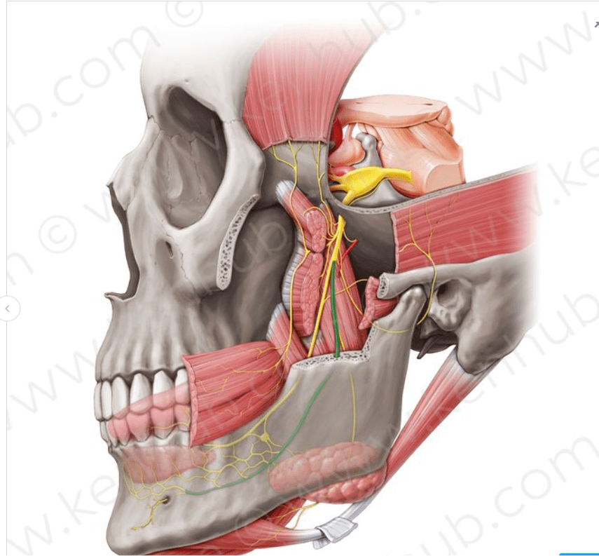
The maxillary artery supplies deep structures of the face.
It branches from the external carotid artery just deep to the neck of the mandible.
Structure:
The maxillary artery, the larger of the two terminal branches of the external carotid artery:
Arises behind the neck of the mandible, and is at first imbedded in the substance of the parotid gland.
It passes forward between the ramus of the mandible and the sphenomandibular ligament, and then runs, either superficial or deep to the lateral pterygoid muscle, to the pterygopalatine fossa.
It supplies the deep structures of the face.
May be divided into:
Mandibular portion (first part / bony part)
Pterygoid portion (second part / muscular part)
Pterygopalatine portions (third part).
Mandibular portion (first part / bony part):
The first or mandibular portion (or bony portion) passes horizontally forward, between the neck of the mandible and the sphenomandibular ligament:
Where it lies parallel to and a little below the auriculotemporal nerve.
It crosses the inferior alveolar nerve, and runs along the lower border of the lateral pterygoid muscle.
Branches include:
Deep auricular artery
Anterior tympanic artery
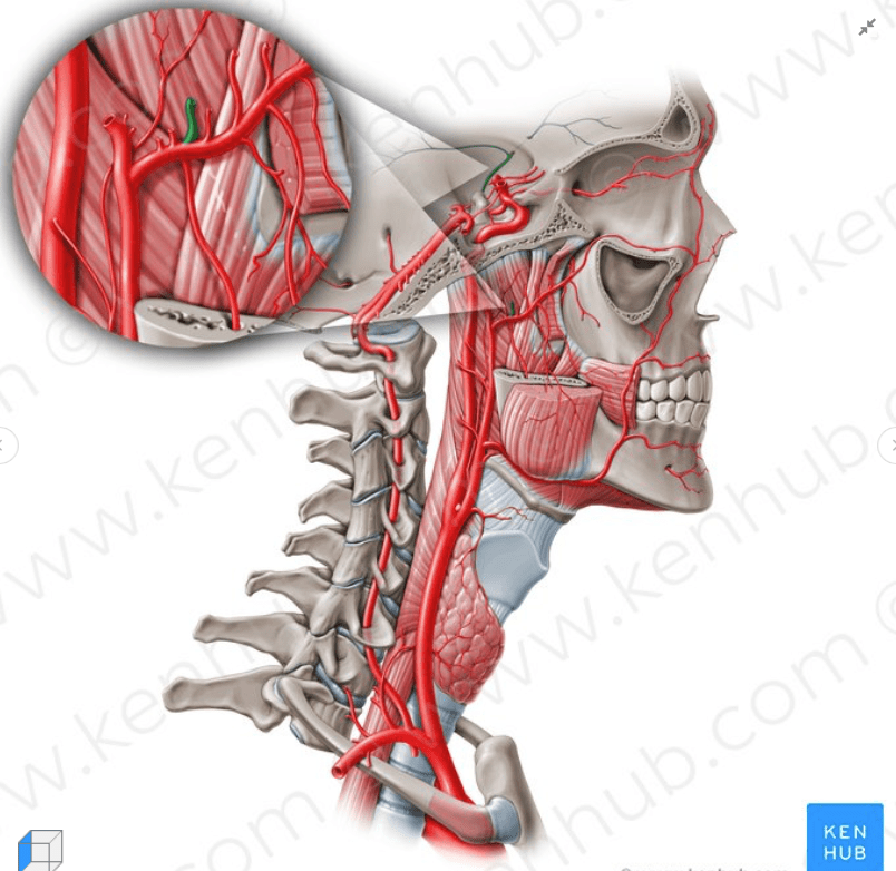
Middle meningeal artery
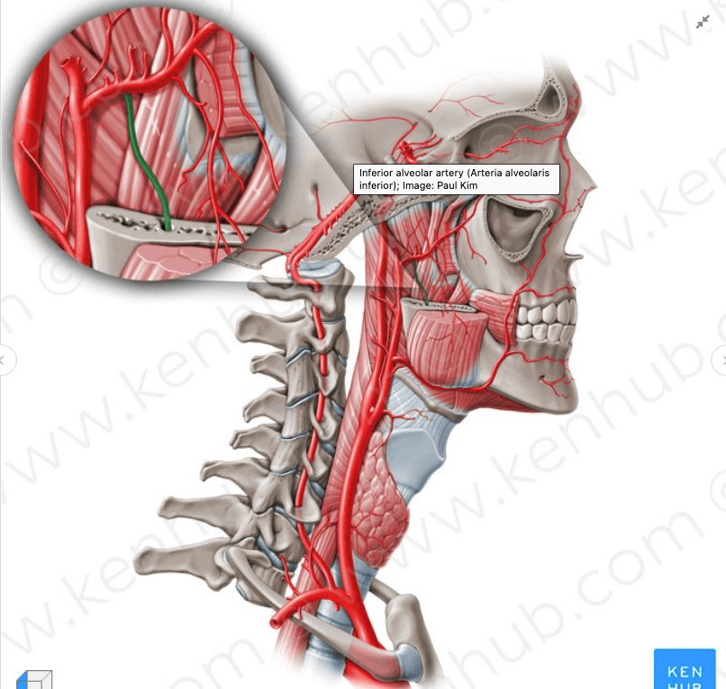
Inferior alveolar artery:
Which gives off its mylohyoid branch just prior to entering the mandibular foramen
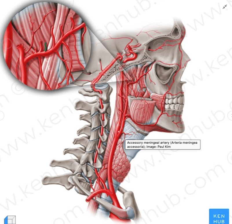
Accessory meningeal artery
Pterygoid portion (second part / muscular part):
The second or pterygoid portion (or muscular portion) runs obliquely forward and upward under cover of the ramus of the mandible and insertion of the temporalis muscle:
On the superficial (very infrequently on the deep) surface of the lateral pterygoid muscle.
It then passes between the two heads of origin of this muscle and enters the fossa.
Branches include:
Masseteric artery
Pterygoid branches
Deep temporal arteries:
Anterior and posterior
Buccal (buccinator) artery
Pterygopalatine portions (third part):
The third or pterygopalatine portion lies in the pterygopalatine fossa in relation with the pterygopalatine ganglion.
This is considered the terminal branch of the maxillary artery.
Branches include:
Sphenopalatine artery:
Nasopalatine artery is the terminal branch of the maxillary artery
Descending palatine artery:
Greater palatine artery
Lesser palatine artery
Infraorbital artery
Posterior superior alveolar artery
Artery of pterygoid canal
Pharyngeal artery
Middle superior alveolar artery (could be a branch of the infraorbital artery)
Anterior superior alveolar arteries (could be a branch of the infraorbital artery)
Rodrigo Arrangoiz MS, MD, FACS a head and neck surgeon at the Braman Comprehensive Cancer Center at Mount Sinai Medical Center in Miami, Florida.
He is first author on some publications on oral cavity cancer:
Oral Tongue Cancer: Literature Review and Current Management
The proximity or direct extension of a primary tumor of the oral cavity to the mandible requires appropriate radiological studies to establish the presence and extent of bone involvement:
Although the absence of radiographic findings does not rule out bone invasion:
Bone destruction as seen on the radiograph confirms tumor invasion
Radionuclide bone scans:
Often are positive before the radiographic appearance of bone destruction:
But they seldom provide accurate information regarding the extent of bone invasion
Bone scans also may be positive in non-neoplastic conditions:
Such as inflammatory lesions
Plain radiographs of the mandible in the antero-posterior and oblique views:
Are not satisfactory as a routine screening test to establish or rule out bone destruction
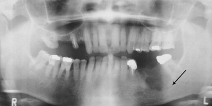
A panoramic view of the mandible (an orthopantomogram):
Is helpful to assess the general architecture of the mandible in relation to the dento-alveolar structures and invasion by the tumor (Figure)
However, for technical reasons:
The midline of the mandible near the symphysis is not adequately evaluated by a panoramic view
In addition, early invasion of the lingual cortex of the mandible is not seen on a panoramic view
Occlusal films of the body of the mandible and intraoral dental films:
Often are most accurate in demonstrating early invasion by a tumor
CT scans of the mandible:
Generally are not optimal for routine screening:
But may be considered in certain circumstances:
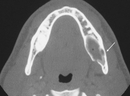
Such as primary tumors of the mandible and lesions where soft tissue extension from tumors involving the ascending ramus of the mandible is suspected (Figure)
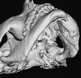
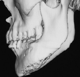
Three-dimensional reconstructions of CT images provide an excellent overview of the mandible or maxilla from any desired angle
A computerized tomogram of the oral cavity and neck:
Is the standard initial radiographic study for assessment of locoregional extent of the tumor
It allows comprehensive evaluation of neck nodes and also the relationship of the primary tumor to adjoining bone:
Especially in situations such as primary tumors of the mandible and lesions where soft-tissue extension from tumors involving the ascending ramus of the mandible is suspected
Three-dimensional reconstructions of the mandible of a patient with an ossifying fibroma of the body of the mandible on the left-hand side causing expansion and involving the lingual cortex are shown in the Figures
A three-dimensional CT scan and a one-to-one reproduction of the CT scan:
Are of great value to the surgeon for mandible reconstruction with a microvascular free flap
The basis and need for elective nodal treatment in head and neck cancer:
Have been based largely on surgical series evaluating pathologic nodal involvement found on elective neck dissection in patients with clinically negative necks
In a consecutive series of 1,081 head and neck cancer patients undergoing radical neck dissection:
The incidence of pathologic node involvement:
Was 33% among those undergoing elective neck surgery
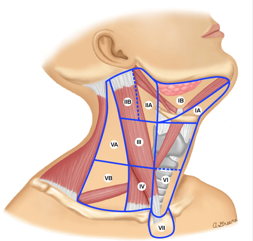
The pathologic findings identified the nodal stations at risk by tumor site:
To establish the rationale for selective neck dissection (SND) as the elective surgical procedure
Several reports have summarized the risk for metastases and nodal stations at risk
Some general observations from such data can be made:
Regarding larynx cancers:
Candela reported the Memorial Sloan Kettering Cancer Center (MSKCC) experience in determining the patterns of cervical nodal metastases in 247 larynx cancer patients undergoing radical neck dissections:
The majority of patients (n = 189) were supraglottic larynx and 58 were glottic
Pathologic nodal involvement:
Was found in 37% undergoing elective neck dissection
It is noted that cervical nodes spread in a similar fashion whether the patients are clinically node negative or positive:
With predominant involvement of:
Level II and III jugular nodes
In clinically node-negative patients:
The incidence of involvement of level I and V:
Is less than 5% with less than 10% involvement of level IV
In node-positive patients:
The incidence of level IV node increases from 15% to 31% with greater involvement of levels II and III
In clinically node-positive patients:
Very rarely did patients present with isolated level I nodal metastases without involvement of the jugular nodes
Shah and Candela reported that among oropharynx or hypopharynx cancers:
Treated with elective radical neck dissection:
Occult metastases are found in 26%
Level I and V were involved in only 1.4%:
Always in association with nodal disease at level II to IV
No skip metastases were reported
Among oropharynx patients:
Levels II to IV were predominantly involved
Among hypopharynx lesions:
The primary levels involved were levels II and III
In patients clinically node positive undergoing therapeutic neck dissection:
The incidence of level I and V involvement increased to about 10% to 15%:
However, levels II to IV were predominantly involved
Level V involvement:
Only occurred in association with nodal involvement at levels II to IV
Whereas the incidence isolated level I involvement without levels II to IV involvement (“skip metastasis”):
Occurred in 0.4%:
Thus, based on these studies, elective treatment of the neck in oropharynx or hypopharynx can be directed at levels II to IV
Among oral cavity patients:
The incidence of nodal disease was 34% on elective evaluation
The majority of metastatic nodes involved:
Levels I to III:
With only 1.5% incidence of skip metastasis to level IV
Level V involvement:
Is found in only 0.5% with occult disease simultaneously involving other levels
Among those undergoing therapeutic neck dissections:
The incidence of level IV involvement increased to 20%
Level V was 4% always restricted to lower gum or floor of mouth primary sites
The need for elective treatment not only relates to the estimated probability of nodal involvement and usually is implemented when the risk is 20% or greater but also relates to the morbidity of such treatment as well as the adequacy of coverage












 Rodrigo Arrangoiz MS, MD, FACS, FSSO