My name is Rodrigo Arrangoiz I am a breast surgeon/ thyroid surgeon / parathyroid surgeon / head and neck surgeon / surgical oncologist that works at Center for Advanced Surgical Oncology in Miami, Florida.
I was trained as a surgeon at Michigan State University from (2005 to 2010) where I was a chief resident in 2010. My surgical oncology and head and neck training was performed at the Fox Chase Cancer Center in Philadelphia from 2010 to 2012. At the same time I underwent a masters in science (Clinical research for health professionals) at the University of Drexel. Through the International Federation of Head and Neck Societies / Memorial Sloan Kettering Cancer Center I performed a two year head and neck surgery and oncology / endocrine fellowship that ended in 2016.
Mi nombre es Rodrigo Arrangoiz, soy cirujano oncólogo / cirujano de tumores de cabeza y cuello / cirujano endocrino que trabaja Center for Advanced Surgical Oncology en Miami, Florida.
Fui entrenado como cirujano en Michigan State University (2005 a 2010 ) donde fui jefe de residentes en 2010. Mi formación en oncología quirúrgica y e n tumores de cabeza y cuello se realizó en el Fox Chase Cancer Center en Filadelfia de 2010 a 2012. Al mismo tiempo, me sometí a una maestría en ciencias (investigación clínica para profesionales de la salud) en la Universidad de Drexel. A través de la Federación Internacional de Sociedades de Cabeza y Cuello / Memorial Sloan Kettering Cancer Center realicé una sub especialidad en cirugía de cabeza y cuello / cirugia endocrina de dos años que terminó en 2016.
Chang SL, Wu TC, Yiu CY. Tornwaldt’s Cyst Formation After Concurrent Chemoradiotherapy for Nasopharyngeal Carcinoma. J Laryngol Otol. 2006.
Sekiya K, Watanabe M, Nadgir RN, et al. Nasopharyngeal Cystic Lesions: Tornwaldt and Mucous Retention Cysts of the Nasopharynx: Findings on MR Imaging. J Comput Assist Tomogr. 2014.
Ikushima I, Korogi Y, Makita O, et al. MR Imaging of Tornwaldt’s Cysts. AJR Am J Roentgenol. 1999.
Turan S, Gürbüz MK, Kaya E, et al. Is Transnasal Endoscopic Marsupialization Sufficient in Thornwaldt Cysts? J Craniofac Surg. 2020.
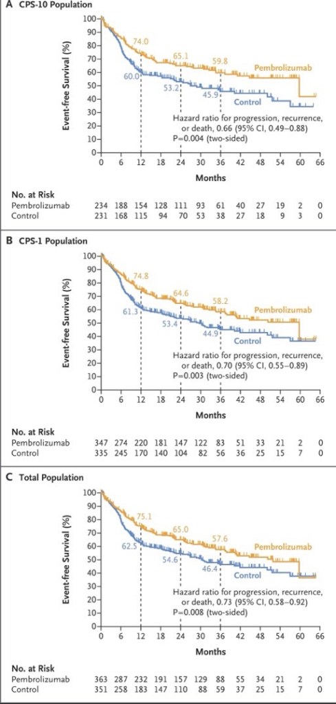
Perioperative Immunotherapy – KEYNOTE-689 Continues to Shape Practice:
The KEYNOTE-689 phase 3 trial:
Which led to the FDA approval of perioperative pembrolizumab (June 2025):
For resectable locally advanced HNSCC with PD-L1 CPS ≥ 1:
Remains a central focus
New ASCO 2026 exploratory analyses demonstrate that the EFS benefit of neoadjuvant / adjuvant pembrolizumab:
Persists across surgical outcome subgroups, including:
Patients with and without extranodal extension or positive margins
Notably, fewer patients in the pembrolizumab arm had ENE or positive margins post-surgery:
Suggesting neoadjuvant pembrolizumab contributes to pathologic downstaging
A pooled meta-analysis of three phase 3 trials (KEYNOTE-689, NIVOPOSTOP, and IMvoke010; n=1,786) presented at ASCO 2026:
Confirmed a significant improvement in event-free / disease-free survival with perioperative or adjuvant PD-1 / PD-L1 blockade (pooled HR 0.79, 95% CI 0.68–0.91):
With no meaningful increase in treatment-related deaths
The following figure from the KEYNOTE-689 trial illustrates the event-free survival benefit across PD-L1 subgroups:
Neoadjuvant Bispecific Antibody Combinations: Ivonescimab Leads the Way:
Several ASCO 2026 abstracts highlight the emerging role of bispecific antibodies in the neoadjuvant setting:
All patients with CPS >30 achieved pCR, and 100% laryngeal / pharyngeal preservation was achieved
A randomized phase II trial comparing ivonescimab (PD-1 / VEGF), cadonilimab (PD-1 /CTLA-4), and penpulimab (PD-1 alone), each combined with chemotherapy:
Showed the highest pCR rate with ivonescimab at 60%, compared to 42.1% with cadonilimab and 40% with single-agent PD-1
An 87.5% ORR and a 95.8% larynx preservation rate in resectable LA-HNSCC:
With all p16-positive and CPS ≥ 20 patients responding
Novel Agents in Recurrent / Metastatic HNSCC:
Bispecific antibodies are generating significant excitement in the recurrent / metastatic setting:
Ficerafusp alfa (EGFR × TGF-β) + pembrolizumab:
Two-year follow-up data showed a confirmed ORR of 54% in HPV-negative R/M HNSCC (21% CR), with a median DOR of 21.7 months and median OS of 21.3 months
The phase 2 / 3 FORTIFI-HN01 trial is now actively enrolling
Petosemtamab (EGFR × LGR5) + pembrolizumab:
Updated phase 2 data showed a 60% ORR (including 5 CRs) in first-line PD-L1+ R/M HNSCC, with median DOR of 11 months
Two phase 3 trials (LiGeR-HN1 and LiGeR-HN2) are recruiting
CRB-701 (Nectin-4 ADC):
A phase 1 / 2 study in heavily pretreated R/M HNSCC (85% refractory to immunotherapy and platinum):
Showed a confirmed ORR of 33.3% at both 2.7 and 3.6 mg/kg doses, regardless of HPV status
Becotatug vedotin (EGFR ADC):
A new randomized phase II trial is evaluating neoadjuvant becotatug vedotin alone or combined with immune checkpoint inhibitors in resectable LA-HNSCC
The deep response rate was 87.9%, with 86% of patients receiving de-escalated definitive therapy
At 23 months median follow-up, 2-year PFS was 86% and OS was 100%
Circulating tumor HPV-DNA was significantly associated with recurrence
Evolving First-Line R / M HNSCC Pipeline:
A landscape analysis presented at ASCO 2026 identified 145 active regimens (111 unique assets) in the first-line R / M HNSCC pipeline
Key trends include a shift toward chemotherapy-free combinations alongside PD-1 blockade:
With bispecific antibodies (10.8%), ADCs (10.8%), and cancer vaccines (9.0%) representing the most common novel modalities
Eight key phase 3 trials are underway:
All incorporating PD-1 as a target, with pembrolizumab as the backbone in 7 of 8
Overall, ASCO 2026 highlights a transformative period in head and neck oncology:
With perioperative immunotherapy now established as a new standard, bispecific antibodies and ADCs showing compelling early efficacy, and biomarker-driven strategies (PD-L1 CPS, HPV status, ctHPV-DNA, MRD) increasingly guiding treatment selection
References
Neoadjuvant and Adjuvant Pembrolizumab in Locally Advanced Head and Neck Cancer. Uppaluri R, Haddad RI, Tao Y, et al. The New England Journal of Medicine. 2025;393(1):37-50. doi:10.1056/NEJMoa2415434.
FDA approves neoadjuvant and adjuvant pembrolizumab for resectable locally advanced head and neck squamous cell carcinoma | FDA. Food and Drug Administration. 2025-06-13.
Neoadjuvant and adjuvant pembrolizumab (pembro) plus standard of care (SOC) for resectable locally advanced head and neck squamous cell carcinoma (LA HNSCC): Efficacy by surgical outcomes in the phase 3 KEYNOTE-689 trial.. Adkins D, Haddad R, Tao Y, et al. Journal of Clinical Oncology. 2026;44(Suppl 16):6057. doi:10.1200/JCO.2026.44.16_suppl.6057.
Perioperative or adjuvant PD-1/PD-L1 blockade with curative-intent multimodality therapy for locally advanced head and neck squamous cell carcinoma: A systematic review and meta-analysis of randomized trials.. Daher S, Daher H, Altal H, et al. Journal of Clinical Oncology. 2026;44(Suppl 16):e18074. doi:10.1200/JCO.2026.44.16_suppl.e18074.
Neoadjuvant ivonescimab (AK112, a PD-1/VEGF bispecific antibody) combined with nab-paclitaxel and cisplatin (AP) for resectable locally advanced head and neck squamous cell carcinoma (LA-HNSCC): An exploratory phase II study.. Kunyu Yang, Xiaomeng Zhang and Lu Wen. Journal of Clinical Oncology. 2026;44(Suppl 16):6014. doi:10.1200/JCO.2026.44.16_suppl.6014.
Neoadjuvant immunotherapy in combination with chemotherapy in resectable locally advanced head and neck squamous cell carcinoma: Updated efficacy and safety data from a randomized phase II trial.. Liu L, Chen F, Li Y, et al. Journal of Clinical Oncology. 2026;44(Suppl 16):6091. doi:10.1200/JCO.2026.44.16_suppl.6091.
Neoadjuvant adebrelimab plus chemotherapy in untreated locally advanced head and neck squamous cell carcinoma: Efficacy and biomarker insights from a single-arm phase 2 trial.. Fang R, Lei W, Huang B, et al. Journal of Clinical Oncology. 2026;44(Suppl 16):6106. doi:10.1200/JCO.2026.44.16_suppl.6106.
Ficerafusp Alfa (BCA101) With Pembrolizumab for Recurrent or Metastatic Head and Neck Squamous Cell Carcinoma: Two-Year Results of an Expansion Cohort of a Phase I/Ib Trial. Hanna GJ, Zandberg DP, Wong DJ, et al. Journal of Clinical Oncology : Official Journal of the American Society of Clinical Oncology. 2026;:JCO2502027. doi:10.1200/JCO-25-02027.
A multicenter, randomized, double-blind, phase 2/3 study of ficerafusp alfa (BCA101) or placebo in combination with pembrolizumab for first-line treatment of HPV-negative, PD-L1–positive, recurrent or metastatic (R/M) head and neck squamous cell carcinoma (HNSCC): FORTIFI-HN01.. Ferrarotto R, Kaczmar J, Spigel D, et al. Journal of Clinical Oncology. 2026;44(Suppl 16):TPS6129. doi:10.1200/JCO.2026.44.16_suppl.TPS6129.
Petosemtamab (MCLA-158) with pembrolizumab as first-line (1L) treatment of PD-L1+ recurrent/metastatic (r/m) head and neck squamous cell carcinoma (HNSCC): Phase 2 trial. Herpen C, Daste A, Arrazubi V, et al. Journal of Clinical Oncology. 2025;43(Suppl 16):6024. doi:10.1200/JCO.2025.43.16_suppl.6024.
LiGeR-HN Phase III Trials of Petosemtamab + Pembrolizumab and Petosemtamab Monotherapy in Recurrent or Metastatic HNSCC. Machiels JP, Fayette J, Haddad R, et al. Future Oncology (London, England). 2025;21(16):2007-2016. doi:10.1080/14796694.2025.2511470.
A phase 1/2 study of the next-generation nectin-4–targeting antibody-drug conjugate CRB-701 (SYS6002) in patients with recurrent or metastatic head and neck squamous cell carcinoma.. Mantia C, Hanna G, Loriot Y, et al. Journal of Clinical Oncology. 2026;44(Suppl 16):6062. doi:10.1200/JCO.2026.44.16_suppl.6062.
A randomized, non-comparative, multicenter phase II trial of neoadjuvant becotatug vedotin alone or combined with immune checkpoint inhibitors (penpulimab/ivonescimab) in resectable locally advanced head and neck squamous cell carcinoma.. Wei X, Xiang Z, Zeng Y, et al. Journal of Clinical Oncology. 2026;44(Suppl 16):TPS6135. doi:10.1200/JCO.2026.44.16_suppl.TPS6135.
Neoadjuvant HPV16-specific viral immunotherapy (HB200) plus chemotherapy with response-adapted de-escalation in HPV16+ oropharyngeal squamous cell carcinoma: TARGET-HPV trial.. Rosenberg A, Juloori A, Cursio J, et al. Journal of Clinical Oncology. 2026;44(Suppl 16):6097. doi:10.1200/JCO.2026.44.16_suppl.6097.
Emerging trends in research strategies in the first-line recurrent or metastatic head and neck cancer (R/M SCCHN) landscape: A top-level analysis by Oncofocus.. Shukla A, Keeshara V, Chamaria M, et al. Journal of Clinical Oncology. 2026;44(Suppl 16):e18029. doi:10.1200/JCO.2026.44.16_suppl.e18029.
Recent Highlights and Breakthroughs in Immunotherapy for Head and Neck Cancers. Vuille JA, Szturz P. Current Opinion in Oncology. 2026;38(3):201-211. doi:10.1097/CCO.0000000000001211.
Immunotherapy in Locally Advanced Head and Neck Squamous Cell Carcinoma: The Current Status and Future Outlook. Köylü B, Selçukbiricik F, Aksoy S, Güven DC. Critical Reviews in Oncology/Hematology. 2026;:105145. doi:10.1016/j.critrevonc.2026.105145.
PART A. HPV-MEDIATED (p16-POSITIVE) OROPHARYNGEAL CANCER — AJCC 8th ed., 2017 (ST-7) – Note: Tis and T4b categories do not exist in this system
PRIMARY TUMOR (T)
T0: No primary identified
T1: Tumor 2 cm or smaller in greatest dimension
T2: Tumor larger than 2 cm but not larger than 4 cm in greatest dimension
T3: Tumor larger than 4 cm in greatest dimension, or extension to lingual surface of epiglottis
T4: Moderately advanced local disease — tumor invades the larynx, extrinsic muscle of tongue, medial pterygoid, hard palate, or mandible, or beyond:
Mucosal extension to the lingual surface of the epiglottis from primary tumors of the base of tongue and vallecula does not constitute invasion of the larynx
CLINICAL REGIONAL LYMPH NODES (cN)
NX: Regional lymph nodes cannot be assessed
N0: No regional lymph node metastasis
N1: One or more ipsilateral lymph nodes, none larger than 6 cm
N2: Contralateral or bilateral lymph nodes, none larger than 6 cm
N3: Lymph node(s) larger than 6 cm
PATHOLOGICAL REGIONAL LYMPH NODES (pN)
NX: Regional lymph nodes cannot be assessed
pN0: No regional lymph node metastasis
pN1: Metastasis in 4 or fewer lymph nodes
pN2: Metastasis in more than 4 lymph nodes
DISTANT METASTASIS (M)
M0: No distant metastasis
M1: Distant metastasis
HISTOLOGIC GRADE (G)
No grading system exists for HPV-mediated oropharyngeal tumors
CLINICAL PROGNOSTIC STAGE GROUPS (p16+)
Stage I:
T0, T1, T2 — N0, N1 — M0
Stage II:
T0, T1, T2 — N2 — M0; or T3 — N0, N1, N2 — M0
Stage III:
T0, T1, T2, T3 — N3 — M0; or T4 — N0, N1, N2, N3 — M0
Stage IV:
Any T — Any N — M1
PATHOLOGICAL PROGNOSTIC STAGE GROUPS (p16+)
Stage I:
T0, T1, T2 — N0, N1 — M0
Stage II:
T0, T1, T2 — N2 — M0; or T3, T4 — N0, N1 — M0
Stage III:
T3, T4 — N2 — M0
Stage IV:
Any T — Any N — M1
PART B. p16-NEGATIVE OROPHARYNGEAL CANCER — AJCC 8th ed., 2017 (ST-4, ST-5, ST-6):
Same system as hypopharynx; excludes p16+ oropharyngeal and nasopharyngeal cancers
PRIMARY TUMOR (T)
TX: Primary tumor cannot be assessed
Tis: Carcinoma in situ
T1: Tumor 2 cm or smaller in greatest dimension
T2: Tumor larger than 2 cm but not larger than 4 cm in greatest dimension
T3: Tumor larger than 4 cm in greatest dimension, or extension to lingual surface of epiglottis
T4a: Moderately advanced local disease — tumor invades the larynx, extrinsic muscle of tongue, medial pterygoid, hard palate, or mandible
T4b: Very advanced local disease — tumor invades lateral pterygoid muscle, pterygoid plates, lateral nasopharynx, or skull base, or encases carotid artery:
Mucosal extension to the lingual surface of the epiglottis from primary tumors of the base of tongue and vallecula does not constitute invasion of the larynx
CLINICAL REGIONAL LYMPH NODES (cN)
NX: Regional lymph nodes cannot be assessed
N0: No regional lymph node metastasis
N1: Metastasis in a single ipsilateral lymph node, 3 cm or smaller, ENE(−)
N2a: Metastasis in a single ipsilateral node larger than 3 cm but not larger than 6 cm, ENE(−)
N2b: Metastases in multiple ipsilateral nodes, none larger than 6 cm, ENE(−)
N2c: Metastases in bilateral or contralateral nodes, none larger than 6 cm, ENE(−)
N3a: Metastasis in a lymph node larger than 6 cm, ENE(−)
N3b: Metastasis in any node(s) with clinically overt ENE(+)
PATHOLOGICAL REGIONAL LYMPH NODES (pN)
NX: Regional lymph nodes cannot be assessed
N0: No regional lymph node metastasis
N1: Metastasis in a single ipsilateral lymph node, 3 cm or smaller, ENE(−)
N2a: Metastasis in a single ipsilateral node 3 cm or smaller with ENE(+); or a single ipsilateral node larger than 3 cm but not larger than 6 cm, ENE(−)
N2b: Metastases in multiple ipsilateral nodes, none larger than 6 cm, ENE(−)
N2c: Metastases in bilateral or contralateral nodes, none larger than 6 cm, ENE(−)
N3a: Metastasis in a lymph node larger than 6 cm, ENE(−)
N3b: Metastasis in a single ipsilateral node larger than 3 cm with ENE(+); or multiple ipsilateral, contralateral, or bilateral nodes any with ENE(+); or a single contralateral node of any size with ENE(+)
Note: A designation of “U” or “L” may be used for any N category to indicate metastasis above (U) or below (L) the lower border of the cricoid
Clinical and pathological ENE should be recorded as ENE(−) or ENE(+)
DISTANT METASTASIS (M)
M0: No distant metastasis
M1: Distant metastasis
HISTOLOGIC GRADE (G)
GX: Grade cannot be assessed
G1: Well differentiated
G2: Moderately differentiated
G3: Poorly differentiated
G4: Undifferentiated
PROGNOSTIC STAGE GROUPS (p16-negative)
Stage 0:
Tis — N0 — M0
Stage I:
T1 — N0 — M0
Stage II:
T2 — N0 — M0
Stage III:
T3 — N0 — M0; or T1, T2, T3 — N1 — M0
Stage IVA:
T1, T2, T3 — N2 — M0; or T4a — N0, N1, N2 — M0
Stage IVB:
T4b — Any N — M0; or Any T — N3 — M0
Stage IVC:
Any T — Any N — M1
KEY DIFFERENCES BETWEEN THE TWO SYSTEMS
p16+ system has no Tis, no T4a/T4b split (single T4 category), and no N2a/b/c or N3a/b subcategories
p16+ cN is based on laterality and size only; extranodal extension is not a cN determinant
p16+ pN is based solely on the number of positive nodes (≤ 4 vs > 4)
p16+ Stage IV is reserved exclusively for M1 disease; there is no Stage IVA/IVB/IVC
p16+ clinical and pathological stage groupings differ from each other and must not be used interchangeably
p16 IHC is required for all oropharyngeal cancers; positivity threshold is ≥ 70% of tumor cells with moderate-to-strong nuclear and cytoplasmic staining
Confirmatory direct HPV testing (PCR or RNA in situ hybridization) is recommended, especially for clinical trials; PCR adds sensitivity, ISH adds specificity:
Patients with p16+/HPV+ tumors have better prognosis than p16+/HPV-negative tumors
Routine HPV/p16 testing of non-oropharyngeal, non-sinonasal head and neck sites is not recommended
AJCC 9th Edition (Version 9) Changes — HPV-Positive Oropharyngeal Carcinoma – NCCN Guidelines, Head and Neck Cancers (v2.2026):
Still base clinical staging definitions on AJCC 8th edition (ST-4 for p16-negative, ST-7 for p16-positive):
Version 9 is not yet incorporated
Version 9 changes apply only to HPV-positive (p16+) oropharyngeal carcinoma:
The p16-negative oropharynx / hypopharynx system is unchanged in the current NCCN tables
PART A. VERSION 9 PATHOLOGICAL CLASSIFICATION (AJCC9V) — HPV-POSITIVE
Rationale for revision:
In AJCC 8th edition, > 85% of cases were pN1 and > 80% were Stage I, producing wide hazard variance within dominant groupings and a disconnect between stage and treatment
Pathological extranodal extension (pENE), widely used in trials and clinical decision making, was absent from the 8th edition system:
Derivation: 14,447 surgically treated patients across 984 US facilities (derivation n = 7,768; validation n = 6,679), treated 2010–2019; median follow-up 52.4 months; 31.5% pENE-positive
Key prognostic findings:
Mortality risk rose with each additional metastatic lymph node (HR 1.20, 95% CI 1.11–1.29, p < 0.0001), with an optimal cutoff at 4.3 nodes
pENE independently associated with mortality (HR 1.47, 95% CI 1.30–1.65, p < 0.0001)
Extent of pENE (minor vs major) showed no significant prognostic difference
VERSION 9 pN CLASSIFICATION (HPV-positive)
pN0: 0 positive lymph nodes
pN1a: 1 positive lymph node, ENE-negative
pN1b:
2 to 4 positive lymph nodes, ENE-negative
pN2:
> 4 positive lymph nodes and ENE-negative; OR 1 to 4 positive lymph nodes and ENE-positive
pN3:
> 4 positive lymph nodes and ENE-positive
Compare AJCC 8th edition: pN1 = 1 to 4 positive nodes; pN2 = > 4 positive nodes; ENE not incorporated
VERSION 9 PATHOLOGICAL STAGE GROUPS (HPV-positive)
Stage I:
T0 to T2, N0 to N1, M0
Stage II:
T0 to T2, N2 to N3, M0; or T3, N0 to N2, M0
Stage III:
T3, N3, M0; or T4, N0 to N3, M0
Stage IV:
Any T, Any N, M1
Compare AJCC 8th edition pathological: Stage I = T0 to 2 N0–1; Stage II = T0 to 2 N2 or T3 to 4 N0 to 1; Stage III = T3 to 4 N2; Stage IV = M1
PERFORMANCE
Stage distribution rebalanced:
AJCC8E 81.4% Stage I / 15.0% Stage II / 3.6% Stage III versus AJCC9V 54.9% Stage I / 41.3% Stage II / 3.8% Stage III
AJCC9V showed superior hazard consistency, outcome prediction, and balance, but NOT superior hazard discrimination, versus AJCC8E
Endorsed by the AJCC Expert Panel on HPV-positive oropharyngeal carcinoma by Delphi consensus
Caveat:
T4N1 cases fit imperfectly within their assigned Stage III grouping, likely reflecting small numbers, as few T4 cases undergo surgery; these were retained in Stage III for clinical consistency
PART B. VERSION 9 CLINICAL CLASSIFICATION — HPV-POSITIVE
Principal change:
Incorporation of imaging-detected extranodal extension (iENE)
Rule:
Each N category is reclassified one stratum higher when iENE is present; iENE-negative cases are unchanged
Derivation: ICON-N dataset, 2,053 patients from 4 institutions, with validation in an independent CHUM cohort (n = 451); median follow-up 5.1 years:
iENE was present in 37.4% of cN-positive patients and was the strongest prognostic nodal feature: adjusted HR 2.43 (95% CI 1.96–3.03) in ICON-N and 2.04 (95% CI 1.28–3.23) in CHUM
The iENE-adjusted schema outperformed AJCC 8th edition for disease-free and overall survival (overall normalized score 2 vs 3)
External validation:
In a retrospective series of 26 node-positive HPV-associated oropharyngeal carcinoma patients treated with definitive chemoradiotherapy, iENE was present in 61.5% and associated with lower 2-year disease-free survival (68.8% vs 100%, p = 0.048)
Reclassification by version 9 improved prognostic discrimination over the 8th edition (p = 0.043)
PART C. PRACTICAL IMPLICATIONS
Report the number of metastatic lymph nodes and ENE status explicitly on pathology, and iENE status on pretreatment CT / MRI, since both now drive N classification under version 9
Version 9 does not alter T categories or M categories for HPV-positive disease
Stage IV remains reserved for M1 disease
References:
National Comprehensive Cancer Network. NCCN Clinical Practice Guidelines in Oncology (NCCN Guidelines): Head and Neck Cancers. Version 2.2026. Staging tables ST-4, ST-5, ST-6 (oropharynx p16-negative and hypopharynx) and ST-7 (HPV-mediated p16-positive oropharynx); ORPH-1, ORPH-B.
Ho AS, Huang SH, O’Sullivan B, et al. Derivation and validation of the AJCC9V pathological stage classification for HPV-positive oropharyngeal carcinoma: a multicentre registry analysis. Lancet Oncol. 2025.
Cramer JD, Hicks KE, Rademaker AW, Patel UA, Samant S. Validation of the eighth edition American Joint Committee on Cancer staging system for human papillomavirus-associated oropharyngeal cancer. Head Neck. 2018.
Hall SR, Neel GS, Chang BA, et al. American Joint Committee on Cancer eighth edition human papilloma virus positive
Nasopharyngeal carcinoma is staged separately from all other head and neck mucosal sites
PRIMARY TUMOR (T):
TX — Primary tumor cannot be assessed
T0 — No tumor identified, but EBV-positive cervical node involvement
Tis — Carcinoma in situ
T1 — Confined to nasopharynx, or extension to oropharynx and / or nasal cavity WITHOUT parapharyngeal involvement
T2 — Extension to parapharyngeal space, and / or adjacent soft tissue (medial pterygoid, lateral pterygoid, prevertebral muscles)
T3 — Unequivocal infiltration of bony structures at skull base, cervical vertebra, pterygoid structures, and / or paranasal sinuses
T4 — Intracranial extension, cranial nerve involvement, hypopharynx, orbit, parotid gland, and / or extensive soft tissue infiltration beyond the anterolateral surface of the lateral pterygoid muscle
REGIONAL LYMPH NODES (N)
Key landmarks:
6 cm size cutoff; caudal border of the cricoid cartilage
N0 — No regional lymph node metastasis
N1 — Unilateral cervical node(s) and / or unilateral or bilateral retropharyngeal node(s), ≤ 6 cm, above the caudal border of the cricoid cartilage, without advanced extranodal extension (ENE)
N2 — Bilateral cervical node(s), ≤ 6 cm, above the caudal border of the cricoid cartilage, without advanced ENE
N3 — Node(s) > 6 cm, and / or extension below the caudal border of the cricoid cartilage, and / or advanced radiologic ENE with involvement of adjacent muscles, skin, and / or neurovascular bundle
DISTANT METASTASIS (M)
M0 — No distant metastasis
M1a — ≤ 3 metastatic lesions
M1b — > 3 metastatic lesions
HISTOLOGIC GRADE
No grading system is applied to nasopharyngeal carcinoma
STAGE GROUPS
Stage 0 — Tis, N0, M0
Stage IA — T1 to T2, N0, M0
Stage IB — T0 to T2, N1, M0
Stage II — T0 to T2, N2, M0
Stage II — T3, N0 to N2, M0
Stage III — T4, Any N, M0
Stage III — Any T, N3, M0
Stage IVA — Any T, Any N, M1a
Stage IVB — Any T, Any N, M1b
STAGING WORKUP:
Nasopharyngeal fiberoptic examination
Biopsy of primary site or FNA of neck node
MRI with and without contrast from skull base to clavicle (± CT of skull base / neck for bony erosion)
FDG-PET/CT and / or contrast-enhanced chest CT for distant disease (bone scan if PET/CT not performed)
Plasma EBV DNA testing:
HPV testing may be considered to inform etiology
KEY CHANGES FROM 8TH EDITION:
Advanced radiologic ENE added as an N3 criterion
Stage I expanded to T1 to 2 N0 to 1 and subdivided into IA (N0) and IB (N1)
Former stage III and IVA down-classified to II and III; all M0 disease now falls within stages I to III
Stage IV reserved exclusively for metastatic disease, subdivided by ≤ 3 cm vs > 3 cm lesions
T3 now requires unequivocal bone involvement
The stage groups, T/N/M definitions, and workup elements above are taken directly from the NCCN-endorsed AJCC 9th edition tables and the nasopharynx workup algorithm
The summarized changes from the 8th edition reflect the published TNM-9 derivation cohort of 4,914 patients:
Note that treatment stratification in the NCCN algorithm keys off TNM categories rather than stage groups, so the stage group alone does not determine whether concurrent chemoradiation or induction chemotherapy is indicated
Plasma EBV DNA, while part of workup, remains outside the anatomic staging system
NCCN Treatment by TNM Category – Nasopharyngeal Carcinoma (v2.2026):
All recommendations are category 2A unless otherwise indicated
Recommendations are based on clinical trial data in EBV-associated nasopharyngeal cancer
NON-METASTATIC DISEASE (M0)
T1, N0, M0:
Definitive RT alone
T2, N0, M0:
Definitive RT ± concurrent systemic therapy if high-risk features
T0 (EBV+) to T2, N1, M0 OR T3, N0, M0:
Concurrent systemic therapy / RT
Consider induction or adjuvant chemotherapy if high-risk features
T3, N1 to 3, M0 OR T4, N0 to 3, M0 OR T0 (EBV+)–T2, N2 to 3, M0:
Clinical trial (preferred)
OR induction chemotherapy followed by concurrent systemic therapy / RT (preferred; category 1)
OR concurrent systemic therapy / RT followed by adjuvant chemotherapy
OR concurrent systemic therapy / RT alone (category 2B)
High-risk features:
Bulky tumor volume
High serum EBV DNA copy number
METASTATIC DISEASE (T1 to 4, N0 to 3, M1):
Oligometastatic disease:
Induction chemotherapy (if PS 0 to 1), followed by one of:
RT to primary and regional nodes, and to oligometastases as indicated
Cisplatin / RT
Maintenance capecitabine
OR concurrent cisplatin + RT (if PS 0–1)
OR systemic therapy (if PS 0–2)
Widely metastatic, good performance status (PS 0 to 2):
Systemic therapy:
If complete or near-complete response:
Consider definitive RT to primary and regional nodes (preferred) and to oligometastases as indicated, or continued systemic therapy
Widely metastatic, poor performance status (PS 3 to 4):
Best supportive care
FOLLOW-UP:
All M0 pathways proceed to post systemic therapy / RT or post-RT neck evaluation, then routine follow-up
Recurrent or persistent disease is managed per the recurrent / advanced disease algorithm
KEY POINT:
Treatment assignment keys off TNM category, not AJCC 9th edition stage group:
For example, stage II under TNM-9 encompasses both T3N0M0 (chemoradiation) and T3N2M0 (induction chemotherapy followed by chemoradiation):
So the stage group alone is insufficient to select therapy
The M0 treatment branches, including the category 1 designation for induction chemotherapy followed by chemoradiation in T3N1 to 3 / T4/N2 to 3 disease and the category 2B designation for concurrent chemoradiation alone in that same group, come directly from the NCCN nasopharynx treatment algorithm
The metastatic branch separates oligometastatic disease — where locoregional RT to the primary and to metastatic sites is incorporated — from widely metastatic disease managed with systemic therapy, with definitive RT considered only after complete or near-complete response
Workup precedes this split, with M0 versus M1 determined by:
FDG-PET/CT and / or contrast chest CT
References:
National Comprehensive Cancer Network. NCCN Clinical Practice Guidelines in Oncology: Head and Neck Cancers, Version 2.2026. AJCC TNM Staging System for the Nasopharynx (9th ed., 2021), page ST-3.
National Comprehensive Cancer Network. NCCN Clinical Practice Guidelines in Oncology: Head and Neck Cancers, Version 2.2026. Staging (nasopharynx excluded from general mucosal head and neck staging systems).
National Comprehensive Cancer Network. NCCN Clinical Practice Guidelines in Oncology: Head and Neck Cancers, Version 2.2026. Cancer of the Nasopharynx: Workup (NASO-1).
National Comprehensive Cancer Network. NCCN Clinical Practice Guidelines in Oncology: Head and Neck Cancers, Version 2.2026. Cancer of the Nasopharynx: Treatment of Primary and Neck by Clinical Staging (NASO-2).
Pan JJ, Mai HQ, Ng WT, et al. Ninth Version of the AJCC and UICC Nasopharyngeal Cancer TNM Staging Classification. JAMA Oncol. 2024.
Pan JJ, Mai HQ, Ng WT, et al. Ninth Version of the AJCC and UICC Nasopharyngeal Cancer TNM Staging Classification. JAMA Oncol. 2024.
Pan JJ, Mai HQ, Ng WT, et al. Ninth Version of the AJCC and UICC Nasopharyngeal Cancer TNM Staging Classification. JAMA Oncol. 2024.
Rai P, Lakhani DA, Agarwal A, Bhatt AA. The 9th Version of the AJCC Staging System for Nasopharyngeal Carcinoma: A Guide for Radiologists. AJR Am J Roentgenol. 2025.
Du XJ, Wang GY, Zhu XD, et al. Refining the 8th Edition TNM Classification for EBV Related Nasopharyngeal Carcinoma. Cancer Cell. 2024.
Cao C, Treechairusame T, Wu Y, et al. Staging of Non-Metastatic Nasopharyngeal Carcinoma With PET/CT. Eur Radiol. 2026.
Lee VH, Kwong DL, Leung TW, et al. The Addition of Pretreatment Plasma Epstein-Barr Virus DNA Into the Eighth Edition of Nasopharyngeal Cancer TNM Stage Classification. Int J Cancer. 2019.
National Comprehensive Cancer Network. NCCN Clinical Practice Guidelines in Oncology: Head and Neck Cancers, Version 2.2026. Cancer of the Nasopharynx: Treatment of Primary and Neck for T1–4, N0–3, M1 (NASO-3).
The decision to extend a supraomohyoid neck dissection (levels I to III) to include level IV (extended supraomohyoid neck dissection):
Hinges on whether pN+ disease is discovered in levels I to III, the number of positive nodes, and the pN classification:
Below is a comprehensive review of the specific scenarios and supporting data
Scenario 1 – cN0 Neck → Intraoperative Discovery of pN+ Disease in Levels I to III:
This is the most clinically relevant scenario
When a patient undergoes END for a cN0 neck and positive nodes are found on frozen section or final pathology:
The risk of concurrent level IV to V disease rises substantially compared to the overall cN0 population
The Haas et al. (2025) study directly addressed this question in 61 cN0 patients who were found to be pN+ in levels I to III [1]:
9.8% (6/61) had metastases in levels IV to V:
Well above the 5% threshold generally used to justify elective dissection
> 1 positive node in levels I to III was the strongest predictor of level IV to V involvement (p = 0.027)
pN classification > pN2b significantly increased the prevalence of level IV to V metastases (p = 0.002)
Extracapsular spread (ECS) showed a trend toward increased IV to V involvement (p = 0.078), though not statistically significant in this cohort
This 9.8% rate stands in stark contrast to the 0.50% true skip metastasis rate in the overall cN0 population:
Underscoring that the discovery of pN+ disease in levels I to III fundamentally changes the risk calculus
Scenario 2 – cN+ Neck (Preoperatively Known Nodal Disease):
For the clinically node-positive neck, both ASCO and NCCN guidelines explicitly recommend including level IV in the dissection [2][3]
The ASCO guideline states:
An ipsilateral therapeutic selective neck dissection for a cN+ neck should include:
Nodal levels Ia, Ib, IIa, IIb, III, and IV with ≥ 18 lymph nodes (evidence-based, intermediate quality, moderate strength) [2]
The NCCN guidelines specify:
cN1 to cN2a–c disease warrants selective or comprehensive neck dissection [3]
The American Head and Neck Society review provides the supporting data:
Occult level IV nodal metastatic disease is reported in 11.1% to 23.7% of cN+ oral tongue cancers, and 15% of all patients undergoing therapeutic neck dissection had level IV metastases [4]:
This rate is high enough that level IV dissection should be considered even in the absence of overt metastatic lymph node involvement at that level [4]
Scenario 3 – When to Add Level V:
Level V dissection carries significant risk to the spinal accessory nerve and is not routinely recommended
However, the data support its inclusion in specific circumstances [4]:
Clinical involvement of levels I to IV:
Was associated with occult metastases in level V in 27% of oral cavity SCCs in one study
Level V should be considered in patients with bulky, multilevel nodal disease
The overall rate of level V metastasis:
In therapeutic neck dissections is only ~4%
Practical Decision Framework – When to Extend to Level IV:
Prognostic Implications of Level IV Disease:
Importantly, the discovery of level IV or V disease is not merely an anatomic finding:
It carries significant prognostic weight
The NCCN guidelines list nodal disease in levels IV or V as an adverse pathologic feature:
Which triggers consideration of adjuvant systemic therapy / RT [3]
A SEER-based study of 8,281 patients:
Found that 5-year disease-specific survival dropped from 42.0% for level I to III disease to 30.6% for level IV [5]
Furthermore, level IV to V metastasis:
Is an independent risk factor for distant metastasis on multivariate analysis [6]
Morbidity Considerations:
Extension to level IV is not without cost
The American Head and Neck Society review notes that dissection of level IV:
Is associated with a low but possible risk of injury to the phrenic nerve or brachial plexus and increases the risk of chylous fistula [4]:
These risks must be weighed against the oncologic benefit, particularly in the cN0 setting where the absolute risk of level IV disease is low
Summary:
For the cN0 neck with no intraoperative evidence of nodal disease:
Levels I to III remain the standard
However, when intraoperative frozen section or final pathology reveals pN+ disease:
Particularly > 1 positive node or pN > pN2b:
The risk of level IV to V involvement rises to ~10%:
Justifying extension to level IV [1]
For any preoperatively cN+ neck:
Level IV should be included as standard practice per ASCO and NCCN guidelines [2][3]
Level V should be reserved for:
Multilevel, bulky nodal disease
References:
1. In Vivo Probability of Metastases in Levels IV-V in Oral Squamous Cell Carcinoma With a cN0/pN+ Situation in Levels I-Iii. Haas L, Mischkowski RA, Knape U, Król KM, Sakkas A. In Vivo (Athens, Greece). 2025 Nov-Dec;39(6):3437-3444. doi:10.21873/invivo.14141.
2. Management of the Neck in Squamous Cell Carcinoma of the Oral Cavity and Oropharynx: ASCO Clinical Practice Guideline. Koyfman SA, Ismaila N, Crook D, et al. Journal of Clinical Oncology : Official Journal of the American Society of Clinical Oncology. 2019;37(20):1753-1774. doi:10.1200/JCO.18.01921.
3. Head and Neck Cancers. National Comprehensive Cancer Network. Updated 2025-12-08.
4. Oral Cavity Cancer Surgical and Nodal Management: A Review From the American Head and Neck Society. Eskander A, Dziegielewski PT, Patel MR, et al. JAMA Otolaryngology– Head & Neck Surgery. 2024;150(2):172-178. doi:10.1001/jamaoto.2023.4049.
5. Impact of Nodal Level Distribution on Survival in Oral Cavity Squamous Cell Carcinoma: A Population-Based Study. Marchiano E, Patel TD, Eloy JA, Baredes S, Park RC. Otolaryngology–Head and Neck Surgery : Official Journal of American Academy of Otolaryngology-Head and Neck Surgery. 2016;155(1):99-105. doi:10.1177/0194599816636356.
6. Risk Factors for Distant Metastasis in Locoregionally Controlled Oral Squamous Cell Carcinoma: A Retrospective Study. Tomioka H, Yamagata Y, Oikawa Y, et al. Scientific Reports. 2021;11(1):5213. doi:10.1038/s41598-021-84704-w.
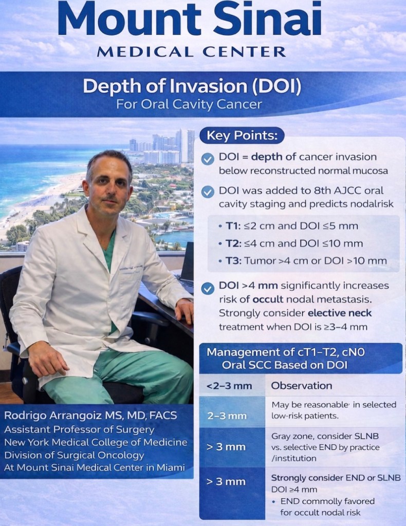
Guiding the decision to perform END in cN0 oral cavity SCC
The NCCN Guidelines stratify the decision as follows [1]:
DOI > 3 mm:
END should be strongly considered if RT is not already planned (supported by level 1 evidence from the D’Cruz / Tata Memorial trial)
The landmark D’Cruz et al. (2015) randomized trial of 500 patients with cT1 to T2 cN0 oral cavity SCC:
Demonstrated that upfront END significantly improved 3-year overall survival (80.0% vs. 67.5%) and disease-free survival (69.5% vs. 45.9%) compared to therapeutic neck dissection [2]
Absolute benefits: 12.5% points for OS and 23.6% point for DFS, corresponding to NNT of 8 to prevent one death and 4 to prevent one relapse
Adverse event rates were 6.6% with END vs. 3.6% with therapeutic dissection
Post hoc analysis showed no benefit for tumors with DOI ≤ 3 mm, while 28% of cN0 necks with DOI > 3 mm harbored occult metastases [3]
A 2025 meta-analysis of 17 studies (2,263 patients):
Confirmed END reduces regional recurrence (RR 0.47) and improves OS (RR 0.75) and DSS (RR 1.32) [4]
A validation study of 300 patients [5]:
Confirmed that DOI ≥ 4 mm is the optimal ROC-derived threshold (95.1% sensitivity, 52.9% specificity) for predicting occult nodal metastasis:
With regional recurrence-free survival significantly higher in the END group for DOI ≥ 4 mm (p = 0.002) [5]
A recent systematic review found:
Diagnostic thresholds converging around 4 mm for mixed oral cavity sites and 3 mm for high-risk subsites (floor of the mouth) [6]
The ASCO Clinical Practice Guideline (2019) recommends [2]:
cT2 to cT4, cN0:
Ipsilateral END should be performed (strong recommendation, high-quality evidence)
cT1, cN0:
Ipsilateral END should be performed:
Alternatively, close surveillance with specialized ultrasound may be offered for selected highly reliable patients
Tumor Size Considerations:
Under AJCC 8th edition staging, both surface size and DOI determine T classification [1]:
T1:
≤ 2 cm with DOI ≤ 5 mm
T2:
≤ 2 cm with DOI > 5 mm
> 2 cm and ≤ 4 cm with DOI ≤ 10 mm
T3 to T4:
T3:
> 4 cm
DOI > 10 mm
Locally advanced tumors carry ~ 40% to 50% risk of occult nodal metastases, making END essentially mandatory [2]
DOI cutoffs are more predictive of overall survival than T category based on tumor surface size alone [3]:
For T1 tumors, those with DOI ≤ 2 mm rarely demonstrate occult neck disease:
While DOI ≥ 2 mm confers at least a 20% risk of pN+ disease [3]
Subsite-Specific Considerations:
Oral Tongue:
The most extensively studied subsite
DOI ≥ 3 mm to 4 mm:
Is the standard threshold for END [3]
Higher propensity for skip metastases:
To level III (17.5% of specimens) compared to other subsites
Anterior tongue tumors at / near midline:
Warrant consideration for bilateral neck dissection [1]
Floor of Mouth (FOM):
FOM tumors cross the critical 20% threshold for nodal metastasis at a lower DOI (≥ 2 mm) compared to oral tongue (≥ 4 mm):
Based on a study of 343 patients:
41.7% nodal metastasis rate for FOM tumors 2.1 mm to 4 mm thick vs. 11.2% for tongue tumors of similar thickness [7]
FOM primaries have a predisposition for contralateral metastases even at earlier T stages:
With a 50% higher risk of contralateral metastasis and 2.6-times higher risk when FOM invasion / extension is present [3][2]
However, a more recent study of 825 patients:
Found no significant difference in the incidence of or correlation between DOI and nodal metastases when FOM was compared to other subsites [8]
SLN biopsy accuracy:
Is lower for FOM than for tongue primaries [1]
Buccal Mucosa:
Behaves more aggressively:
With occult metastasis rates as high as 32% and up to 43% presenting with clinical / radiographic nodal disease [3]
END is recommended for all buccal cases:
Irrespective of DOI given higher rates of nodal metastases even in early T-category disease [3]
A multicenter study of 101 patients confirmed END:
Improved survival in pT2, cN0 buccal SCC:
123 months vs. 26 months, p = 0.009) [9]
END demonstrated improved:
Locoregional recurrence-free rates (61% vs. 38%, p = 0.042)
5-year DFS (75% vs. 63%, p = 0.019) [10]
Other Subsites (Alveolar Ridge, Retromolar Trigone, Hard Palate):
Generally follow the same DOI-based principles
Hard palate and upper gingiva:
May not lend themselves well to SLN biopsy [1]
Levels of Dissection – I to III vs. I to IV:
This is the NCCN treatment algorithm for cT1 to cT2, N0 oral cavity SCC:
For the cN0 neck:
The standard END is a supraomohyoid neck dissection (SOHND) encompassing levels I to III
Both the NCCN and ASCO guidelines recommend at minimum:
Levels Ia, Ib, II, and III, with an adequate yield of ≥ 18 lymph nodes [1][2]
Two randomized trials comparing SOHND (levels I to III) versus modified radical neck dissection (levels I to V):
Showed the adequacy of SOHND, with more extensive dissection increasing morbidity (particularly shoulder dysfunction) without survival or recurrence benefit:
Only 3.7% of occult metastases were detected in levels IV and V [2]
The American Head and Neck Society review consolidates the recommendation:
Level I to III dissection in the cN0 setting, with consideration of level IV inclusion in higher-risk tumors [3]
Skip Metastasis to Level IV:
The Key Data:
The Warshavsky et al. (2019) systematic review and meta-analysis in JAMA Otolaryngology (13 studies, 1,359 patients) provides the most comprehensive data [11]:
Overall rate of level IV involvement in cN0 patients:
2.53% (95% CI, 1.64%–3.55%)
True skip metastasis rate:
Level IV positive without levels I to III involvement): 0.50% (95% CI, 0.09%–1.11%)
Subgroup by T stage:
Level IV involvement was 0% for stages I to II and 0% for stages III to IV in the skip metastasis analysis
Oral tongue subsite:
Level IV involvement was notably higher at 3.60% (95% CI, 2.09%–5.42%):
The only subsite with significant findings
A large audit of 1,004 cases:
Confirmed skip metastases to level IV were 2.2% for tongue and 1.2% for gingivobuccal primaries [12]
A 2024 study of 544 early-stage oral tongue SCC patients:
Found only 1.3% had nodal involvement of level IV or V, concluding that END of levels I to III is sufficient [13]
A recent 2026 study directly comparing outcomes of END levels I to III vs. I to IV in 120 oral tongue SCC patients found [14]:
Only 1 of 33 patients (3%) undergoing level I to IV dissection had level IV metastasis
No significant difference in regional recurrence (21.8% vs. 18.2%, p = 0.66), level IV recurrence (3.5% vs. 3%, p = 0.91), or 5-year OS (69.3% vs. 61.1%, p = 0.7)
However, one study of 111 T1 / T2 N0 oral tongue SCC patients:
Found a 6.3% occult metastasis rate to level IV:
Arguing that extended SOHND covers this risk [15]
Additionally, when levels I to III are already pN+:
The risk of level IV to V metastases rises to 9.8%:
Particularly with > 1 positive node or pN classification > pN2b [16]
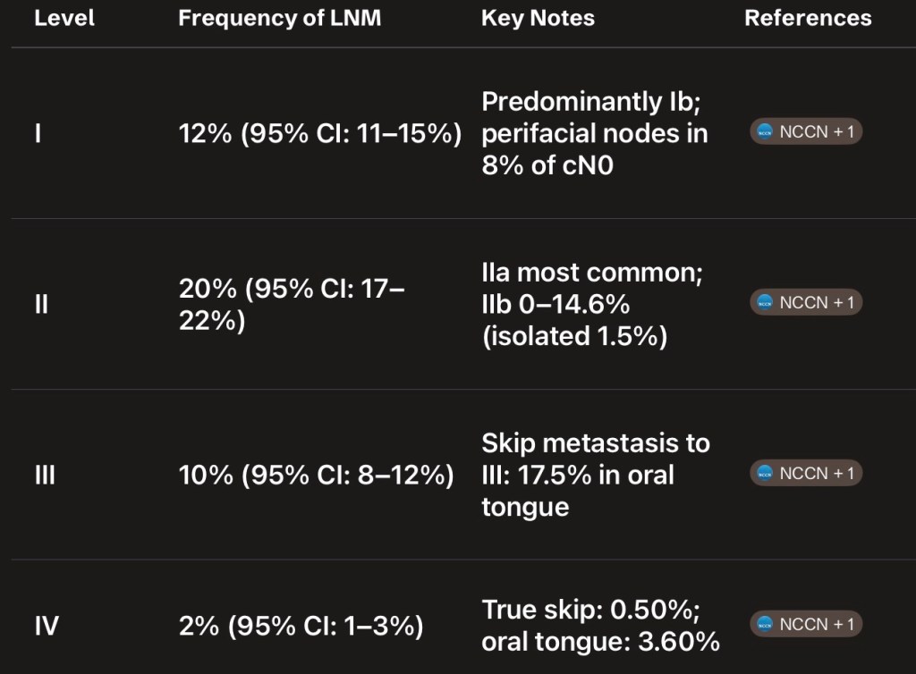
Summary of Frequency of Nodal Metastasis by Level:
A meta-analysis of 17 studies found the following frequencies of lymph node metastasis across all OSCC [17] – Figure
Clinical Synthesis:
For the cN0 neck, levels I to III dissection is the standard of care across all oral cavity subsites
The evidence does not support routine inclusion of level IV given the extremely low rate of true skip metastasis (0.50% overall)
For oral tongue SCC specifically:
The slightly higher rate of level IV involvement (3.60%) has prompted some authors to advocate for level IV inclusion:
But direct comparative studies show no survival or recurrence benefit from extending to level IV [14]
Level IV should be considered in higher-risk scenarios:
pN+ disease in levels I to III (especially multistation), greater DOI, or adverse pathologic features
For the cN+ neck:
Therapeutic dissection should include levels I to IV:
With level V considered in multistation disease [2]
References:
1. Head and Neck Cancers. National Comprehensive Cancer Network. Updated 2025-12-08.
2. Management of the Neck in Squamous Cell Carcinoma of the Oral Cavity and Oropharynx: ASCO Clinical Practice Guideline. Koyfman SA, Ismaila N, Crook D, et al. Journal of Clinical Oncology : Official Journal of the American Society of Clinical Oncology. 2019;37(20):1753-1774. doi:10.1200/JCO.18.01921.
3. Oral Cavity Cancer Surgical and Nodal Management: A Review From the American Head and Neck Society. Eskander A, Dziegielewski PT, Patel MR, et al. JAMA Otolaryngology– Head & Neck Surgery. 2024;150(2):172-178. doi:10.1001/jamaoto.2023.4049.
4. Impact of Elective Cervical Dissection on the Prognosis of Patients With Oral Squamous Cell Carcinoma cT1/T2N0: A Systematic Review and Meta-Analysis. Binda NC, Lavareze L, de Souza Vieira G, et al. Critical Reviews in Oncology/Hematology. 2025;:104982. doi:10.1016/j.critrevonc.2025.104982.
5. Depth of Invasion in Early Stage Oral Cavity Squamous Cell Carcinoma: The Optimal Cut-Off Value for Elective Neck Dissection. van Lanschot CGF, Klazen YP, de Ridder MAJ, et al. Oral Oncology. 2020;111:104940. doi:10.1016/j.oraloncology.2020.104940.
6. Elective Neck Dissection Strategies Guided by AJCC-8 Depth-of-Invasion (DOI) in cT1-T2N0 Oral Cavity Cancer-a Systematic Review. Abdul NS, Shivakumar S, Alreshaid L, et al. Cancers. 2026;18(4):697. doi:10.3390/cancers18040697.
7. Tumour Thickness as a Predictor of Nodal Metastases in Oral Cancer: Comparison Between Tongue and Floor of Mouth Subsites. Balasubramanian D, Ebrahimi A, Gupta R, et al. Oral Oncology. 2014;50(12):1165-8. doi:10.1016/j.oraloncology.2014.09.012.
8. The Relative Propensity for Regional Metastasis in Floor of Mouth Squamous Cell Carcinoma Versus Other Oral Cavity Subsites. Wicks C, Zubair F, Ogunbowale A, McMahon J. The British Journal of Oral & Maxillofacial Surgery. 2022;60(9):1276-1278. doi:10.1016/j.bjoms.2022.07.012.
9. Management of the Neck in T1 and T2 Buccal Squamous Cell Carcinoma. Nicholson OA, Van Lanschot CGF, van den Besselaar BN, et al. International Journal of Oral and Maxillofacial Surgery. 2024;53(4):259-267. doi:10.1016/j.ijom.2023.07.004.
10. What Is the Role of Elective Neck Dissection in the Management of Patients With Buccal Squamous Cell Carcinoma and Clinically Negative Neck?. Dillon JK, Villing AS, Jones RS, et al. Journal of Oral and Maxillofacial Surgery : Official Journal of the American Association of Oral and Maxillofacial Surgeons. 2019;77(3):641-647. doi:10.1016/j.joms.2018.10.021.
11. Assessment of the Rate of Skip Metastasis to Neck Level IV in Patients With Clinically Node-Negative Neck Oral Cavity Squamous Cell Carcinoma: A Systematic Review and Meta-analysis. Warshavsky A, Rosen R, Nard-Carmel N, et al. JAMA Otolaryngology– Head & Neck Surgery. 2019;145(6):542-548. doi:10.1001/jamaoto.2019.0784.
12. Nodal Yield and Topography of Nodal Metastases From Oral Cavity Squamous Cell Carcinoma – An Audit of 1004 Cases Undergoing Primary Surgical Resection. Roy P, Mallick I, Arun I, et al. Oral Oncology. 2021;113:105115. doi:10.1016/j.oraloncology.2020.105115.
13. Risk Factors and Impact of Occult and Skip Metastasis in Early-Stage Oral Tongue Squamous Cell Carcinoma. Yang X, Xiang W, Sun Y, et al. Clinical Oral Investigations. 2024;28(9):510. doi:10.1007/s00784-024-05897-8.
14. The Prognostic Significance of Elective Level 4 Neck Dissection in Oral Tongue Cancer. Yosefof E, Edri N, Ritter A, et al. Journal of Surgical Oncology. 2026;133(4):459-464. doi:10.1002/jso.70191.
15. How Adequate Is Supraomohyoid Neck Dissection for Node-Negative Oral Tongue Squamous Cell Carcinoma?. Sharma R, Singh N, Joshi KD, Sr A, Patrikar S. Journal of Cranio-Maxillo-Facial Surgery : Official Publication of the European Association for Cranio-Maxillo-Facial Surgery. 2025;:S1010-5182(25)00102-7. doi:10.1016/j.jcms.2025.03.006.
16. In Vivo Probability of Metastases in Levels IV-V in Oral Squamous Cell Carcinoma With a cN0/pN+ Situation in Levels I-Iii. Haas L, Mischkowski RA, Knape U, Król KM, Sakkas A. In Vivo (Athens, Greece). 2025 Nov-Dec;39(6):3437-3444. doi:10.21873/invivo.14141.
17. Frequency of Lymph Node Metastases at Different Neck Levels in Patients With Oral Squamous Cell Carcinoma: A Systematic Review and Meta-Analysis. Yu YF, Cao LM, Li ZZ, et al. International Journal of Surgery (London, England). 2025;111(1):1285-1300. doi:10.1097/JS9.0000000000001953.
A meta-analysis of 17 studies found the following frequencies of lymph node metastasis across all OSCC [17]#Arrangoiz #Doctor #Surgeon #SurgicalOncologist #HeadandNeckSurgeon #CancerSurgeon #Miami #MSMC #MountSinaiMedicalCenter #Mexico
The 15.8% skip metastasis rate reported by Byers et al. (1997):
Is widely considered an overestimate due to several important methodological issues that have been carefully dissected in subsequent literature:
Most notably by Warshavsky et al. in their 2019 JAMA Otolaryngology meta-analysis [1][2]
What Byers Reported:
Byers et al. reviewed 277 previously untreated patients with oral tongue SCC (1970 to 1990):
Who underwent glossectomy and neck dissection
They reported that 15.8% of all patients had either:
Level IV metastasis as the only manifestation of neck disease
Level III as the only positive node without disease in levels I to II
Subsequent level IV recurrence after initial dissection that did not include level IV
Based on this, they recommended routine dissection of levels I to IV for all oral tongue SCC [2]
Why the 15.8% Figure Is Misleading:
Warshavsky et al. performed a careful reanalysis of the Byers data and identified several critical flaws that inflated the rate [1]:
Conflation of skip metastasis definitions:
Byers combined true level IV skip metastasis with level III skip metastasis (level III positive without levels I to II involvement):
These are fundamentally different clinical scenarios — level III disease is already captured by a standard supraomohyoid neck dissection (levels I to III)
By lumping both together, the rate was artificially elevated
Inclusion of neck recurrences as “skip metastases”:
Nine patients (9.9%) who developed level IV recurrences after an initial dissection that did not include level IV were counted toward the 15.8% figure:
As Warshavsky et al. noted, counting neck recurrence as a missed pathological lymph node is problematic because the neck has lost its normal anatomical lymphatic drainage and, in many cases, has been irradiated
This makes it impossible to determine whether these were true skip metastases or recurrences from altered lymphatic flow [1]
True level IV skip metastasis rate was only 5.5%:
When the data are restricted to patients with cN0 disease who had level IV metastasis found in the initial neck dissection specimen (i.e., true pathologic skip metastasis):
The rate drops to 5.5% — roughly one-third of the reported figure [1]
Even the most generous calculation yields only 4.8%:
When accounting for all cases mentioned in the study (both initial pathologic findings and subsequent recurrences), the combined incidence of skip metastasis or subsequent recurrence in level IV was only 4.8% (13 of 270), not 15.8% [1]
Mixed cN0 and cN+ populations:
The study included patients across all clinical N stages, and the data were not clearly stratified by preoperative nodal status, making it difficult to isolate the true elective (cN0) skip metastasis rate
Era of the study (1970 to 1990):
Preoperative imaging was far less sophisticated, meaning some patients classified as cN0 may have had undetected nodal disease, further confounding the results
What Modern Data Shows:
The Warshavsky et al. meta-analysis of 13 studies (1,359 cN0 patients):
Found the true skip metastasis rate to level IV is only 0.50% (95% CI, 0.09%–1.11%):
With an overall level IV involvement rate of 2.53% [1]
Even for oral tongue specifically — the highest-risk subsite:
The level IV involvement rate was 3.60% [1]
A large Tata Memorial audit of 761 early-stage cN0 patients:
Found skip metastasis to level IV in only 0.3% [3]
In summary, the Byers 15.8% figure resulted from a broad definition of “skip” that included level III skips (already addressed by SOHND), conflation of initial pathologic findings with subsequent recurrences, and lack of stratification by preoperative nodal status:
Modern evidence overwhelmingly supports that true skip metastasis to level IV in cN0 oral tongue SCC is a rare event (~0.5%), and supraomohyoid neck dissection (levels I to III) remains adequate for the elective setting
References:
1. Assessment of the Rate of Skip Metastasis to Neck Level IV in Patients With Clinically Node-Negative Neck Oral Cavity Squamous Cell Carcinoma: A Systematic Review and Meta-analysis. Warshavsky A, Rosen R, Nard-Carmel N, et al. JAMA Otolaryngology– Head & Neck Surgery. 2019;145(6):542-548. doi:10.1001/jamaoto.2019.0784.
2. Frequency and Therapeutic Implications of “Skip Metastases” in the Neck From Squamous Carcinoma of the Oral Tongue. Byers RM, Weber RS, Andrews T, et al. Head & Neck. 1997;19(1):14-9. doi:10.1002/(sici)1097-0347(199701)19:13.0.co;2-y.
3. Incidence and Impact of Skip Metastasis in the Neck in Early Oral Cancer: Reality or a Myth?. Gurmeet Singh A, Sathe P, Roy S, et al. Oral Oncology. 2022;135:106201. doi:10.1016/j.oraloncology.2022.106201.
For patients diagnosed with asymptomatic primary hyperparathyroidism (PHPT – 5% of the cases):
Additional evaluation is necessary to make subsequent management decisions
In order to make management recommendations for patients with asymptomatic PHPT, I usually send the following studies:
Urinary calcium excretion
Serum 25-hydroxyvitamin D
Serum creatinine and estimated glomerular filtration rate (eGFR):
To assess for renal compromise
Bone density to determine if it is low
I some situations I will also get the following imaging studies:
Ultrasound of the kidneys to assess for occult kidney stones
Spine to assess for asymptomatic vertebral compression fracture
The Fourth International Workshop guidelines recommended further assessment to help in the decision regarding surgery for PHPT:
This includes renal imaging (plain radiographs or ultrasound) to determine if the patient has a clinically silent kidney stone and vertebral imaging (spine radiograph or vertebral fracture assessment [VFA] of dual-energy x-ray absorptiometry [DXA] image) for subclinical vertebral fracture
Urinary stone risk profile is also recommended:
But only if urinary calcium excretion is extremely elevated (> 400 mg/day) and a patient is unsure about parathyroid surgery
However, these tests are more expensive, may not be readily available to all clinicians, and there are few data to support their role in managing patients with PHPT
Urinary calcium excretion:
In asymptomatic patients:
The urinary calcium excretion is helpful to assess the risk of renal complications (when urine calcium is high) and thus determine subsequent management
The Fourth International Workshop on Asymptomatic Primary Hyperparathyroidism guidelines recommended surgical intervention as opposed to observation in asymptomatic patients who have:
A 24-hour urinary calcium excretion greater than 400 mg/day (> 10 mmol/day)
It should be noted that urinary calcium levels are considered to be elevated at a significantly lower level of excretion (250 mg/24 hours in women and 300 mg/24 hours in men):
There are no specific data assessing the cut-point of 400 mg calcium excretion daily
This particular recommendation reflected expert opinion regarding a level above which there is consensus that patients should be sent for surgery even in the absence of other criteria
Serum vitamin D:
I measure serum 25(OH)D in all patients with suspected or diagnosed PHPT
Measurement of 25(OH)D is important:
To identify patients with PHPT and vitamin D deficiency (which is a significant proportion of patients)
Due to the significant prevalence of vitamin D insufficiency in individuals with PHPT:
The Fourth International Workshop on Asymptomatic Primary Hyperparathyroidism recommended measuring 25(OH)D in all patients with the disease and repleting those with low levels (defined as ≤ 20 ng/mL [50 nmol/L]) prior to making any management decisions:
Specially if calcium levels are within the normal range
In some cases, the decision regarding surgery will be clear despite the low vitamin D level:
In such patients, it is still advisable to replete vitamin D if this can be done safely:
In order to mitigate postoperative hypocalcemia
Serum creatinine:
The serum creatinine concentration provides information about renal function:
Which can be diminished by hypercalcemia
Rather than using serum creatinine alone, the eGFR can be estimated in patients with a stable serum creatinine concentration:
An eGFR of 60 mL/min is the threshold of chronic kidney disease for deciding:
Which asymptomatic individuals with PHPT may benefit from early surgical treatment
Renal imaging:
In several studies, clinically silent kidney stones were reported in 7%to 21% of patients with PHPT:
Patients with undiagnosed (subclinical) nephrocalcinosis or calcium kidney stones:
Are regarded as having symptomatic disease:
Regardless of the absence of symptoms
Thus, these patients meet criteria for surgical intervention
For the few patients who do not have other overt indications for surgery:
Obtaining renal imaging (ultrasound, computed tomography [CT], or abdominal radiograph):
To look for nephrocalcinosis or asymptomatic nephrolithiasis at the time of the original evaluation for PHPT
Ultrasound is typically the imaging modality used
Bone mineral density:
Patients with PHPT may have decreased bone mineral density (BMD):
In particular at more cortical sites (forearm and hip) as compared with more trabecular (cancellous) sites (spine)
Although measurement of BMD is not required for the diagnosis of PHPT:
It is an essential part of the management of the disease:
BMD must be measured at the:
Spine, hip, and distal one-third forearm sites
The degree of bone loss:
Is reflective of the severity of hyperparathyroidism and is useful for making recommendations for parathyroid surgery or observation with monitoring in asymptomatic patients (minority of the cases)
Assessment for vertebral fracture:
Patients with subclinical, idiopathic vertebral compression fractures are regarded as having osteoporosis:
Independent of BMD findings
Vertebral compression fractures are underdiagnosed in all populations:
Many studies have demonstrated an increased risk of asymptomatic vertebral fractures in patients with PHPT
In order to diagnose asymptomatic vertebral compression fractures:
The Fourth International Workshop guidelines recommended imaging to assess for vertebral fracture in asymptomatic patients who do not have osteoporosis on BMD testing (patients with clinical osteoporotic fractures or with BMD in the osteoporosis range already meet criteria for surgery):
If a vertebral fracture is present by VFA or radiograph:
Parathyroidectomy is recommended
Vertebral assessment can be performed with plain radiographs or with VFA by DXA:
The latter can be done at the time of BMD testing, at greater patient convenience, less cost, and lower radiation exposure than conventional radiography of the spine
Other tests:
Biochemical renal stone prediction:
For asymptomatic patients with hypercalciuria (> 400 mg/day [10 mmol/day]):
The Fourth International Workshop Guidelines on the Management of Asymptomatic Primary Hyperparathyroidism recommended:
Assessment of the urine composition to identify patients at highest risk for nephrolithiasis [32]:
There are no specific data to suggest that urine stone risk profiling can predict risk of nephrolithiasis in patients with PHPT
Some experts believe that urinary calcium excretion > 400 mg/day alone raises sufficient enough concern about long-term renal complications to warrant a recommendation for parathyroidectomy
Serum phosphorus — The serum phosphorus concentration may be decreased but typically is in the lower range of normal. Some patients have mild hyperchloremic acidosis. (See “Primary hyperparathyroidism: Clinical manifestations”.)
Markers of bone turnover — Biochemical markers of bone turnover (collagen crosslinks, osteocalcin, bone-specific alkaline phosphatase) are often at the upper end of normal or mildly elevated in asymptomatic PHPT (see “Bone physiology and biochemical markers of bone turnover”). In those with more severe disease, they are typically high. They are only occasionally helpful in the management of hyperparathyroidism and should not be routinely measured [8].
Localization studies — The diagnosis of PHPT is established by appropriate biochemical testing. Localization studies with ultrasonography, technetium-99m sestamibi, CT, or magnetic resonance imaging (MRI) scanning should not be used to establish the diagnosis of PHPT or to determine management. Localization studies should be performed only after a decision for surgery has been made. Their utility is questionable when bilateral neck exploration is planned. However, they are commonly used now, along with intraoperative parathyroid hormone (PTH) monitoring, to facilitate unilateral exploration and minimally invasive surgery in those with probable single gland disease. (See “Preoperative localization for parathyroid surgery in patients with primary hyperparathyroidism”.)
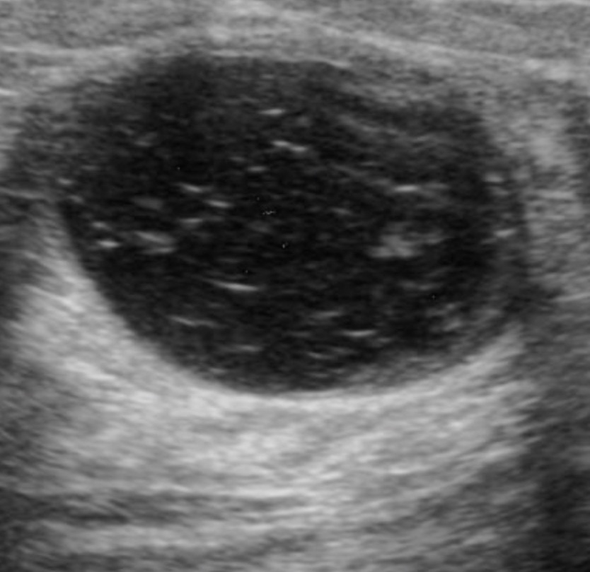
Sonographic mass with a benign mammographic imaging:
Is managed based on ultrasound features
Ultrasound imaging of a palpable breast lesion. Marked hypoechogenicity with immobile echogenic foci (representing calcifications)
References
Evaluation and imaging features of malignant breast masses. In: Cardenosa G. Clinical Breast Imaging: The Essentials. Philadelphia, PA: Wolters Kluwer; 2015:234-282.
Stavros AT. Breast Ultrasound. Philadelphia, PA: Lippincott Williams & Wilkins; 2004.