Radiation Therapy for Invasive Lobular Carcinoma of the Breast

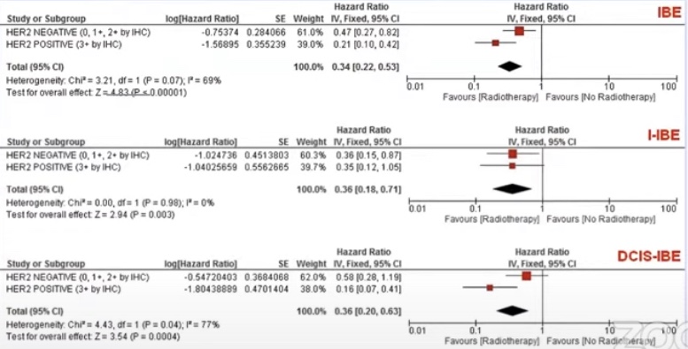
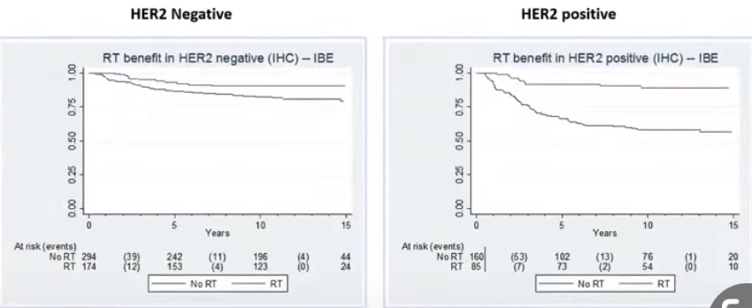
  • Considerations for radiation therapy (RT) in locoregional control, once more generally common among the ductal and lobular cancer types, are summarized here (Figure)
  • Adjuvant whole breast RT:
    • Reduces the risk of both local regional recurrence (LRR) and death from breast cancer after BCS and is a necessary element of BCT
  • Additional regional nodal irradiation:
    • May also be indicated for those with involved lymph nodes or high-risk tumors
  • It is noteworthy that it may be acceptable to omit RT:
    • Among elderly women with select low-risk, ER-positive tumors:
      • Data to support this include the Cancer and Leukemia Group B (CALBG) 9343 randomized trial of women age 70 years or older with stage I ER-positive cancers treated with lumpectomy and tamoxifen with or without RT:
        • Which demonstrated no advantage in
          overall survival
          :
          • Although there was a small improvement in LRR among those treated with RT
  • Accelerated partial breast irradiation (APBI):
    • Is a newer technique involving more focused RT
      delivered in higher doses over a shorter time span
    • Notably, the recent American Society for
      Radiation Oncology (ASTRO) guideline update cites lobular histology as a criterion for “cautionary” use of APBI outside of a clinical trial
  • Postmastectomy RT:
    • May also benefit selected patients, a decision generally made by consideration of the presence of:
      • Macrometastatic nodal involvement
      • Large tumor size
      • High-risk disease features
    • It is important to note that the implications of margins at mastectomy remain controversial among radiation oncologists, and there are no data to support a definite benefit of postmastectomy RT in patients with close margins
    • Similar to surgical and systemic therapy trials, ILC patients comprise a minority in postmastectomy RT trials
    • A recent study using Survival, Epidemiology, and End Results (SEER) data including 12,703 ILC patients treated from 2004 to 2009, of which 26% had a definite indication for postmastectomy RT:
      • Found an improvement in 5-year overall survival and disease-specific survival from 80.9% to 84.7% (p = .0003) among ILC patients, a benefit to the same degree as IDC:
        • These data support continued decision
          making for radiotherapy using existing criteria, regardless of cancer histology

Hereditary Breast Cancer

  • The list of cancer-associated genes continues to expand, and it is therefore increasingly important to obtain a thorough family history to assess any potential for hereditary cancer syndromes:
    • The BRCA1 and 2 genes account for the majority of hereditary breast cancer cases
  • The BRCA1 gene is located on chromosome 17q21:
    • It is part of the DNA repair pathway:
      • Functioning as a tumor suppressor gene
    • Presence of a deleterious BRCA1 mutation is associated with:
      • A lifetime breast cancer risk of:
        • 72% by age 80
      • A lifetime ovarian cancer risk of:
        • 44%
    • In addition, BRCA1 mutations have been associated with:
      • An increased risk of pancreatic cancer and melanoma
    • BRCA1 associated breast cancers:
      • Tend to occur at younger ages and are more likely to have aggressive phenotypes compared to non-BRCA-associated tumors
  • Lynch syndrome, also known as hereditary nonpolyposis colorectal cancer syndrome:
    • Is caused by genetic mutations in the mismatch repair system:
      • With the most common associated gene mutations being MLH1, MSH2, MSH6, and PMS2
    • Lynch syndrome is the most common hereditary form of colorectal cancer, and is also associated with an increased risk of:
      • Endometrial, urogenital, pancreatic, biliary tract and ovarian cancers:
        • Women with Lynch syndrome have a 20% to 60% lifetime risk of endometrial cancer
  • Germline mutations in the PTEN gene:
    • Are associated with Cowden syndrome:
      • Characterized by the formation of multiple hamartomas as well as an increased risk of:
        • Breast, endometrial, non-medullary thyroid, and renal cell cancers
  • Hereditary diffuse gastric cancer syndrome:
    • Is associated with a mutation in the CDH1 gene
    • It leads to an increased risk of early onset gastric cancer and lobular breast cancer
  • PALB2 is a breast cancer susceptibility gene:
    • With an estimated breast cancer risk of 45%:
    • PALB2 mutations have also been reported to increase the risk of:
      • Ovarian cancer and possibly pancreatic and prostate cancer
  • BRIP1 mutations:
    • Have been shown to confirm a high-risk of ovarian cancer (OR 20.97), but no increase in breast cancer risk
  • References
    • Shulman LP. Hereditary breast and ovarian cancer (HBOC): clinical features and counseling for BRCA1 and BRCA2, Lynch syndrome, Cowden syndrome, and Li-Fraumeni syndrome. Obstet Gynecol Clin North Am. 2010;37(1):109-133, Table of Contents.
    • Kuchenbaecker KB, Hopper JL, Barnes DR, et al. Risks of Breast, Ovarian, and Contralateral Breast Cancer for BRCA1 and BRCA2 Mutation Carriers. Jama. 2017;317(23):2402-2416.
    • Mersch J, Jackson MA, Park M, et al. Cancers associated with BRCA1 and BRCA2 mutations other than breast and ovarian. Cancer. 2015;121(2):269-275.
    • Southey MC, Winship I, Nguyen-Dumont T. PALB2: research reaching to clinical outcomes for women with breast cancer. Hered Cancer Clin Pract. 2016;14:9.
    • Weber-Lassalle N, Hauke J, Ramser J, et al. BRIP1 loss-of-function mutations confer high risk for familial ovarian cancer, but not familial breast cancer. Breast Cancer Res. 2018;20(1):7.
#Arrangoiz #Doctor #Surgeon #CancerSurgeon #SurgicalOncologist #BreastSurgeon #MountSiniaMedicalCenter #MSMC #Miami #Mexico #BreastCancer

Lymph Node Dissection in Thyroid Cancer

  • Papillary thyroid cancer (PTC):
    • Has a high predilection for spread to locoregional lymph nodes (LNs):
      • Occurring in up to 40% to 90% of cases:
        • When prophylactic nodal dissection is performed:
        • Though such high rates of metastatic disease may prove enticing to recommend routine prophylactic node dissection:
          • Recurrence-free survival is not effected by the removal of sonographically normal, microscopically diseased nodes
        • Instead, prophylactic central neck dissection may be individually considered for those patients with:
          • T3 or T4 tumors, or in the presence of lateral neck metastases
        • Clinically suspicious or biopsy-proven nodal disease warrants a “therapeutic” dissection of the involved compartments
          • “Berry picking,” or selective removal of suspicious LN metastases, is not recommended:
            • As it is associated with significantly higher recurrence rates and does not lower the rate of postoperative complications compared with systematic compartmental dissections
  • The risk of surgical complications with nodal dissection should be weighed against the benefit of LN removal:
    • Central neck dissections may result in temporary or permanent injury to the RLN and hypoparathyroidism
    • Surgeon case volume predicts patient outcomes:
      • Those performing less than 10 cases compared with those performing more than 100 cases per year had complications in 24% and 14.5% of cases, respectively
    • Although dissection of the lateral neck is less often associated with adverse events:
      • Injury to the spinal accessory nerve may occur with dissection of level II or V
    • Similarly, chyle leaks may be seen after removal of nodes in level IV:
      • Particularly on the left side
#Arrangoiz #Surgeon #Doctor #Surgeon #CancerSurgeon #ThyroidSurgeon #ParathyroidSurgeon #HeadandNeckSurgeon #SurgicalOncologist #Miami #Mexico #MountSinaiMedicalCenter #MSMC #ThyroidCancer

Thyroid-Stimulating Hormone Suppression Therapy after Thyroid Cancer Surgery

  • Historically:
    • Almost all patients were given thyroid hormone:
      • To fully suppress serum thyroid-stimulating hormone (TSH)
    • The rationale for this approach:
      • Was based on the theory that TSH is a stimulant for thyroid cell proliferation and suppression of thyrotropin will inhibit tumor growth:
        • Indeed, early studies supported the role of TSH suppression in reducing the likelihood of disease progression and improving survival:
          • Particularly in those with high-risk disease
        • More recent analyses, however, have failed to demonstrate a benefit of such suppressive therapy in those with low-risk tumors:
          • In fact, such treatment may prove harmful
        • A long-term observational study showed a three-fold increased risk of cardiovascular death for each ten-fold reduction in mean TSH level
        • Patients with subclinical thyrotoxicosis:
          • Are also at increased risk of atrial fibrillation, ventricular hypertrophy, diastolic dysfunction, and impaired cardiac reserve
          • Additionally, bone turnover may be adversely affected by suppressive doses of levothyroxine:
            • Higher rates of osteoporosis may be seen in thyroid cancer patients:
              • There is an increased risk of fracture when suppressive doses of levothyroxine are used
    • As a consequence of the myriad negative effects of excess levothyroxine:
      • The target TSH range should be determined on an individual basis
      • It is also worthy of note that lowering TSH to undetectable levels probably does not confer additional benefit beyond that seen with less aggressive suppression below 0.1 mU/L
      • The optimal TSH range should consider the initial risk for recurrence, the response to therapy, and the risk for thyrotoxicosis-related morbidities in the individual patient
      • Furthermore, this target TSH for the individual patient may evolve over time, depending on the response to therapy

Data from Haugen BR, Alexander EK, Bible KC, et al. 2015 American Thyroid Association management guidelines for adult patients with thyroid nodules and differentiated thyroid cancer: the American Thyroid Association Guidelines Task Force on Thyroid Nodules and Differentiated Thyroid Cancer. Thyroid. 2016;26(1):1–133.
#Arrangoiz #ThyroidCancer #ThyroidSurgeon #CancerSurgeon #Doctor #Surgeon #HeadandNeckSurgeon #SurgicalOncologist #EndocrineSurgery #Miami #Mexico #MountSianiMedicalCenter #MSMC

American Thyroid Association Response to Therapy Classification in Thyroid Cancer

  • Excellent Response to Therapy:
    • Patients with no biochemical (unstimulated serum thyroglobulin (Tg) < 0.2 or stimulated Tg < 1.0 ng/mL) or radiographic evidence of disease are classified as having an excellent response to therapy
    • Patients with an initial low to intermediate risk of recurrence who meet these criteria:
      • Are recommended to have serum Tg monitored every 12 to 24 months
    • Patients with initially high-risk disease:
      • Should continue to have a serum Tg measurement at least every 6 to 12 months
  • Biochemical Incomplete Response to Therapy:
    • Patients who have undergone total thyroidectomy and remnant ablation and have an unstimulated serum Tg > 1 ng/mL or a stimulated Tg > 10 ng/mL or a rising thyroglobulin antibody (TgAb) titer with negative imaging:
      • Are classified as having a biochemical incomplete response to therapy
    • Such patients should undergo imaging with sonography of the neck:
      • If the disease is unable to be located:
        • Cross-sectional imaging of the neck and chest should be performed
    • Serum Tg should be followed at least every 6 to 12 months.
  • Structural Incomplete Response to Therapy:
    • Those patients with structurally or functionally (on diagnostic whole-body scan [DxWBS] or 18(FDG-PET) evident disease are classified as:
      • Having a structural incomplete response to therapy
    • Unfortunately, the majority of patients in this category will have persistent disease in spite of additional treatments
    • Disease-specific death rates are high in this group:
      • 11% with locoregional metastases
      • 50% with distant metastases
  • Indeterminate Response to Therapy:
    • Patients with biochemical or structural findings that cannot be confidently classified as either excellent response or persistent disease:
      • Are deemed as having an indeterminate response to therapy
    • Such patients may be carefully followed with biochemical testing and serial imaging to better delineate which category is ultimately appropriate
    • It is estimated that up to 20% of these patients will eventually develop conclusive evidence of disease requiring additional therapy
#Arrangoiz #Doctor #Surgeon #ThyroidSurgeon #ParathyroidSurgeon #HeadandNeckSurgeon #CancerSurgeon #MountSinaiMedicalCenter #MSMC #Miami #Mexico #ThyroidCancer

Invasive Encapsulated Follicular Variant of Papillary Thyroid Carcinoma (IEFVPTC)

  • Invasive encapsulated follicular variant of papillary thyroid carcinoma (IEFVPTC):
    • Is now considered a separate entity and no longer a subtype of PTC
  • IEFVPTC has a RAS-like mutational and transcriptomic profile:
    • Similar to that of follicular adenoma (FA) and follicular thyroid carcinoma (FTC) (Figure):
      • Whereas classic PTC and the infiltrative follicular subtype of PTC:
        • Have BRAF V600E-like molecular profiles
  • IEFVPTCs:
    • Have a fibrous capsule or well-defined border and lack the histologic features of infiltrative follicular PTC
  • Like FTC:
    • IEFVPTC can invade vessels in the capsule and develop distant metastasis
#Arrangoiz #ThyroidSurgeon #CancerSurgeon #HeadandNeckSurgeon #SurgicalOncologist #MountSinaiMedicalCenter #MSMC #Miami #Mexico #ThyroidCancer

Risk Stratification in Differentiated Thyroid Cancer

  • Risk stratification in differentiated thyroid cancer has traditionally used a relatively small set of clinical and pathological factors to create models that predict disease-specific mortality or overall survival:
    • Although clinically useful, these models provided static estimates of risk with information available within the first few months of initial therapy and demonstrated suboptimal, long-term outcome predictions for any individual patient
  • Over the last decade, additional models have been developed that provide predictive information with regard to other clinically relevant outcomes, such as:
    • The risk of having persistent disease after initial therapy
    • The risk of structural or biochemical disease recurrence
    • The likelihood of going into remission following initial therapy in adult patients with thyroid cancer
  • Furthermore, rather than using information that is only available at one particular point in time, these new models emphasize the importance of dynamic risk assessment:
    • Where the initial risk assessment is modified over time as new data become available
  • These dynamic risk assessments allow us to:
    • Integrate response to therapy assessments with the underlying individual tumor biology:
      • To provide real-time risk assessments at any point in the course of the patient’s disease
  • Thus, the modern view of risk stratification begins with:
    • The identification of a suspicious nodule (peri-diagnostic period) and continues through the phases of:
      • Diagnosis
      • Treatment
      • Adjuvant therapy
      • Follow-up

#Arrangoiz #ThyroidSurgeon #CancerSurgeon #HeadandNeckSurgeon #SurgicalOncologist #Teacher #Miami #MountSinaiMedicalCenter

Risk Stratification in Active Surveillance of Papillary Microcarcinoma

  • Asymptomatic, small thyroid nodules (usually ≤ 1 cm maximal diameter, 1 cm3, or 1 mL volume) confined to the thyroid and surrounded by normal thyroid parenchyma:
    • Can be followed with active surveillance:
      • With or without cytologic confirmation:
        • In patients who value their normal thyroid function and who desire avoidance of thyroid surgery
  • Patients who demonstrate tumors larger than 1.5 to 2.0 cm; tumors in subcapsular locations adjacent to important structures, such as the trachea and recurrent laryngeal nerve; or tumors with documented growth rate doubling times of < 2 years:
    • Are generally considered inappropriate for observation and would be considered to have actionable disease
  • If the tumor growth rate is unknown at the time of nodule detection:
    • Then this can be established with serial ultrasound evaluations done approximately every 6 months for 1 to 2 years
  • The frequency of ultrasound evaluations and long-term follow-up:
    • Depends on the tumor size, location, and established growth rate
  • With the use of this paradigm:
    • Active surveillance continues until:
      • There is a 3-mm increase in tumor diameter:
        • Which corresponds to a 100% increase in tumor volume
      • Identification of metastatic disease
      • Direct invasion into surrounding structures of the thyroid
      • A decision to discontinue active surveillance based on patient preference
  • This risk-stratified, minimalistic management approach to very low-risk thyroid cancers has been shown to be safe and effective over 5 to 10 years of follow-up in studies from Japan, Korea, and the United States:
    • In the first 10 years of active surveillance follow-up:
      • Only 2% to 8% of papillary microcarcinomas:
        • Increase ≥ 3 mm in maximum diameter
      • 12% to 14% demonstrate an increase in tumor volume of > 50%:
        • The smallest change in nodule volume that can be reproducibly measured
      • Novel lymph node metastases:
        • Are detected in 2% to 4%
    • The likelihood of disease progression is higher in younger patients than in older patients
  • Importantly, at the time of disease progression:
    • Deferred surgical intervention is quite effective with excellent outcomes and no disease-specific mortality
  • References:
    • Ito Y, Miyauchi A. Active surveillance as first-line management of papillary microcarcinoma. Annu Rev Med. 2019;70:369–379.
    • Ito Y, Miyauchi A, Kudo T, Oda H, Yamamoto M, Sasai H, Masuoka H, Fukushima M, Higashiyama T, Kihara M, Miya A.. Trends in the implementation of active surveillance for low-risk papillary thyroid microcarcinomas at Kuma Hospital: gradual increase and heterogeneity in the acceptance of this new management option. Thyroid. 2018;28(4):488–495.
    • Tuttle RM, Zhang L, Shaha A. A clinical framework to facilitate selection of patients with differentiated thyroid cancer for active surveillance or less aggressive initial surgical management. Expert Rev Endocrinol Metab. 2018;13(2):77–85. 
    • Tuttle RM, Fagin JA, Minkowitz G, Wong RJ, Roman B, Patel S, Untch B, Ganly I, Shaha AR, Shah JP, Pace M, Li D, Bach A, Lin O, Whiting A, Ghossein R, Landa I, Sabra M, Boucai L, Fish S, Morris LGT. Natural history and tumor volume kinetics of papillary thyroid cancers during active surveillance. JAMA Otolaryngol Head Neck Surg. 2017;143(10):1015–1020. 
    • Tuttle RM, Zhang L, Shaha A. A clinical framework to facilitate selection of patients with differentiated thyroid cancer for active surveillance or less aggressive initial surgical management. Expert Rev Endocrinol Metab. 2018;13(2):77–85.
    • D’Agostino TA, Shuk E, Maloney EK, Zeuren R, Tuttle RM, Bylund CL. Treatment decision making in early-stage papillary thyroid cancer. Psychooncology. 2018;27(1):61–68.
    • Groopman J, Hartzband P.. Your Medical Mind. How to Decide What Is Right for You. New York, NY: Penguin Books.
    • Ito Y, Miyauchi A. Prognostic factors and therapeutic strategies for differentiated carcinomas of the thyroid. Endocr J. 2009;56(2):177–192.