My name is Rodrigo Arrangoiz I am a breast surgeon/ thyroid surgeon / parathyroid surgeon / head and neck surgeon / surgical oncologist that works at Center for Advanced Surgical Oncology in Miami, Florida.
I was trained as a surgeon at Michigan State University from (2005 to 2010) where I was a chief resident in 2010. My surgical oncology and head and neck training was performed at the Fox Chase Cancer Center in Philadelphia from 2010 to 2012. At the same time I underwent a masters in science (Clinical research for health professionals) at the University of Drexel. Through the International Federation of Head and Neck Societies / Memorial Sloan Kettering Cancer Center I performed a two year head and neck surgery and oncology / endocrine fellowship that ended in 2016.
Mi nombre es Rodrigo Arrangoiz, soy cirujano oncólogo / cirujano de tumores de cabeza y cuello / cirujano endocrino que trabaja Center for Advanced Surgical Oncology en Miami, Florida.
Fui entrenado como cirujano en Michigan State University (2005 a 2010 ) donde fui jefe de residentes en 2010. Mi formación en oncología quirúrgica y e n tumores de cabeza y cuello se realizó en el Fox Chase Cancer Center en Filadelfia de 2010 a 2012. Al mismo tiempo, me sometí a una maestría en ciencias (investigación clínica para profesionales de la salud) en la Universidad de Drexel. A través de la Federación Internacional de Sociedades de Cabeza y Cuello / Memorial Sloan Kettering Cancer Center realicé una sub especialidad en cirugía de cabeza y cuello / cirugia endocrina de dos años que terminó en 2016.
Was a phase 3 clinical trial that randomized postmenopausal women with ER positive DCIS (n = 3,104) to either five years of anastrozole or tamoxifen following breast-conserving surgery and radiation:
The trial sought to determine how effective anastrozole was compared to tamoxifen in preventing a breast cancer occurrence
With a median follow-up of 9 years:
Investigators found significantly fewer breast cancer events in the anastrozole group (n = 90) than in the tamoxifen group (n = 122)
The 10-year breast cancer event rate was lower among women randomized to anastrozole compared to tamoxifen:
6.9% [anastrozole] vs. 10.9% [tamoxifen], HR 0.73, p=0.02
This recorded difference in breast cancer events was attributable almost entirely to:
Younger postmenopausal women less than 60 years of age who received tamoxifen
Women less than 60 receiving tamoxifen had nearly twice the events as those receiving anastrozole:
Events on tamoxifen: 63 vs events on anastrozole: 34, HR 0.53 (0.35-0.80), p=0.0026)
Interestingly, the difference between treatments did not become apparent until after 5 years of follow-up:
Likely due to the low number of events in both groups
There was no difference in overall survival (OS) between the two treatment groups:
The 10-year estimates for overall survival were 92.1% for the tamoxifen group and 92.5% for the anastrozole group (p=0.38)
In B-35, anastrozole was found to further significantly reduce the rate of contralateral invasive cancer compared with tamoxifen
Based on this trial and others:
Aromatase inhibitors are the preferred adjuvant hormonal therapy for ER positive disease in post-menopausal women with either DCIS or invasive breast cancer:
Provided they have no contraindications to taking an aromatase inhibitor
References
Margolese RG, Cecchini RS, Julian TB, Ganz PA, Constantino JP, Vallow LA, et al. Anastrozole versus tamoxifen in postmenopausal women with ductal carcinoma in situ undergoing lumpectomy plus radiotherapy (NSABP B-35): a randomised, double-blind, phase 3 clinical trial. Lancet. 2016;387(10021):849-856
M Baum, AU Budzar, J Cuzick, et al., ATAC Trialists’ Group. Anastrozole alone or in combination with tamoxifen versus tamoxifen alone for adjuvant treatment of postmenopausal women with early breast cancer: first results of the ATAC randomized trial. Lancet. 2002;359(9324):2131-2139.
Is a rare benign inflammatory breast disease first described in 1972 by Kessler and Wolloch
The most common presenting symptom is:
A unilateral, firm, and discrete breast mass:
Which may be accompanied by overlying skin changes and / or possible lymph node involvement
The average duration of symptoms is around 3.9 months with:
The most common signs and symptoms including:
Discrete mass
Tenderness to palpation
Erythema
Swelling
The pain could be out of proportion to findings:
Suggestive of a localized ischemic etiology
The pain could be a motivating factor in prompting all symptomatic patients to seek consultation
The lesion may occur in any quadrant of the breast:
But often extends radially from the retroareolar region
The disease often presents in women:
Of childbearing age:
With a recent history of pregnancy or ongoing lactation:
The mean age of diagnosis is around:
31.7 years and all within reproductive years
The high majority of women are:
Hispanic or African / African-American ancestry
A history of previous granulomatous disease (tuberculosis, sarcoidosis, autoimmune disease, or granulomatous disease) is very rare
The overlap of presenting symptoms with other disease processes such as:
Malignancy, acute or chronic infections, and chronic inflammatory diseases:
Makes definitive diagnosis difficult
Because the differential may include malignancy:
The patient may experience significant anxiety during the evaluation
In addition, the broad differential and the lack of pathognomonic features make definitive diagnosis difficult:
Often resting as a diagnosis of exclusion on a clinical basis
The typical mammographic and ultrasonographic findings of granulomatous mastitis are:
Mammogram:
Ill-defined mass to an asymmetric density without specific margins
It is usually not accompanied by microcalcifications or architectural distortion
Ultrasound:
Heterogeneously hypoechoic lesion
Segmental masses with ill-defined margins, with tubular structures extending from the mass:
The tubular structures may be clustered, separate or contiguous
Malignant features or findings:
Suspicious microcalcifications, architectural distortion, or intra-ductal mass:
Would warrant appropriate histologic diagnosis and subsequent management
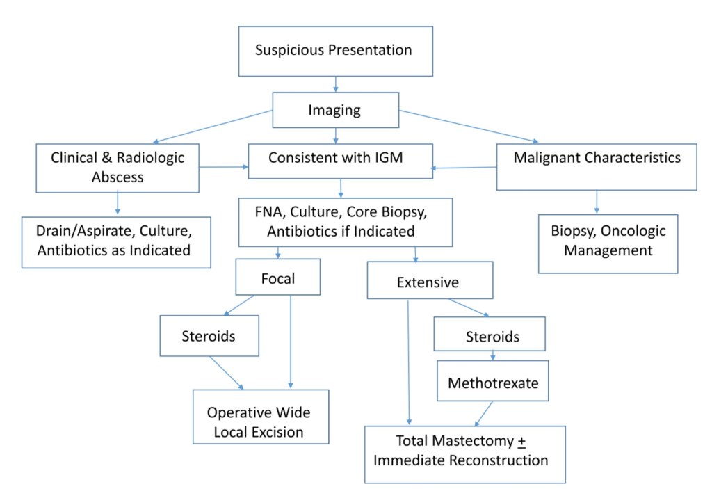
If the findings clinically and radiographically indicate an abscess:
They warrant treatment with antibiotics along with drainage by aspiration or incision, if the abscess is larger
Idiopathic granulomatous mastitis is usually regarded as a sterile process:
However, there is evidence to suggest a link to Corynebacterium kroppenstedtii infection or colonization:
This would then mandate routine cultures
One-third of the patients with granulomatous mastitis who are evaluated:
Have findings suggestive of an abscess
Because diagnosis is difficult, patients typically have received:
Prolonged courses of antibiotics, frequent biopsies, or surgical procedures
With a lack of pathognomonic imaging findings associated with granulomatous mastitis:
Histopathology is key to confirming a diagnosis
Histologically:
The disease has been described as non-caseating granulomas among epithelioid histiocytes and multinucleated giant cells surrounded by lymphocytes and plasma cells
Histologic differentials begin with the use of:
Hematoxylin and eosin stains, gram stain, and may also include acid fast stains and Grocott’s methenamine silver to evaluate for the possibility of sarcoidosis or mycobacterium infection
Once the diagnosis is made, treatment strategies are not clearly delineated but are often supportive
Steroid treatment has been shown to decrease the extent of involvement, and enable complete resection with removal of less breast tissue should resection be pursued:
However, lower doses have also proven to be efficacious and help to avoid adverse effects of weight gain, hyperglycemia, and Cushingoid symptoms
Regimens of 16 mg of prednisone twice a day for two weeks with a slow taper over a two-month period are described in the literature, as is prednisolone 30 mg/day for eight weeks, with taper
If symptoms do not improve, a course of methotrexate could be utilized:
Since there is a contingent who feel that this disease is an abnormal immune response
Success with methotrexate, including administration in steroid refractory cases:
Is limited to small studies of 3 to 5 patients
In consideration of the duration of symptoms patients experienced, along with the lack of durable evidence favoring treatment with methotrexate, our institution favors proceeding directly to surgery in steroid-refractory cases
We recommend the surgical options of:
Wide local excision for focal involvement and
Total mastectomy with the option of reconstruction for diffuse involvement
A 15% to 20% recurrence rate has been reported for surgery alone:
And inversely proportional to negative surgical margins
In light of the fact that these masses are painful and may lead to fistula formation and deformity:
Intervention may be the preferred path to avoid progression or static disease
Our experience allowed for a trial of “conservative” medical management for two months:
Followed by treatment escalation for lack of progress
A course of methotrexate may be entertained in patients who desire continued attempts at medical management, to help preserve breast tissue
However, it is not included in the first line of conservative management
DCIS is a proliferation of malignant cells that have not breached the ductal basement membrane:
They arise from ductal epithelium:
In the region of the terminal ductal–lobular unit (TDLU)
DCIS had previously been considered one stage in the continuum of histologic progression from ADH to invasive carcinoma:
But, in fact, DCIS comprises a heterogeneous group of lesions:
With variable histologic architecture, molecular and cellular characteristics, and clinical behavior
Malignant cells proliferate:
Until the ductal lumen is obliterated
There is an associated breakdown of the myoepithelial celllayer of the basement membrane surrounding the ductal lumen
DCIS has also been linked with changes in the surrounding stroma resulting in:
Fibroblast proliferation
Lymphocyte infiltration
Angiogenesis
Thus, although the process is poorly understood:
Most but not all invasive ductal carcinomas are believed to arise from DCIS:
Which is considered a nonobligate precursor of invasive breast carcinoma
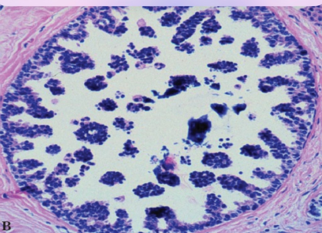
Architectural heterogeneity is a common feature of DCIS. Even in the same lesion, DCIS may show different growth patterns. Image is showing a cribriform DICSMicropapillary DCISPapillary DCISSolid DCIS
Most patients with papillary thyroid cancer (PTC) have an excellent prognosis:
But predicting which patients do not do well has been an ongoing area of interest
Ideally, identifying those at higher risk of cancer recurrence:
Would potentially allow the more aggressive therapies to be utilized when appropriate for patients with high risk papillary thyroid cancer
A lot of work has identified molecular markers, which are mutations in cancer-related genes that can help in the diagnosis of thyroid cancer on thyroid biopsy specimens
Two specific molecular markers, BRAFv600E and TERT promotor mutations:
Have been associated with aggressive tumor behavior and worse outcomes in papillary thyroid cancer
The BRAFv600E mutation is quite common in papillary thyroid cancer:
So using this mutation alone to predict outcome has been challenging, though it has been associated with poor prognosis
The TERT promoter mutation alone was not shown to cause adverse outcomes in some previous studies, though other studies suggested it was associated with a more aggressive clinical picture
A study by Moon S et al. aimed to determine the prognosis of papillary thyroid cancer in patients with either of these mutations alone or in combination by a review of the current studies:
Moon S et al. Effects of coexistent BRAFV600E and TERT promoter mutations on poor clinical outcomes in papillary thyroid cancer: a meta-analysis
Summary of the Study:
A literature review was done to identify studies that included BRAFV600Eand TERT promoter mutations in thyroid cancer
A total of 13 studies were identified
Data was extracted and reviewed for clinical information to include the number of males and females, age at diagnosis, cancer stage, spread to lymph nodes, extrathyroidal extention, spread outside of the neck, cancer recurrence and death
A total of 4347 patients with papillary thyroid cancer were evaluated in the study and 283 patients had both BRAFv600E and TERT promoter mutations
A BRAFv600E mutation alone:
Was related to advanced age at time of diagnosis, advanced cancer stage, extrathyroidal extension of tumor, and spread to lymph nodes, compared with no mutation
A TERT promoter mutation alone:
Was associated with older age at diagnoses, spread to lymph node and spread outside of the neck
The combination of BRAFv600E and TERT promoter mutations together when compared with no mutations:
Was associated with older age at diagnosis, male gender, advanced cancer staging, extrathyroidal extension, spread to lymph node and spread outside of the neck
Overall, the combination of BRAF600E and TERT mutations:
Was associated with high recurrence rate when compared with no mutations
Further, it was noted that the combination of mutations also had a higher risk of death than no mutations or BRAFv600E alone, although few patients were in this group
What are the implications to this study:
This study shows that molecular marker analysis can be used to identify patients that have more aggressive thyroid cancer
The combination of BRAFv600E and TERT promotor mutations worsens the prognosis for papillary thyroid cancer
Additionally, a limited data set suggested higher risk of death with the combination of BRAF600E and TERT promoter mutations
As we improve our understanding of the molecular changes in thyroid cancer, we will improve our ability to identify patients that have a more aggressive thyroid cancer
Ultimately this knowledge will lead to improved treatment options
Future studies must aim to determine if identifying these mutations at the time of diagnosis can lead to improved outcomes for patients at higher risk
Cancers derived from thyroid follicular cells are classified into five main types:
Papillary thyroid carcinoma (PTC:
Representing 65% to 93% of all thyroid cancers worldwide
Follicular thyroid carcinoma (FTC):
6% to 10% of cases
Oncocytic thyroid carcinoma (OC):
3% to 7% of cases
Poorly differentiated thyroid carcinoma (PDTC):
0.5% to 2%
Anaplastic thyroid carcinoma (ATC)
1% of cases
PTC, FTC and OC:
Are generically termed differentiated thyroid carcinomas (DTCs):
Most patients present with localized disease and have a 5-year survival rates of greater than 98% by contrast to PDTC, which has a 5-year survival rate of 76%
The 2022 WHO classification of thyroid tumors:
Introduced a new intermediate clinical entity:
Differentiated high-grade thyroid carcinoma:
To define DTCs with a high mitotic rate and / or tumor necrosis:
As these have a 5-year survival rate comparable with that of PDTC
ATC is an extremely aggressive form of the disease:
Until recently, patients with ATC had a dismal median overall survival of 4 months:
Although this has improved markedly since 2018 with the FDA approval of new oncoprotein-targeted treatments coupled to evidence that immunotherapies may confer additional benefit
Thyroid parafollicular or C cells are a neuroendocrine lineage that gives rise to medullary thyroid cancers:
Which account for less than 5% of all thyroid cancers
Is a malignant intra-ductal proliferation of epithelial cells within the tubular-lobular system of the breast:
With no microscopic evidence of permeation across the basement membrane
There appears to be a progression between:
Flat epithelial atypia, atypical ductal hyperplasia (ADH), and DCIS:
In which DCIS is final step prior to the development of invasive disease
The clinical risk factors and molecular aberrations related with malignant transformation:
Are almost indistinguishable between DCIS and invasive cancer
The concurrence of DCIS and invasive carcinoma within one lesion suggests that:
DCIS is a precursor lesion to invasive carcinoma
Evidence of the ability of DCIS to progress is that:
50% of all recurrences after breast-conserving surgery (BCS) for DCIS, with or without adjuvant treatment, are invasive
Data is sparse on the natural history of DCIS:
But some series have reported the outcomes for women many years after undergoing a surgical biopsy that was interpreted as benign that contained an unrecognized area of DCIS:
These data identified that approximately 20% to 53% of these women developed:
Ipsilateral invasive carcinoma
Sanders et al. reported on 28 women with unrecognized low-grade DCIS in the surgical biopsy specimen:
Of which 11 developed invasive carcinoma:
All of these cancers developed in the same breast and quadrant as the biopsy containing the DCIS
The vast majority of these invasive cancers developed within 10 years, but three were diagnosed after 20 years
Collins et al, in the Nurses’ Health Study, singled out 13 women who were found to have DCIS on reexamination of the surgical biopsies that were previously diagnosed as benign:
Ten of these women subsequently developed breast cancer
All were ipsilateral
Four were DCIS and six were invasive
The interval between the biopsy and the progression to invasive cancer was on average nine years
Approximately one in eight women (12%) in the United States (US) will be diagnosed with breast cancer in her lifetime:
20% to 25% of these newly diagnosed cases will be DCIS (Siegel 2015, CA Cancer J Clin)
In 2020, an estimated 51, 400 cases of DCIS will be diagnosed in US
Universal screening mammography:
Has resulted in a 10-fold increase in the incidence of DCIS since the mid-1980s:
But since 2003:
The incidence of DCIS has decreased in women age 50 years and older:
Conceivably secondary to decline in the use of hormone replacement therapy
While the incidence in women younger than 50 continues to increase:
Altekruse SF, Kosary CL, Krapcho M, et al.: SEER Cancer Statistics Review, 1975-2007. Bethesda, Md: National Cancer Institute, 2010. Also available online. Last accessed April 3, 2020
Roughly one in every 1,300 mammograms performed in US:
Will lead to a diagnosis of DCIS:
Representing 17% to 34% of all mammographically detected breast cancers
Before the institution of widespread screening mammography in the mid-1980s:
Most of the cases of DCIS were not identified until a palpable tumor developed:
But today:
80% to 85% of DCIS cases are screen detected
The incidence of DCIS in necropsy studies is higher than in the general population:
Proposing that not all DCIS lesions become clinically significant:
Supporting concerns that most of the increase in DCIS incidence is due to the detection of non-aggressive subtypes:
That are unlikely to progress to invasive cancer
Most women with DCIS are diagnosed at a median age:
That ranges from 47 to 63 years:
Similar to that reported for patients with invasive carcinoma
However, the age of peak incidence for DCIS (96.7 per 100,000 women):
Occurs between the ages of 65 and 69 years:
Which is younger than that for invasive breast cancer:
For which peak incidence (453.1 per 100,000 women):
Occurs between the ages of 75 and 79 years
The incidence of first-degree relatives having breast cancer (i.e., 10% to 35%) as well as deleterious mutations in the breast cancer associated (BRCA) genes:
Are similar for patients with DCIS as for women with invasive breast cancer
Other risk factors for DCIS include:
Older age
Proliferative breast disease
Increased breast density
Nulliparity
Older age at first live birth
History of breast biopsy
Early menarche
Late menopause
Long-term use of postmenopausal hormone replacement therapy
Elevated body mass index in postmenopausal women
Are the same as those for invasive breast cancer, but in many cases:
The relationship between a given characteristic and invasive cancer is stronger than the relationship between that characteristic and DCIS
Is the mucosal lining of the inner surface of the cheek
The area extends from:
The oral commisure anteriorly to the retromolar trigone posteriorly:
The junction between the buccal mucosa and retromolar trigone:
Is an arbitrary line drawn from the maxillary tuberosity to the distobuccal aspect of the mandibular third molar (or its anticipated position if not present)
The inferior and superior boundaries of the area are delineated by:
The mandibular and maxillary gingivobuccal sulci, respectively
The buccal mucosa is not exposed to masticatory loads:
So is covered by a lining mucosa with nonkeratinizing stratified squamous epithelium:
The mucosa is firmly attached to the underlying buccinator muscle
Minor salivary glands are located within the cheek (submucosa)
The parotid duct:
Pierces the buccinator muscle to enter the oral cavity adjacent to the second maxillary molar tooth
Sensory innervation to the area:
Is via the buccal branch of the mandibular division of the trigeminal nerve
Lymphatic drainage of the site:
Is via the ipsilateral facial and submandibular nodes:
To the deep cervical chain
The thickness of the cheek, from mucosal lining to external skin:
Is 1 cm to 3 cm
Epidemiology
The buccal mucosa is the most common site for oral cancer:
In South East Asia:
Up to 40% of oral cancers arising at this site
This contrasts with North America and Western Europe:
Where buccal carcinoma only accounts for 2% to 10% of oral carcinomas
The consumption of betel quid:
Is socially and culturally embedded in the countries of South East Asia:
It is responsible for the difference in site predilection
The ingredients of betel quid (paan / paan masala) varies throughout South East Asia:
The main ingredients include:
The piper betel leaf
Slaked lime
Spices
Tobacco
Areca nut
For many years, the tobacco content alone was credited as being the carcinogenic agent in betel quid:
However it is now recognized that the areca nut is also carcinogenic:
As well as being the main etiological agent in:
Oral submucous fibrosis
Individuals who consume betel quid frequently have a preference regarding which side they chew betel:
This corresponding to the side of tumor development
There is a strong association with smoking and alcohol consumption:
In populations where betel chewing is not prevalent
The male-to-female ratio:
In Western countries approximates 1:1:
However in South East Asia the ratio reflects the consumption of betel quid
In India, the male-to-female ratio:
Is approximately 4:1
In the Taiwanese population, where betel quid use occurs primarily in the male population:
The ratio may be as high as 27:1
Buccal carcinoma typically occurs over the age of 40 years:
Although it may occur in younger patients:
Particularly when associated with the habit of betel chewing
Presentation:
Buccal carcinoma may be described as:
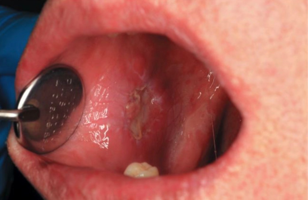
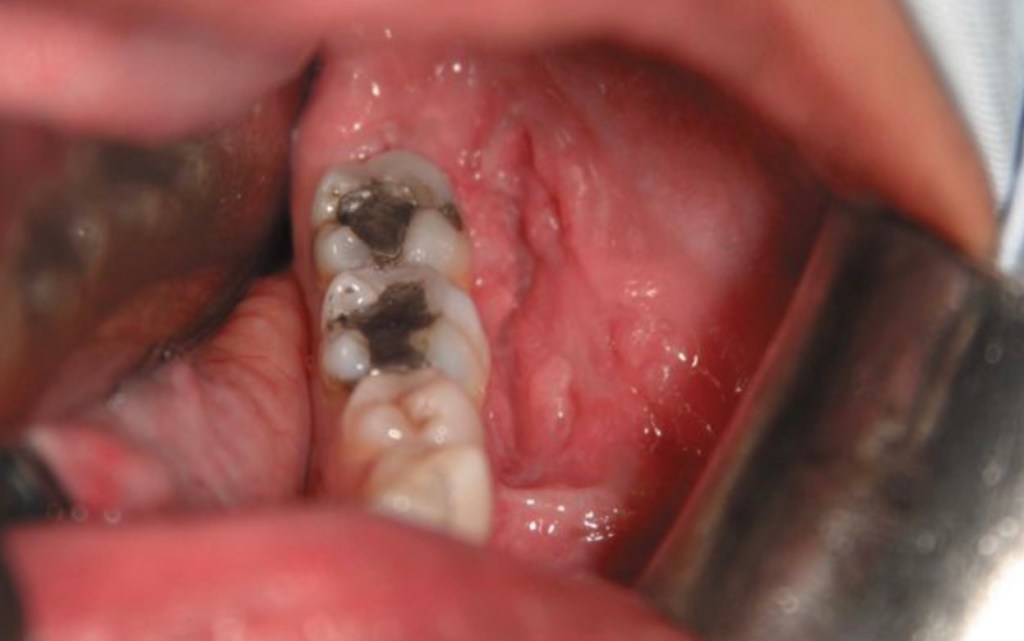
Verrucous, exophytic or ulceroinfiltrative in character
Squamous cell carcinoma buccal mucosa of verrucous appearanceSquamous cell carcinoma buccal mucosa of ulceroinfiltrative appearance
Presentation of buccal carcinoma of the oral cavity:
Patients may present with:
Pain
An intraoral mass
Ulceration
Trismus
Patients who chew betel often have areas of:
Erythroleukoplakia of the buccal mucosa or submucous fibrosis and consequent trismus:
Making the detection of invasive squamous cell carcinoma difficult
Advanced buccal carcinomas may extend into adjacent sites to include:
External skin, mandible or maxilla
It is not unusual for patients to present with advanced disease:
40% or more presenting with stage III / IV disease
Palpable lymphadenopathy on presentation:
May be as high as 57% for T3 / T4 lesions
Occult nodal metastasis:
May be present in 26% of those who are clinically N0 at presentation:
Tumors greater than T2, are poorly differentiated, have a poor lymphocytic response or are thicker than 5 mm:
Are more likely to demonstrate cervical metastasis
Tumors are usually well differentiated
Work up:
Biopsies of buccal carcinomas should be of sufficient depth to help the pathologist give an indication of depth of invasion:
Since this will help decide on management of the neck
Buccal carcinoma may rapidly extend to adjacent sites:
Thus accurate imaging is required:
Most patients will require MRI / CT imaging:
Augmented with ultrasound scan if necessary to help in the assessment of depth of primary and cervical lymphadenopathy
Treatment
Primary site:
Traditional treatment of buccal carcinoma is:
Surgery with postoperative radiation therapy (PORT) for selected patients
T1 / T2 disease:
Can typically be resected perorally
T3 / T4 disease:
May require facial access incisions and bony resection of the maxilla and / or mandible
The primary tumor should be resected with:
A 1 cm margin and up to 2 cm if skin is involved
The buccinator muscle:
Should be included as the deep margin at the very least
The parotid duct:
May need to be repositioned or ligated
External skin should be taken with the specimen:
If there is any evidence clinically or on imaging that it is involved
Partial maxillectomy or mandibular resection (rim (marginal) or segmental) may be required.
Small T1 tumors:
May be resected and reconstructed by primary closure
Healing by secondary intention may be considered:
However postoperative trismus may be anticipated:
Unless vigorous mouth opening exercises are conducted
Split thickness skin grafts may be used:
The use of silicone sheets to stabilize the graft being useful
The use of a skin graft to reconstruct deeper resections:
May leave a very thin cheek with potentially poor aesthetics
Local flaps such as:
The buccal fat pad or temporoparietal fascial flap:
May be used for reconstruction if tumor extension does not compromise their use
Microvascular free flap reconstruction with a radial free forearm flap or anterolateral thigh flap:
Restores the thickness of the cheek and if external skin is involved:
The flaps can be bipaddled to provide reconstruction of mucosal and skin surfaces
T4 tumors requiring segmental resection of the mandible:
May require composite free flap reconstruction
Reconstruction with a radial free forearm flap:
Has been shown to give better postoperative mouth opening than reconstruction with a split skin graft or buccal fat pad
As a single treatment modality for T1 / T2 tumors has been advocated:
However, a change of practice from radiotherapy to surgery at Memorial Sloan Kettering Cancer Center was associated with improved prognosis
Brachytherapy or external beam irradiation may be considered
Management of the Neck:
Regional spread of disease in buccal carcinoma is usually to:
The ipsilateral level I and II lymph nodes
Patients with palpable lymphadenopathy or pathological nodes on imaging:
Should have a comprehensive neck dissection:
Although if pathological nodes are only located in level I, a level I to III selective neck dissection (SND) may be considered
Nodes in the region of the facial artery as it crosses the mandible:
Should be removed with the neck dissection specimen
Patients with a cN0 neck:
With a T2 or greater primary tumors or tumors with a thickness greater than 5 mm:
Should have an elective neck dissection:
Some institutions will conduct an elective neck dissection (END) if the tumor is 3 to 4 mm thick or if histological examination of the tumor demonstrates lymphatic infiltration
PORT:
The indications for postoperative radiotherapy to the loco-regional area are similar to other sites:
Notably two or more nodes in the neck, extracapsular spread (ECS), positive margins or stage III / IV disease
The beneficial role of PORT in selected patients with buccal carcinoma has been demonstrated by several authors:
Some authors suggest that PORT should be considered even in stage I and II disease, or tumors greater than 10 mm thick
Recurrence:
Recurrence rates for buccal carcinoma are 26% to 80%:
Usually occurring within two years
Involvement of the parotid duct and buccinatormuscle:
Have not been found to be significant indicators of recurrence
Outcomes and prognosis in ILC are generally favorable:
Consistent with the luminal A phenotype
The majority of evidence supporting similar or better survival as IDC:
These include a large SEER study of 263,408 women (27,639 with ILC and 235,769 with IDC) treated between 1993 and 2003:
A stage-matched analysis showed that ILC was more likely to be:
Greater than 2 cm
Lymph node positive
ER positive
The 5-year disease-free survival was significantly better for ILC than for IDC after matching for stage:
With an overall 14% survival benefit (HR 0.86) identified on multivariable analysis
As such, although overall stage-corrected prognosis appears to be favorable, some propose that this may be offset by a higher stage at presentation and higher rates of late metastatic recurrences, often occurring in atypical sites
The pleomorphic subtype of ILC is:
Also a known exception to the generally favorable prognosis, having been shown in retrospective series to more frequently develop metastatic disease than other nonpleomorphic ILCs
Currently, there are no unique specifications for surveillance of ILC:
For all treated nonmetastatic breast cancers, NCCN guidelines recommend a history and physical examination one to four times per year as clinically appropriate for 5 years and then annually
Annual mammography should be performed for patients treated with BCT
The role of MRI in surveillance is unclear and presently recommended only for those with a lifetime risk greater than 20% of developing a second primary breast cancer
Adherence to hormonal therapy should be encouraged for those prescribed and yearly gynecologic assessment arranged for those without a previous hysterectomy
Signs of disease recurrence, either locoregional or systemic, should prompt evaluation with appropriate laboratory work and diagnostic imaging, which may include diagnostic CT or fluorodeoxyglucose PET/CT scans followed by biopsy to prove first recurrence of disease
It should be noted that the generally low-grade nature of ILC may limit the sensitivity of traditional PET/CT scans, and studies are ongoing for the use of alternative radiotracers using ER ligands for increased sensitivity
Confirmed LRRs (those of the breast / chest wall and / or regional lymph nodes alone):
Can be managed with complete surgical resection and systemic therapy
Distant metastatic disease (stage IV) is managed with individualized systemic therapy












 Rodrigo Arrangoiz MS, MD, FACS, FSSO