Vigilancia con Resonancia Magnetica

La Sociedad Americana del Cáncer (American Cancer Society) respalda el examen con resonancia magnetica (RM) para cualquier persona con riesgo de cáncer de mama de por vida superior al 20%.

  • Esto incluiría a la mayoría de las personas con una mutación patógena en cualquiera de los genes de predisposición al cáncer de mama establecidos incluidos en los modernos paneles de pruebas genéticas.

Las directrices NCCN han respaldado específicamente la vigilancia mejorada con MRI para BRCA1, BRCA2, ATM, CDH1, CHEK2, NBN, NF1, PALB2, PTEN, STK11 y TP53.

Rodrigo Arrangoiz MS, MD, FACS

Cirugía General y Gastrointestinal
Michigan State University
Cirugía Oncológica
Fox Chase Cancer Center
Tumores de Cabeza y Cuello / Cirugía Endocrina
Fox Chase Cancer Center
Tumores de Cabeza y Cuello / Cirugía Endocrina
IFHNOS / Memorial Sloan Kettering Cancer Center
Maestría en Ciencias de Investigación
Drexel University
Certificado por el Colegio Americano de Cirugía
Fellow del Colegio Americano de Cirugía
Fellow de la Sociedad de Cirugia Oncológica
Sociedad Quirúrgica S.C.
Hospital ABC Santa Fé
Av. Carlos Graef Fernández #154

Consultorio 515
Col. Tlaxala, Delg. Cuajimalpa
México, D.F. 05300
Tel: 1103 – 1600 Ext 4515 a la 4517
Fax:1664 – 7164
 

ENHANCED SURVEILLANCE

The American Cancer Society supports screening MRI for anyone with lifetime breast cancer risk greater than 20%.

  • This would include all most anyone with a pathogenic mutation in any of the established breast cancer predisposition genes included in the modern genetic testing panels.

The NCCN guidelines have specifically endorsed enhanced surveillance with MRI for BRCA1, BRCA2, ATM, CDH1, CHEK2, NBN, NF1, PALB2, PTEN, STK11, and TP53.


Rodrigo Arrangoiz MS, MD, FACS

Cirugía General y Gastrointestinal
Michigan State University
Cirugía Oncológica
Fox Chase Cancer Center
Tumores de Cabeza y Cuello / Cirugía Endocrina
Fox Chase Cancer Center
Tumores de Cabeza y Cuello / Cirugía Endocrina
IFHNOS / Memorial Sloan Kettering Cancer Center
Maestría en Ciencias de Investigación
Drexel University
Certificado por el Colegio Americano de Cirugía
Fellow del Colegio Americano de Cirugía
Fellow de la Sociedad de Cirugia Oncológica
Sociedad Quirúrgica S.C.
Hospital ABC Santa Fé
Av. Carlos Graef Fernández #154

Consultorio 515
Col. Tlaxala, Delg. Cuajimalpa
México, D.F. 05300
Tel: 1103 – 1600 Ext 4515 a la 4517
Fax:1664 – 7164
 

Estimating Breast Cancer Risk

The average probability of developing breast cancer by age 70 is estimated at 57% to 59% for BRCA1 gene mutation carriers and 49% to 51% for BRCA2 gene mutation carriers.

  • Emphasis is on the word “average” because these estimates are based on data from families with a lot of breast cancer cases as well as families with very few breast cancers.
    • Previous studies that had only included families with many breast cancer cases had concluded that lifetime breast cancer risk was more in the range of 80% to 85%.

This illustrates an important point:

  • The risk conferred by a pathologic mutation in any breast cancer predisposition gene will vary widely by family:
    • This is most likely due to gene-gene interactions, which are the modifying effects of small differences (e.g., polymorphisms) in many different genes.

Average Lifetime Brest Cancer Risk for the Syndromes

Rodrigo Arrangoiz MS, MD, FACS
Cirugía General y Gastrointestinal
Michigan State University
Cirugía Oncológica
Fox Chase Cancer Center
Tumores de Cabeza y Cuello / Cirugía Endocrina
Fox Chase Cancer Center
Tumores de Cabeza y Cuello / Cirugía Endocrina
IFHNOS / Memorial Sloan Kettering Cancer Center
Maestría en Ciencias de Investigación
Drexel University
Certificado por el Colegio Americano de Cirugía
Fellow del Colegio Americano de Cirugía
Fellow de la Sociedad de Cirugia Oncológica  

Sociedad Quirúrgica S.C.
Hospital ABC Santa Fé
Av. Carlos Graef Fernández #154

Consultorio 515
Col. Tlaxala, Delg. Cuajimalpa
México, D.F. 05300
Tel: 1103 – 1600 Ext 4515 a la 4517
Fax:1664 – 7164

Multigene Panel Testing

Multi-gene panel testing will identify a pathogenic mutation in 4% to 12% of familial high-risk patients who have tested negative for BRCA1 and BRCA2 mutation.

  • BRCA1 and BRCA2 are still the genes most frequently implicated in hereditary breast cancer predisposition:
    • PALB2, CHEK2, and ATM are the next most common.

Variants of uncertain clinical significance are reported for 20% to 40% of tests.

  • This is likely to improve as data accumulate.

Average cancer risk can be estimated for most of the genes, but this should be adjusted up or down based on family history for management decisions.

Most patients with a pathologic mutation in any of the breast cancer predisposition genes meet the criteria for enhanced surveillance with magnetic resonance imaging.

Risk-reducing surgery is considered for the highest-risk patients.

The results of a genetic test can influence management decisions for the newly diagnosed breast cancer patient.

Breast-Cancer-Genetics-Orange-County-California
Rodrigo Arrangoiz MS, MD, FACS
Cirugía General y Gastrointestinal
Michigan State University
Cirugía Oncológica
Fox Chase Cancer Center
Tumores de Cabeza y Cuello / Cirugía Endocrina
Fox Chase Cancer Center
Tumores de Cabeza y Cuello / Cirugía Endocrina
IFHNOS / Memorial Sloan Kettering Cancer Center
Maestría en Ciencias de Investigación
Drexel University
Certificado por el Colegio Americano de Cirugía
Fellow del Colegio Americano de Cirugía
Fellow de la Sociedad de Cirugia Oncológica
Sociedad Quirúrgica S.C.
Hospital ABC Santa Fé
Av. Carlos Graef Fernández #154

Consultorio 515
Col. Tlaxala, Delg. Cuajimalpa
México, D.F. 05300
Tel: 1103 – 1600 Ext 4515 a la 4517
Fax:1664 – 7164
 

El Modelo de Gail

El modelo de Gail es una herramienta de evaluación de riesgo de cáncer de mama validada que se basa principalmente en factores de riesgo de cáncer de seno no modificables.

Es un modelo estadístico multivariable que utiliza la edad, la edad de la menarca, la edad del primer nacimiento vivo, los antecedentes familiares de cáncer de mama y el número de biopsias de mama para calcular el riesgo de cáncer de mama en personas sin antecedentes de cáncer de mama.

Se ha demostrado que calcula con precisión la proporción de mujeres en las que se desarrollará cáncer de mama cuando se usan en grupos grandes. Sin embargo, tiene un rendimiento bajo al discriminar entre mujeres individuales en las que el cáncer de mama se desarrollará o no.

_BreastCancer-T1
Rodrigo Arrangoiz MS, MD, FACS
Cirugía General y Gastrointestinal
Michigan State University
Cirugía Oncológica
Fox Chase Cancer Center
Tumores de Cabeza y Cuello / Cirugía Endocrina
Fox Chase Cancer Center
Tumores de Cabeza y Cuello / Cirugía Endocrina
IFHNOS / Memorial Sloan Kettering Cancer Center
Maestría en Ciencias de Investigación
Drexel University
Certificado por el Colegio Americano de Cirugía
Fellow del Colegio Americano de Cirugía
Fellow de la Sociedad de Cirugia Oncológica
Sociedad Quirúrgica S.C.
Hospital ABC Santa Fé
Av. Carlos Graef Fernández #154

Consultorio 515
Col. Tlaxala, Delg. Cuajimalpa
México, D.F. 05300
Tel: 1103 – 1600 Ext 4515 a la 4517
Fax:1664 – 7164
 

The Gail Model

The Gail model is a validated breast cancer risk assessment tool that is primarily based on non-modifiable breast cancer risk factors.

It is a multivariable statistical model that uses age, age at menarche, age at first live birth, family history of breast cancer, and number of breast biopsies to estimate breast cancer risk in individuals without a previous history of breast cancer.

It has been shown to accurately estimate the proportion of woman in whom breast cancer will develop when used in large groups. However, it performs poorly in discriminating between individual women in whom breast cancer will and will not develop.

_BreastCancer-T1

Rodrigo Arrangoiz MS, MD, FACS

Cirugía General y Gastrointestinal
Michigan State University
Cirugía Oncológica
Fox Chase Cancer Center
Tumores de Cabeza y Cuello / Cirugía Endocrina
Fox Chase Cancer Center
Tumores de Cabeza y Cuello / Cirugía Endocrina
IFHNOS / Memorial Sloan Kettering Cancer Center
Maestría en Ciencias de Investigación
Drexel University
Certificado por el Colegio Americano de Cirugía
Fellow del Colegio Americano de Cirugía
Fellow de la Sociedad de Cirugia Oncológica
Sociedad Quirúrgica S.C.
Hospital ABC Santa Fé
Av. Carlos Graef Fernández #154

Consultorio 515
Col. Tlaxala, Delg. Cuajimalpa
México, D.F. 05300
Tel: 1103 – 1600 Ext 4515 a la 4517
Fax:1664 – 7164


Vascular Anatomy of the Thyroid Gland

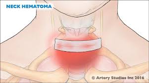
The thyroid gland as well as the adrenal glands have the greatest blood supply per gram of tissue. The significance is that hemostasis is a major problem of thyroid surgery, especially in patients with goiter.

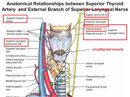
The arterial irrigation to the thyroid gland comes from two paired arteries, the superior and inferior thyroid arteries and, sometimes, from the thyroidea ima. These arteries have abundant collateral anastomoses with each other, ipsilaterally and contra-laterally (Figure). The thyroid ima is a single vessel that, when present (1% to 4% of individuals), originates from the aortic arch or the innominate artery and enters the thyroid gland at the inferior border of the isthmus. Its position anterior to the trachea makes it important in  tracheostomy.

The superior thyroid artery is the first anterior branch of the external carotid artery. In rare cases, it may arise from the common carotid artery just before its bifurcation. The superior thyroid artery passes downward and anteriorly to reach the superior pole of the thyroid gland under the cover of the superior belly of the omohyoid and sternohyoid muscles. In part of its track, the artery parallels the external branch of the superior laryngeal nerve (SLN) which supplies the cricothyroid muscle and the cricopharyngeus muscle, the lowest voluntary part of the pharyngeal musculature. The artery runs superficially on the anterior border of the lateral lobe, dividing into anterior and posterior branches at the apices of the thyroid lobes. These branches send branches (terminal) deep into the thyroid gland before curving toward the isthmus, where they anastomose with the contralateral artery (usually the anterior branch).

The superior thyroid artery has six branches: the infrahyoid, sternocleidomastoid, superior laryngeal, cricothyroid, inferior pharyngeal constrictor, and terminal branches of the artery for the supply of the thyroid and parathyroid glands. There are usually two branches to the thyroid gland (anterior and posterior) but in rare instances there may be a third branch, the so-called lateral  branch. The anterior branch anastomoses with the contralateral artery, the posterior branch  anastomoses with the ipsilateral inferior thyroid artery. The posterior branch gives off a small parathyroid artery which supplies the superior parathyroid gland in 45% of the cases. Nobori et al. noted that in 67% of the cases the superior parathyroid glands received their blood supply from a single vessel, and in 1/3 of the cases two or more branches reached the superior parathyroid glands.

High ligation of the superior thyroid artery during thyroidectomy places the external branch of the SLN at risk of inadvertent injury, which would produce dysphonia by altering pitch regulation. The cricothyroid artery, a potentially troublesome branch of the superior thyroid artery, runs superior to the upper pole and runs toward the midline on the cricothyroid ligament. This vessel can be lacerated during emergent cricothyroidotomy.

The inferior thyroid artery usually arises from the thyrocervical trunk (Figure 5), a branch of the subclavian artery, but in 15% of individuals it may arise from the subclavian artery. The inferior thyroid artery ascends vertically behind the carotid sheath curving medially and posteriorly on the anterior surface of the longus coli muscle before entering the tracheoesophageal groove. After penetrating the prevertebral fascia, the artery divides into two or more branches as it crosses the RLN. The lowest branch sends a tinny branch to supply the inferior parathyroid gland and supplies the lower pole of the thyroid gland [2]. The upper branch of the inferior thyroid artery supplies the posterior surface of the gland, usually anastomosing with the descending  branch of the superior thyroid artery (posterior  branch). On the right-hand side the inferior thyroid artery is absent in approximately 2% of individuals, on the left-hand side it is absent in 5% of the cases. A duplicated artery is a rare occurrence.

The RLN ascends in the tracheoesophageal groove and enters the larynx between the inferior cornu of the thyroid cartilage and the arch of the cricoid cartilage (Figure). The RLN can be found after it emerges from the superior thoracic outlet, in a triangle bounded laterally by the common carotid artery, medially by the trachea, and superiorly by the thyroid lobe. The relationship between the inferior thyroid artery and the RLN is highly variable, as established by the work of Reed A.F., who in 1943 described 28 variations in this relationship. The RLN can be found deep to the inferior thyroid artery (40% of the cases), superficial to the RLN (20% of the cases), or between the branches of the inferior thyroid artery (35% of the cases).  Notably, the association between the RLN and the inferior thyroid artery on one side of the neck is comparable to that found on the other side in only 17% of individuals. Furthermore, at the level of the inferior thyroid artery, branches of the RLN that are extra laryngeal may be present in 5% of the cases. Preservation of all of those branches is very important during thyroid surgery.

Veins of the thyroid gland form a plexus of vessels lying in the substance and on the surface of the gland. The plexus is drained by three pairs of veins that provide venous drainage for the thyroid (Figure). The superior thyroid vein accompanies the superior thyroid artery. As it emerges from the superior pole of the thyroid gland, the vein passes superiorly and laterally across the  superior belly of the omohyoid muscle and the common carotid artery to enter  the internal jugular vein alone or with the common facial vein. The middle thyroid vein arises on the lateral surface of the thyroid gland at about two-thirds of its anteroposterior extent. It crosses the common carotid artery following a direct course to the internal jugular vein (no artery escorts it). This vein may be absent or, in very rare occurrences it may be double. The extra vein is inferior to the normal vein (fourth thyroid vein). The importance of these thyroid veins is in their vulnerability during thyroid surgery. The inferior thyroid veins are the largest and most variable of the thyroid veins (the right and left side are usually asymmetric. They follow different pathways on each side. The right inferior thyroid vein passes anterior to the innominate artery to drain into the right brachiocephalic vein, rarely in may cross anterior to the trachea and drain into the left brachiocephalic vein. The left vein crosses the trachea to enter the left brachiocephalic vein. Sporadically, both inferior thyroid veins form a common trunk called the thyroid ima vein, which empties into the left brachiocephalic vein.


Rodrigo Arrangoiz MS, MD, FACS
Cirugía Oncológica
Fox Chase Cancer Center
Tumores de Cabeza y Cuello / Cirugía Endocrina
Fox Chase Cancer Center
Tumores de Cabeza y Cuello / Cirugía Endocrina
IFHNOS / Memorial Sloan Kettering Cancer Center
Maestría en Ciencias de Investigación
Drexel University
Certificado por el Colegio Americano de Cirugía
Fellow del Colegio Americano de Cirugía
Fellow de la Sociedad de Cirugia Oncológica 

Sociedad Quirúrgica S.C.
Hospital ABC Santa Fé
Av. Carlos Graef Fernández #154

Consultorio 515
Col. Tlaxala, Delg. Cuajimalpa
México, D.F. 05300
Tel: 1103 – 1600 Ext 4515 a la 4517
Fax:1664 – 7164
 

Complications of Thyroid Surgery

A. Hypocalcemia secondary to hypoparathyroidism

Reported rates of transient hypocalcemia vary in the medical literature from 5% to 50%, but the rate of permanent hypocalcemia secondary to hypoparathyroidism (ie, lasting more than 6 months) is below 3% (0.5% to 2%).

  • The pathophysiology behind transient hypoparathyroidism and hypocalcemia is not well understood but is thought to be related to a transient ischemia to the parathyroid glands or perhaps an increased release of the acute phase reactant endothelin 1.
  • A systematic review of predictors of post-thyroidectomy hypocalcaemia found perioperative parathyroid hormone (PTH), preoperative vitamin D and postoperative changes in calcium to be biochemical predictors.
  • Patients who are at increased risk for this complication are those with Graves disease or malignancy or those undergoing total thyroidectomy, or total thyroidectomy with central compartment neck dissection.
 

Patients may initially be asymptomatic while hypocalcemic. Classic presenting symptoms include numbness and tingling of the digits or perioral area, carpopedal spasm, or the presence of a Chvostek sign or a Trousseau sign. In severe cases, patients may also experience tetany, EKG changes (QT prolongation), seizures, mental status changes, or cardiac arrest secondary to hypocalcemia.

  • The Chvostek sign can be reproduced by tapping on the face just anterior to the ear, causing contraction of the ipsilateral facial muscles.
  • A patient with a positive Trousseau sign will have spasm of the wrist, fingers, or thumb with inflation of a sphygmomanometer above the systolic blood pressure.
    • Either sign is indicative of neuromuscular excitability associated with hypocalcemia.

Patients who are noted to have postoperative hypocalcemia should be managed with calcium supplementation. By following the trend of serum calcium levels, oral calcium supplementation can be titrated accordingly.

  • If patients are receiving 2 grams of elemental calcium and continue to have decreasing or low serum calcium, calcitriol supplementation between 0.25 mcg to 1 mcg per day can be considered.
  • Additionally, intravenous calcium replacement may be necessary for patients refractory to oral management or those with severe symptomatic hypocalcemia.
    • Endocrinology consultation should be considered in these patients.
  • Of note, serum calcium levels should be corrected for concurrent hypoalbuminemia and any hypomagnesemia should be medically corrected.

Patients who develop hypocalcemia should be discharged with calcium and vitamin D supplementation and if necessary calcitriol supplementation. After a few months, weaning from the calcium supplementation can be considered.

B. Injury to the recurrent laryngeal nerve

  • Injury to the recurrent laryngeal nerve (RLN) can yield vocal fold paresis or paralysis. The implementation of nerve monitoring has not been proven to lower this risk, but may provide prognostic value.
    • Studies show that identifying the RLN is associated with lower rates of injury (knowledge of anatomy). 
 Permanent RLN paralysis occurs in 1% to 2% of thyroidectomies in experienced hands.
  • These cases may be underestimated, as not all patients undergo postoperative laryngeal evaluation.
  • Should an injury occur, the patient usually presents with postoperative persistent hoarseness.
  • Patients may also describe dysphagia or aspiration with thin liquids.
  • Patients who undergo total thyroidectomy are at risk for bilateral vocal fold paralysis, a devastating complication.
    • This usually manifests in the immediate postoperative period with airway obstruction, biphasic stridor, or respiratory distress.

Patients with suspected recurrent laryngeal nerve injury should be evaluated with flexible laryngoscopy or videostroboscopy to confirm the position and movement of the vocal folds.

  • Should they have aspiration or dysphagia symptoms, they should be evaluated by a speech language pathologist.
  • Patients with suspected bilateral vocal fold paralysis may require urgent and definitive airway management with a tracheotomy.
 Permanent corrective procedures for vocal fold paralysis are not entertained until 9 to 12 months have passed.
  • At this point, any persistent injury may be considered permanent.
 C. Injury to the superior laryngeal nerve

The superior laryngeal nerve has both an internal and external branch.

  • The internal branch provides sensory innervation to the larynx (supra glottic larynx), while the external branch innervates the cricothyroid muscle.
  • This posterior laryngeal muscle assists with lengthening of the vocal fold.
  • Estimates of this complication vary, and are likely underestimated.
Often this injury is relatively asymptomatic. Patients may occasionally experience hoarseness or vocal fatigue.
  • Voice professionals, however, can be significantly affected by this injury, as it affects the ability to produce higher-pitched sounds and thus may affect a singer’s upper register.

This injury too may be evaluated videostroboscopy, as well as laryngeal electromyography (EMG):

  • Some slight bowing of the affected vocal cord may be present, and the affected vocal cord may be lower than the normal cord.
  • Additionally, EMG shows a deficit in the cricothyroid muscle.
 Unknown

D. Neck hematoma

A rare but but dangerous complication of thyroidectomy, neck hematomas can form secondary to inadequate hemostasis or a coagulopathy.

  • Incidence of this complication is approximately 1%, but its occurrence can lead to asphyxiation and airway compromise.
  • When identified on physical examination, the patient must be taken back to the operating room for exploration and achieving hemostasis.
  • If the patient is in respiratory distress, the surgical wound should be opened and the hematoma evacuated immediately (even at the bedside) and then the patient should be taken to the operating room.

Unknown

E. Infection

The rates of infection after thyroidectomy have significantly decreased with improvements in technology and aseptic technique and are currently estimated between 1% to 2%. 

  • The usual presentation is a superficial cellulitis with warmth, erythema, and tenderness surrounding the surgical incision.
  • If fluctuance is present, a superficial abscess may also be present.
  • Other signs of infection, such as fever and leukocytosis, without an overlying cellulitis, may point to a deep space neck infection or abscess.
CT imaging can be helpful in evaluating the deep spaces of the neck.
  • Abscess needs to be drained, and the aspirate should be sent for cultures.
  • Patients with a superficial cellulitis need to be on antibiotics that cover gram-positive organisms, while those with abscesses should be placed on broad-spectrum antibiotics until cultures yield specific bacteria.
Rodrigo Arrangoiz MS, MD, FACS
Cirugía Oncológica
Fox Chase Cancer Center
Tumores de Cabeza y Cuello / Cirugía Endocrina
Fox Chase Cancer Center
Tumores de Cabeza y Cuello / Cirugía Endocrina
IFHNOS / Memorial Sloan Kettering Cancer Center
Maestría en Ciencias de Investigación
Drexel University
Certificado por el Colegio Americano de Cirugía
Fellow del Colegio Americano de Cirugía
Fellow de la Sociedad de Cirugia Oncológica  

Sociedad Quirúrgica S.C.
Hospital ABC Santa Fé
Av. Carlos Graef Fernández #154

Consultorio 515
Col. Tlaxala, Delg. Cuajimalpa
México, D.F. 05300
Tel: 1103 – 1600 Ext 4515 a la 4517
Fax:1664 – 7164


Embriologia de la Glándula Tiroides

La glándula tiroides es la primera de las glándulas endocrinas del cuerpo que se desarrolla, se origina como una bolsa externa del intestino anterior primitivo alrededor de la tercera semana de gestación (aproximadamente al día 24).

  • Se origina en la base de la lengua en el foramen ciego.

La tiroides se origina en dos estructuras principales:

  • La faringe primitiva y la cresta neural.

La glándula tiroides se forma como una proliferación de células epiteliales endodérmicas en la superficie media del piso faríngeo en desarrollo:

  • El sitio de este desarrollo se encuentra entre 2 estructuras clave, el tuberculum impar y la cópula, y se conoce como foramen ciego.
    • La glándula tiroides inicialmente se origina en la parte caudal del tubérculo impar, que también se conoce como brote mediano de la lengua:
      • Esta inflamación embrionaria se desarrolla desde el primer arco faríngeo y se produce en la línea media en el suelo de la faringe en desarrollo, y finalmente ayuda a formar la lengua a medida que las dos tumefacciones linguales laterales la sobrecreden.
  • El foramen ciego comienza rostral a la cópula, también conocida como la eminencia hipobranquial:
    • Esta hinchazón embriológica mediana consiste en un mesodermo que surge de la segunda bolsa faríngea (aunque también están involucradas la tercera y la cuarta bolsa):
      • La glándula tiroides, por lo tanto, se origina entre la primera y la segunda bolsa.

Los anlages laterales emparejados se originan a partir de la cuarta bolsa branquial y se fusionan con el anlage tiroideo mediano aproximadamente a la quinta semana de gestación:

  • Los anlages laterales son de origen neuroectodérmico (cresta neural) (cuerpos ultimobranquiales) y producen las células C o parafoliculares que son productoras de calcitonina, que así se encuentran en la región posterior superior de la glándula.
    • El anlage tiroideo lateral rudimentario se desarrolla a partir de las células de la cresta neural, mientras que el angio tiroideo medio, que forma la mayor parte de la glándula, surge de la faringe primitiva.

El precursor tiroideo inicial, el primordio tiroideo, comienza como un simple engrosamiento de la línea media (endodermo) y se desarrolla para formar el divertículo tiroideo. Esta estructura es inicialmente hueca, aunque luego se solidifica y se vuelve bilobulada.

  • El tallo generalmente tiene un lumen, el conducto tirogloso, que no desciende a los lóbulos laterales.
  • Los dos lóbulos se encuentran a ambos lados de la línea media y están conectados a través de un istmo
  • Las células foliculares tiroideas se desarrollan a partir del anlage tiroideo mediano: Se hacen aparentes alrededor de la octava semana de gestación y comienzan a producir coloides alrededor de la semana 11 de gestación.


Rodrigo Arrangoiz MS, MD, FACS

Cirugía Oncológica
Fox Chase Cancer Center
Tumores de Cabeza y Cuello / Cirugía Endocrina
Fox Chase Cancer Center
Tumores de Cabeza y Cuello / Cirugía Endocrina
IFHNOS / Memorial Sloan Kettering Cancer Center
Maestría en Ciencias de Investigación
Drexel University
Certificado por el Colegio Americano de Cirugía
Fellow del Colegio Americano de Cirugía
Fellow de la Sociedad de Cirugia Oncológica
Sociedad Quirúrgica S.C.
Hospital ABC Santa Fé
Av. Carlos Graef Fernández #154

Consultorio 515
Col. Tlaxala, Delg. Cuajimalpa
México, D.F. 05300
Tel: 1103 – 1600 Ext 4515 a la 4517
Fax:1664 – 7164
 

Thyroid Embryology

The thyroid gland is the first of the body’s endocrine glands to develop, arising as an out-pouching of the primitive foregut around the third week of gestation (on approximately the 24th day). It originates at the base of the tongue at the foramen cecum.

  • The thyroid originates from two main structures:

  • The primitive pharynx and the neural crest.

The thyroid gland forms as a proliferation of endodermal epithelial cells on the median surface of the developing pharyngeal floor:

  • The site of this development lies between 2 key structures, the tuberculum impar and the copula, and is known as the foramen cecum.

    • The thyroid gland initially arises caudal to the tuberculum impar, which is also known as the median tongue bud:

      • This embryonic swelling develops from the first pharyngeal arch and occurs midline on the floor of the developing pharynx, eventually helping form the tongue as the two lateral lingual swellings overgrow it.

  • The foramen cecum begins rostral to the copula, also known as the hypobranchial eminence:

    • This median embryologic swelling consists of mesoderm that arises from the second pharyngeal pouch (although the third and fourth pouches are also involved):

      • The thyroid gland, therefore, originates from between the first and second pouches.

The paired lateral anlages originate from the fourth branchial pouch and fuse with the median thyroid anlage at approximately the fifth week of gestation:

  • The lateral anlages are neuroectodermal (neural crest) in origen (ultimobranchial bodies) and produce the calcitonin producing parafolicullar or C cells, which thus come to lie in the superior posterior region of the gland.

    • The rudimentary lateral thyroid anlage develops from neural crest cells, while the median thyroid anlage, which forms the bulk of the gland, arises from the primitive pharynx.

The initial thyroid precursor, the thyroid primordium, starts as a simple midline thickening (endoderm) and develops to form the thyroid diverticulum.

  • This structure is initially hollow, although it later solidifies and becomes bilobed.
  • The stem usually has a lumen, the thyroglossal duct, that does not descend into the lateral lobes.
  • The two lobes are located on either side of the midline and are connected via an isthmus

The thyroid follicular cells develop from the median thyroid anlage:

  • The become apparent around the 8th week of gestation and start producing colloid around the 11th week of gestation.


Rodrigo Arrangoiz MS, MD, FACS

Cirugía Oncológica
Fox Chase Cancer Center
Tumores de Cabeza y Cuello / Cirugía Endocrina
Fox Chase Cancer Center
Tumores de Cabeza y Cuello / Cirugía Endocrina
IFHNOS / Memorial Sloan Kettering Cancer Center
Maestría en Ciencias de Investigación
Drexel University
Certificado por el Colegio Americano de Cirugía
Fellow del Colegio Americano de Cirugía
Fellow de la Sociedad de Cirugia Oncológica
Sociedad Quirúrgica S.C.
Hospital ABC Santa Fé
Av. Carlos Graef Fernández #154
Consultorio 515
Col. Tlaxala, Delg. Cuajimalpa
México, D.F. 05300
Tel: 1103 – 1600 Ext 4515 a la 4517
Fax:1664 – 7164
 
#cirujanotiroides
#cirujanotumoresdecabezaycuello
#cirujanoendrocrino