Rodrigo Arrangoiz MS, MD, FACS – Breast Surgeon / Cirujano de Mama

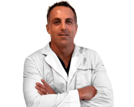
👉Rodrigo Arrangoiz MS, MD, FACS – cirujano oncólogo y miembro de Sociedad Quirúrgica S.C en el America British Cowdray Medical Center en la ciudad de Mexico:

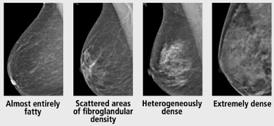
  • Es experto en el manejo del patología de mama:

prof_739_20190417135234

Training:

• General surgery:

• Michigan State University:

• 2004 al 2010

• Surgical Oncology / Head and Neck Surgery / Endocrine Surgery:

• Fox Chase Cancer Center (Filadelfia):

• 2010 al 2012

• Masters in Science (Clinical research for health professionals):

• Drexel University (Filadelfia):

• 2010 al 2012

• Surgical Oncology / Head and Neck Surgery / Endocrine Surgery:

• IFHNOS / Memorial Sloan Kettering Cancer Center:

• 2014 al 2016

  • Quick Facts:
    • I went to medical school at the Anahuac University in Mexico City, which is one of the most prestigious medical schools in Mexico:
      • I graduated Suma Cum Laude from this medical school and was the president of the student medical council.
    • I trained in general surgery at Michigan State University where I was named chief resident during my fifth year of residency which was a great honor.
    • My complex surgical oncology fellowship which included a head and neck training was performed at the Fox Chase Cancer Center in Philadelphia, Pennsylvania.
    • At the same time, I undertook a Masters in Science (Clinical Research for Health Care Professionals) at Drexel University in Philadelphia, Pennsylvania.  
    • I also performed a two-year global online fellowship in Head and Neck Surgery and Oncology with the International Federations of Head and Neck Societies / Memorial Sloan Kettering Cancer Center.
      • I encountered patients with very complex problems, and the greatest lesson I learned was there are always treatment options, utilizing all different types of techniques including radiation, chemotherapy and surgery.
        • This comprehensive training has provided me with an extensive understanding of the multidisciplinary approach to treating patients with cancer.
    • I have developed a particularly strong interest in the surgical and multimodal treatment of patients with breast cancer, head and neck cancer (including thyroid and parathyroid cancer), and endocrine diseases (benign and malignant thyroid and parathyroid diseases), using traditional surgery, regional therapies, and minimally invasive techniques.
      • I am an expert in the treatment of thyroid cancer including; active surveillance for early, small papillary thyroid cancers, minimally invasive thyroid surgery, selective and comprehensive neck dissections.  
      • For the management of parathyroid disease, I offer a minimally invasive radio-guided technique called MIRP (minimally invasive radio-guided parathyroidectomy) through a 2 cm incision which will allow the patient to have a great cosmetic result and quick return to normal life after the operation.
      • I am extremely aware of the impact that a breast cancer diagnosis has on a patient. I do my best to promote a positive atmosphere in which to start my patients’ course of treatment and take the time to explain the pros and cons of each treatment option, so that they can make an informed decision.
        • My management philosophy also includes, not just an emphasis on successful treatment, but also preserving a good cosmetic outcome. I feel fortunate to be a fellowship trained, very highly specialized clinician, because this combination of factors allows me, and our treatment team to focus on one thing all day, every day, and do it well: curing cancer. I think there is nothing more rewarding that I could do as a clinician.
    • I hold my patients as my number one priority. I will spend as much time as necessary educating, answering questions and providing guidance for each individual patient to help them throughout each stage of their management. I believe in honest discussions, where both the patients and family’s goals and expectations are openly communicated. We will work together as a team to put together an evidence based personalized treatment plan. My personal goal is to treat and care for every patient with the same compassion and honesty as if they were a friend or family member.

#Arrangoiz

#Surgeon

#Cirujano

#SurgicalOncologist

#CirujanoOncologo

#BreastSurgeon

#CirujanodeMama

#CancerSurgeon

#CirujanodeCancer

Why Should You Get a Screening Mammogram Starting at Age 40?

img_3760

  • The Swedish Two-County Trial was the first breast screening trial to demonstrate a reduction in breast cancer mortality from screening mammography alone:
    • Showing a 30% reduction in mortality among women aged 40 to 74 years invited to screening:
      • Three decades of follow-up on this study have shown a stable effect upon mortality reduction and an increased absolute benefit in terms of lives saved.
  • Mammographic screening:
    • Has been shown to be associated with a reduction in breast cancer mortality across a range of study designs:
      • Including randomized controlled trials and observational studies.
    • Although specific estimates vary, a meta-analysis of eight randomized trials demonstrated:
      • A 14% to 32% mortality reduction among women invited to screening compared with women who were not invited.
    • A recent Norwegian prospective cohort study:
      • Found invitation to modern mammography screening resulted:
        • In a 28% decrease in mortality.
    • An Australian case-control study and meta-analysis of women participating in organized clinical screening programs showed:
      • A 49% mortality reduction.
    • The Swedish Organised Service Screening Evaluation Group:
      • In an incidence-based mortality study:
        • Demonstrated a mortality reduction of 40% to 45% in women screened.
  • According to these mammography screening trials:
    • The overall reduction in breast cancer mortality gained in populations screened with mammography:
      • Is between 14% to 49%
  • References:
    1. Helvie MA, Chang JT, Hendrick RE, Banerjee M. Reduction in late-stage breast cancer incidence in the mammography era: implications for overdiagnosis of invasive cancer. Cancer. 2014;120:2649-2656.
    2. Nelson HD, Tyne K, Naik A, Bougatsos C, Chan BK, Humphrey L; U.S. Preventive Services Task Force. Screening for breast cancer: an update for the U.S. Preventive Services Task Force. Ann Intern Med. 2009;151:727-737.
    3. Nickson C, Mason KE, English DR, Kavanagh AM. Mammographic screening and breast cancer mortality: a case-control study and meta-analysis. Cancer Epidemiol Biomarkers Prev. 2012;21:1479-1488.
    4. Oeffinger KC, Fontham ET, Etzioni R, et al; American Cancer Society. Breast cancer screening for women at average risk: 2015 guideline update from the American Cancer Society. JAMA. 2015;20;314:1599-1614.
    5. Swedish Organised Service Screening Evaluation Group. Reduction in breast cancer mortality from organized service screening with mammography: 1. Further confirmation with extended data. Cancer Epidemiol Biomarkers Prev. 2006;15:45-51.
    6. Tabár L, Fagerberg CJ, Gad A, et al. Reduction in mortality from breast cancer after mass screening with mammography. Randomised trial from the Breast Cancer Screening Working Group of the Swedish National Board of Health and Welfare. Lancet. 1985;13:829-832.
    7. Tabár L, Vitak B, Chen TH, et al. Swedish two-county trial: impact of mammographic screening on breast cancer mortality during 3 decades. Radiology. 2011;260:658-663.
    8. Weedon-Fekjær H, Romundstad PR, Vatten LJ. Modern mammography screening and breast cancer mortality: population study. BMJ. 2014;348:g3701.


#Arrangoiz

#Surgeon

#Cirujano

#SurgicalOncologist

#CirujanoOncologo

#BreastSurgeon

#CirujanodeMama

#CancerSurgeon

#CirujanodeCancer

Benefits from the addition of tomosynthesis to conventional digital mammography alone

👉Debate on adjunct screening in women with dense breasts has resulted from legislation mandating that women be informed if their mammograms show dense breast tissue, including informing them that other screening modalities are available.
👉In addition to MRI and molecular imaging, both tomosynthesis and breast ultrasound are additional techniques for enhanced screening in patients with dense breasts.

👉Multiple studies have now shown significant benefits from the addition of tomosynthesis to conventional digital mammography alone in screening programs.

👉Ciatto et al. found an increase in detection rate of invasive breast cancer from 5.3/1000 to 8.1/1000, while also decreasing the recall rate by 17%.
👉Skaane et al. found a 40% increase in the detection of invasive cancers with a 15% reduction in false negatives.
👉Rose et al. and Haas et al. showed statistically significant relative reductions in recall rates of 37% and 30%, respectively.
👉A recent retrospective review of 454,850 examinations in 13 screening centers in the United States demonstrated a 41% increase in invasive cancer detection, a 15% reduction in call backs, and a 49% increase in the positive predictive value for recall.
👉Implementation of tomosynthesis did not lead to a significant reduction in biopsy rates as compared to digital mammography screening.
👉As yet, there are no data that show a reduction in mortality with enhanced screening in dense breasts.

👉A prospective multicenter study compared tomosynthesis with bilateral physician hand-held ultrasound screening in 3231 asymptomatic patients with mammography-negative dense breasts. 

– In all, 24 additional cancers were detected, 23 of which were invasive.
– Tomosynthesis detected 13 cancers, and ultrasound detected 23.
– These data suggest that even though tomosysthesis significantly increases the number of cancers found in dense breasts, in the hands of a skilled breast radiologist, ultrasound may be even better. 

👉REFERENCES

  1. Ciatto S, Houssami N, Bernardi D, et al. Integration of 3D digital mammography with tomosynthesis for population breast-cancer screening (STORM): a prospective comparison study. Lancet Oncol. 2013;14:583-589.
  2. Friedewald SM, Rafferty EA, Rose SL, et al. Breast cancer screening using tomosynthesis in combination with digital mammography. JAMA. 2014;311:2499-2507.
  3. Haas BM, Kalra V, Geisel J, Raghu M, Durand M, Philpotts LE. Comparison of tomosynthesis plus digital mammography and digital mammography alone for breast cancer screening. Radiology. 2013;269:694-700.
  4. Rose SL, Tidwell AL, Bujnoch LJ, Kushwaha AC, Nordmann AS, Sexton R Jr. Implementation of breast tomosynthesis in a routine screening practice: an observational study. AJR Am J Roentgenol. 2013;200:1401-1408.
  5. Skaane P, Bandos AI, Gullien R, et al. Comparison of digital mammography alone and digital mammography plus tomosynthesis in a population-based screening program. Radiology. 2013;267:47-56.
  6. Tagliafico AS, Calabrese M, Mariscotti G, et al. Adjunct screening with tomosynthesis or ultrasound in women with mammography-negative dense breasts: interim report of a prospective comparative trial. J Clin Oncol. 2016 Mar 9. pii: JCO634147. [Epub ahead of print]

#Arrangoiz

#BreastSurgeon

#CancerSurgeon

Breast Density

👉According to the 5th edition of the ACR BI-RADS® Atlas, visual estimation of breast fibroglandular composition should be defined by one of the following four descriptions:

  • Almost entirely fatty
  • Scattered areas of fibroglandular density
  • Heterogeneously dense
  • Extremely dense


👉The U.S. population distribution of breast density is as follows:

  • 10% almost entirely fatty
  • 40% scattered areas of fibroglandular density
  • 40% heterogeneously dense
  • 10% extremely dense

👉Women with heterogeneously dense or extremely dense breasts are considered to have “dense breasts.” 

👉Sensitivity of mammography decreases as breast density increases.

👉Increased breast density not only has a masking effect which may obscure masses, but also serves as an independent risk factor for breast cancer.

👉It has been reported that the increased risk may be as much as 4- to 6-fold.

👉Estimates this high are obtained when comparing women with dense breasts to those with fatty replaced breasts.

👉Since only 10% of women have fatty replaced breasts, it makes more sense to make the comparison with women of average breast density.

👉The relative risk for cancer in women with heterogeneously dense breasts compared with the average woman is approximately 1.2, and the relative risk for cancer in women with extremely dense breasts compared with the average woman is approximately 2.1.

👉In general, breast density decreases with increasing age and increasing body mass index, so it is not the absolute density that is a risk factor, but the difference in the observed and expected density—the so called “residual density.”

👉Several states have recently passed legislation requiring women with dense breasts to be specifically informed of their breast density.

👉Women are informed of the limitations of mammography in dense breasts and are instructed to discuss further management with their physicians.

👉An informed decision regarding potential use of supplemental screening options, in addition to mammography, should be discussed, factoring in elements such as overall breast cancer risk as well as the positives and negatives of additional screening.

👉Despite a lack of consensus, algorithms for screening women with dense breasts are available and often emphasize breast cancer risk.

👉Women with heterogeneously dense breasts (or extremely dense breasts) with no additional risk factors require only annual mammography.

👉The decision to pursue additional imaging in patients with elevated risk should supplement but never entirely replace mammography.

👉Biannual screening mammography is not considered appropriate, and there is no indication for thermography.

👉Use of screening ultrasound or MRI are appropriate for women at increased risk, but the benefit remains to be determined in women of average risk for breast cancer.


#Arrangoiz

#BreastSurgeon

#SurgicalOncologist

Feliz Día de la Enfermera

👉MUCHAS FELICIDADES A TODAS LAS ENFERMERAS Y ENFERMEROS QUE HACEN POSIBLE NUESTRO TRABAJO, SON EL CORAZÓN DE LOS HOSPITALES.

👉Sociedad Quirúrgica les agradece toda sus ayuda que es indispensable

#Arrangoiz

#Surgeon

#Teacher

#CancerSurgeon

Rodrigo Arrangoiz MS, MD, FACS

  • I went to medical school at the Anahuac University in Mexico City, which is one of the most prestigious medical schools in Mexico:
    • I graduated Suma Cum Laude from this medical school and was the president of the student medical council.
  • I trained in general surgery at Michigan State University where I was named chief resident during my fifth year of residency which was a great honor.
  • My complex surgical oncology fellowship which included a head and neck training was performed at the Fox Chase Cancer Center in Philadelphia, Pennsylvania.
  • At the same time, I undertook a Masters in Science (Clinical Research for Health Care Professionals) at Drexel University in Philadelphia, Pennsylvania.  
  • I also performed a two-year global online fellowship in Head and Neck Surgery and Oncology with the International Federations of Head and Neck Societies / Memorial Sloan Kettering Cancer Center.
    • I encountered patients with very complex problems, and the greatest lesson I learned was there are always treatment options, utilizing all different types of techniques including radiation, chemotherapy and surgery.
      • This comprehensive training has provided me with an extensive understanding of the multidisciplinary approach to treating patients with cancer.
  • I have developed a particularly strong interest in the surgical and multimodal treatment of patients with breast cancer, head and neck cancer (including thyroid and parathyroid cancer), and endocrine diseases (benign and malignant thyroid and parathyroid diseases), using traditional surgery, regional therapies, and minimally invasive techniques.
    • I am an expert in the treatment of thyroid cancer including; active surveillance for early, small papillary thyroid cancers, minimally invasive thyroid surgery, selective and comprehensive neck dissections.  
    • For the management of parathyroid disease, I offer a minimally invasive radio-guided technique called MIRP (minimally invasive radio-guided parathyroidectomy) through a 2 cm incision which will allow the patient to have a great cosmetic result and quick return to normal life after the operation.
    • I am extremely aware of the impact that a breast cancer diagnosis has on a patient. I do my best to promote a positive atmosphere in which to start my patients’ course of treatment and take the time to explain the pros and cons of each treatment option, so that they can make an informed decision.
      • My management philosophy also includes, not just an emphasis on successful treatment, but also preserving a good cosmetic outcome. I feel fortunate to be a fellowship trained, very highly specialized clinician, because this combination of factors allows me, and our treatment team to focus on one thing all day, every day, and do it well: curing cancer. I think there is nothing more rewarding that I could do as a clinician.
  • I hold my patients as my number one priority. I will spend as much time as necessary educating, answering questions and providing guidance for each individual patient to help them throughout each stage of their management. I believe in honest discussions, where both the patients and family’s goals and expectations are openly communicated. We will work together as a team to put together an evidence based personalized treatment plan. My personal goal is to treat and care for every patient with the same compassion and honesty as if they were a friend or family member.

#Arrangoiz #Surgeon #CancerSurgeon #HeadandNeckSurgeon #BreastSurgeon #SurgicalOncologist #PalmettoGeneralHospital #CenterforAdvancedSurgicalOncology

Active Surveillance in Low Risk Papillary Thyroid Cancer

👉Thinking about active surveillance for a patient with low-risk PTC? This meta-analysis from #AAES2019 showing the safety of this strategy.

https://www.surgjournal.com/article/S0039-6060(19)30561-6/fulltext

#Arrangoiz

#ThyroidSurgeon

#HeadandNeckSurgeon

#EndocrineSurgeon

Stimulated postoperative thyroglobulin (Tg) level before radioactive iodine ablation (RAI)

👉Stimulated postoperative thyroglobulin (Tg) level before radioactive iodine ablation (RAI) can be used to quantify risk of papillary thyroid cancer (PTC) recurrence.

👉Levels equal or greater than 2 ng/mL was associated with a 30% chance of recurrence based on this paper presented at #AAES2019

👉https://www.sciencedirect.com/science/article/pii/S0039606019305100?via%3Dihub


Rodrigo Arrangoiz MS, MD, FACS cirujano de tumores de cabeza y cuello y cirugía endocrina de Sociedad Quirúrgica S.C. es experto en el manejo de nódulos tiroides y del cáncer de tiroides.

Articulo publicado por el Dr. Arrangoiz sobre el manejo de nódulos tiroides:

https://file.scirp.org/pdf/IJOHNS_2018072717023407.pdf

Es pionero en México:

  • Cirugia tiroidea minimamente invasiva
  • La cirugia minimamente invasiva radio-guiada de paratiroides

Su entrenamiento fue el siguiente:

• Cirugia general y gastrointestinal:
• Michigan State University:
• 2004 al 2010image-48• Cirugia oncológica / tumores de cabeza y cuello / cirugia endocrina:
• Fox Chase Cancer Center (Filadelfia):
• 2010 al 2012image-39• Maestria en ciencias (Clinical research for healthprofessionals):
• Drexel University (Filadelfia):
• 2010 al 2012image-50• Cirugia de tumores de cabeza y cuello / cirugiaendocrina
• IFHNOS / Memorial Sloan Kettering Cancer Center:
• 2014 al 2016

image-6image-51 

http://www.sociedadquirurgica.com

http://www.hiperparatiroidismo.info

http://www.cirugiatiroides.com

#Arrangoiz

#CirugiadeTumoresdeCabezayCuello

#CirugiaEndocrina

#CirugiaOncologica

#HeadandNeckSurgery

#EndocrineSurgery

#SurgicalOncology