HEREDITARY BREAST AND OVARIANCANCER SYNDROME

  • In the year 2018 in the United States:
    • Approximately 22 000 women received a new diagnosis of ovarian cancer, and there were 14 000 ovarian cancer deaths
  • Inherited germline mutations:
    • That increase the risk for ovarian cancer:
      • Are present in approximately 7% to 25% of these women
  • Hereditary breast and ovarian cancer syndrome (HBOC):
    • Is the most common genetic syndrome:
      • Linked to ovarian cancer:
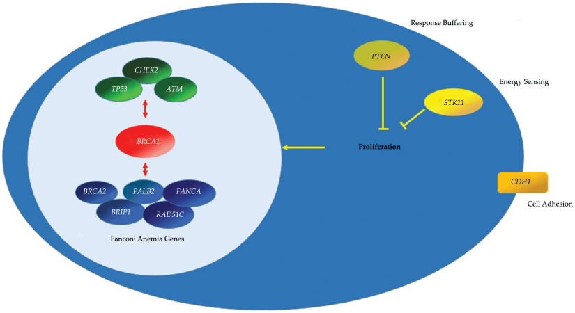
        • Is associated with mutations in the BRCA genes
    • BRCA1 and BRCA2 are:
      • Tumor suppressor genes:
        • Involved in the repair of double‐stranded DNA breaks
    • Certain populations are at significantly higher risk for BRCA mutations than the general population including:
      • Ashkenazi Jewish, French Canadian, and Icelandic populations:
        • This is because of the founder effect:
          • Which causes a decrease in genetic diversity in a population that is geographically or reproductively isolated 
          • For example, in the general United States non‐Jewish population:
            • The risk of inheriting a BRCA mutation is 1 in 400 compared with theAshkenazi Jewish population, who carry an increased risk of 1 in 40
    • The inheritance pattern of the BRCA mutations is:
      • Autosomal dominant:
        • However, tumor suppressor genes are known to be recessive, requiring both copies of the gene to be affected
        • Therefore, a germline BRCA mutation is inherited in one copy of the gene, the second copy then undergoes a somatic mutation:
          • The second hit and the carrier is phenotypically affected
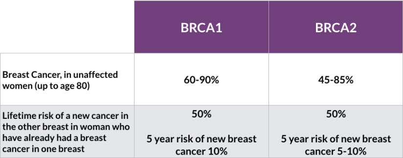
  • The most recently estimated cumulative cancer risk by age 80 for:
    • BRCA1 carriers is:
      • 72% 
    • BRCA2 carriers is:
      • 69%
  • By comparison, the lifetime risk of ovarian and breast cancer in the general population is:
    • 1.3% and 12.4%, respectively
  • Furthermore, the lifetime risk for ovarian cancer in:
    • BRCA1 carriers is:
      • 44%
    • BRCA2 carriers is:
      • 17%
  • BRCA carriers are also at risk for other cancers including:
    • Pancreatic, prostate, melanoma, and colon cancer
  • Recently, findings by Shu et al8 suggest that BRCA1 carriers:
    • Have an increased risk for uterine papillary serous carcinoma:
      • rare but aggressive type of uterine cancer
      • In a prospective cohort of 627 BRCA1 carriers:
        • Four women developed serious uterine cancer:
          • Which was double the expected incidence in the general population
  • References:
    • Siegel RL, Miller KD, Jemal A. Cancer statistics, 2019. CA Cancer J Clin. 2019;69(1):7‐34. 
    • Noone AM, Howlader N, Krapcho M, et al. “SEER Cancer Statistics Review, 1975‐2015,” National Cancer Institute, Bethesda, MD, 2018. 
    • Morgan RD, Burghel GJ, Flaum N, et al Prevalence of germline pathogenic BRCA1/2 variants in sequential epithelial ovarian cancer cases. J Med Genet. 2019;56:301‐307. 
    • McClainMR,PalomakiGE,NathansonKL,etalAdjustingtheestimated proportion of breast cancer cases associated with BRCA1 and BRCA2 mutations: public health implications. Genet Med. 2005;7(1):28‐33. 
    • Gudmundsson J, Johannesdottir G, Bergthorsson JT, et al Different tumor types from BRCA2 carriers show wild‐type chromosome deletions on 13q12‐q13. Cancer Res. 1995;55(21):4830‐4832. 
    • Kuchenbaecker KB, Hopper JL, Barnes DR, et al Risks of breast, ovarian, and contralateral breast cancer for BRCA1 and BRCA2 mutation carriers. JAMA. 2017;317(23):2402‐2416. 
    • Mersch J, Jackson MA, Park M, et al Cancers associated with BRCA1 and BRCA2 mutations other than breast and ovarian. Cancer. 2015;121(2):269‐275. 

#Arrangoiz #BreastSurgeon #CancerSurgeon #SurgicalOncologist #BreastCancer #HereditaryBreastCancer #BRCA #CASO #CenterforAdvancedSurigcalOncology #Miami


Axillary Sentinel Node Biopsy Has Been Shown To Be Feasible For Axillary Staging

  • Axillary sentinel node biopsy has been shown to be feasible for axillary staging:
    • In patients with in-breast recurrence or ipsilateral breast second primary tumors
  • Limited prior axillary sampling (less than nine nodes) has been shown to have greater success in localization
  • Preoperative lymphoscintigraphy should be considered:
    • Given the possibility of aberrant lymphatic drainage due to alterations secondary to prior surgery and radiation
  • References
  • Tokmak H, Kaban K, Muslumanoglu M, Demirel M, Aktan S. Management of sentinel node re-mapping in patients who have second or recurrent breast cancer and had previous axillary procedures. World J Surg Oncol. 2014;12:205.
  • Kothari MS, Rusby JE, Agusti AA, MacNeill FA. Sentinel lymph node biopsy after previous axillary surgery: a review. Eur J Surg Oncol. 2012;38(1):8-15.

#Arrangoiz #BreastSurgeon #BreastCancer #CancerSurgeon #SurgicalOncologist #Miami #CASO #CenterforAdvancedSurgicalOncologist

Contralateral Breast Cancer and Risk Reducing Mastectomy

  • In the last 10 years:
    • There has been a significant focus on the role of contralateral prophylactic mastectomy and the risk of contralateral breast cancer
  • Single-institution studies and Surveillance, Epidemiology, and End Results program (SEER) data:
    • Have found contralateral prophylactic mastectomy rates:
      • To be as high as 25%, which continue to increase
      • Further, this trend seems to be limited to the United States
  • Nichols et al. reviewed rates of contralateral breast cancer between 1976 and 2006 using the SEER database:
    • Overall, they found the rate of contralateral breast cancer:
      • To be declining approximately 3% per year since 1985
    • From 1990 forward:
      • They found this benefit to be restricted to those presenting with an ER+ breast cancer:
        • With annual incidence rates of contralateral breast cancer of 0.25% to 0.37% when the index cancer was diagnosed after age 30 years
      • The rates of contralateral breast cancer were higher in patients with ER negative breast cancers:
        • But still remained reasonably low with annual incidence rates of contralateral breast cancer of 0.45% to 0.65% when the index cancer was diagnosed after age 40 years
  • The use of adjuvant endocrine therapy has contributed to this decline in contralateral breast cancer rates:
    • As it reduces the risk of contralateral breast cancer by approximately 50%
  • Contralateral prophylactic mastectomy:
    • Is attributed with approximately a 90% to 94% overall reduction in the risk of contralateral breast cancer, not 100%
  • Contralateral prophylactic mastectomy:
    • Does not improve overall survival
    • Recent modeling suggests the added absolute benefit of contralateral prophylactic mastectomy to overall survival is less than 1.45%
  • The American Society of Breast Surgeons published a consensus statement with indications and suggestions for patient management and decision making with regard to contralateral prophylactic mastectomy:
    • Discouraging the procedure for women with unilateral breast cancer at average risk for contralateral cancer
  • References
  • Nichols HB, Berrington de Gonzalez A, Lacey JV, Rosenberg PS, Anderson WF. Declining incidence of contralateral breast cancer in the United States from 1975 to 2006. J Clin Oncol. 2011;29(12):1564-1569.
  • Early Breast Cancer Trialists’ Collaborative Group, Davies C, Godwin J, et al. Relevance of breast cancer hormone receptors and other factors to the efficacy of adjuvant tamoxifen: patient-level meta-analysis of randomised trials. Lancet. 2011;378(9793):771-784.
  • King TA, Sakr R, Patil S, et al. Clinical management factors contribute to the decision for contralateral prophylactic mastectomy. J Clin Oncol. 2011;29(16):2158-2164.
  • Metcalfe K, Lynch HT, Ghadirian P, et al. Contralateral breast cancer in BRCA1 and BRCA2 mutation carriers. J Clin Oncol. 2004;22(12):2328-2335.
  • Portschy PR, Kuntz KM, Tuttle TM. Survival outcomes after contralateral prophylactic mastectomy: a decision analysis. J Natl Cancer Inst. 2014;106(8).
  • Boughey JC, Attai DJ, Chen SL, et. al.. Contralateral prophylactic mastectomy consensus statement from the American Society of Breast Surgeons: additional considerations and a framework for shared decision making. Ann Surg Oncol. 2016 Oct;23(10):3106-3111.

#Arrangoiz #BreastSurgeon #CancerSurgeon #SurgicalOncologist #Miami #BreastCancer #ProphylacticMastectomy #RiskReducingSurgery #CASO #CenterforAdvancedSurgicalOncology

The Annual Risk for BRCA Mutation Carriers

  • The annual risk for BRCA mutation carriers:
    • Is estimated to be 2.1%
  • The risk of contralateral breast cancer at 20 years post initial breast cancer diagnosis is:
    • 41% in BRCA1 mutation carriers:
      • Age at diagnosis does affect risk of contralateral breast cancer:
        • Women under 40 are at highest risk
    • Tamoxifen has been shown to decrease the risk of contralateral breast cancer overall
  • Phillips et al:
    • Report that estrogen receptor status of the initial breast cancer diagnosis did not affect contralateral breast cancer risk:
      • However the status was missing in over 50% of the cohort
    • In addition, women found to be at the highest risk:
      • Were those diagnosed with initial breast cancer under age 50
      • With two or more family members with history of breast cancer
      • Intact ovaries
        • The 15-year risk of contralateral breast cancer was 68% in these women
    • These women should be counseled about risk reducing contralateral mastectomy and salpingo-oopherectomy
    • The strongest predictor of contralateral risk was oophorectomy
    • Contralateral prophylactic mastectomy is a controversial area in breast surgical oncology, and patient decision-making around this process continues to be studied
  • References
    • Metcalfe K, Gershman S, Lynch HT, et al. Predictors of contralateral breast cancer in BRCA1 and BRCA2 mutation carriers. Br J Cancer. 2011;104(9):1384–1392.
    • Phillips KA, Milne RL, Rookus MA, et al. Tamoxifen and risk of contralateral breast cancer for BRCA1 and BRCA2 mutation carriers. J Clin Oncol. 2013;31(25):3091-3099.
    • Kuchenbaecker KB, Hopper JL, Barnes DR, et al. Risks of breast, ovarian, and contralateral breast cancer for BRCA1 and BRCA2 mutation carriers. JAMA. 2017;317(23):2402-2416.

#Arrangoiz #BreastSurgeon #BreastCancer #CancerSurgeon #SurgicalOncologist #CASO #Miami #BRCA #CenterforAdvancedSurgicalOncology

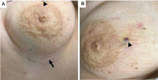
Radiation-Associated Angiosarcoma

  • Radiation-associated angiosarcoma:
    • Is a rare complication from prior radiation
    • The average time from radiation to presentation:
      • Is 10 years
    • The mainstay of treatment remains:
      • Surgical excision with negative margins:
        • However, local recurrence and distant recurrence remains quite high:
          • Close monitoring is recommended
  • The role of chemotherapy:
    • Is unclear
      • Therefore, surgery should remain the primary treatment of choice
  • Preoperative radiation:
    • With hyperfractionated and accelerated radiation therapy:
      • Has also been identified as a potential alternative to surgery alone, with improved survival and should be considered:
        • More trials are needed to improve outcomes for this aggressive but rare complication of radiation
  • References
    • Torres, K.E., Ravi, V., Kin, K. et al. Long-term outcomes in patients with radiation-associated angiosarcomas of the breast following surgery and radiotherapy for breast cancer. Ann Surg Oncol. 2013;20(4):1267-1274.
    • Palta M , Morris CG, Grobmyer SR, Copeland EM, Mendenhall NP. (2010), Angiosarcoma after breast‐conserving therapy. Cancer. 116(8):1872-1878.
    • Smith TL, Morris CG, Mendenhall NP. Angiosarcoma after breast-conserving therapy: long-term disease control and late effects with hyperfractionated accelerated re-irradiation (HART). Acta Oncol. 2014;53(2):235-241.
    • Penel N, Bui BN, Bay JO, et al. Phase II trial of weekly paclitaxel for unresectable angiosarcoma: the ANGIOTAX Study. J Clin Oncol. 2008;26(32):5269-5274.
    • Palta M, Morris CG, Grobmyer SR, Copeland EM 3rd, Mendenhall NP. Angiosarcoma after breast-conserving therapy: long-term outcomes with hyperfractionated radiotherapy. Cancer. 2010;116(8):1872-1878.

#Arrangoiz #BreastCancer #BreastSurgeon #CASO #AngiosarcomaoftheBreast #RadiationInducedAngiosarcoma #CancerSurgeon #SurgicalOncologist #CenterforAdvancedSurgicalOncology #Miami

Occult Primary Breast Cancer

  • Three important conclusions are agreed upon regarding this clinical entity: 
    • Prognosis of occult primary breast cancer is the same or slightly better than women with classic stage IIA disease (T0, N1, M0)
    • An exhaustive workup for the non-breast primary is usually not fruitful
    • Treatment of the breast in some manner decreases the risk of local failure over time
  • Modified radical mastectomy has been the traditional surgical treatment for many years
  • Previously, the primary breast cancer was found in the mastectomy specimen:
    • 40% to 80% of the time, but with the advent of much better mammography and ultrasound along with breast MRI, this rate is much lower now
  • However, what was true then and still holds today:
    • Is that no treatment to the breast itself results in an unacceptably high local recurrence rate
  • An alternative to a modified radical mastectomy:
    • Is complete ALND followed by whole-breast irradiation
  • Axillary dissection:
    • Provides local control while also fine tuning staging
  • Theoretically the whole-breast radiation:
    • Should control any subclinical disease in the breast not detected on imaging
  • Primary radiation to the breast, axilla, and supraclavicular area without any surgery of the breast or axilla:
    • Results in higher local and regional recurrence compared to surgery and radiation combined
  • Axillary node dissection and whole-breast irradiation:
    • Has been found to have equivalent survival as a modified radical mastectomy
  • A recent meta-analysis of 7 studies and more than 240 patients with occult primary breast cancers (0.3% to 0.8% of all breast cancers):
    • Found 39% were treated with ALND and radiation while 47% had modified radical mastectomy and 15% had ALND alone
    • With a mean follow-up of 5 years:
      • The study found no difference in local regional recurrence (12.7% vs 9.8%), distant metastasis (7.2% vs 12.7%), or mortality (9.5% vs 17.9%) between ALND and radiation vs modified radical mastectomy (all p>0.16)
  • ALND with radiation was superior to ALND alone in terms of local regional recurrence (12.7% vs 34.3%, p < 0.01) and trended towards improved survival but this was not statistically significant (P=0.09)
  • References:
    • Barton SR, Smith IE, Kirby AM, Ashley S, Walsh G, Parton M. The role of ipsilateral breast radiotherapy in management of occult primary breast cancer presenting as axillary lymphadenopathy. Eur J Cancer. 2011;47(14):2099-2106.
    • Macedo FI, Eid JJ, Flynn J, Jacobs MJ, Mittal VK. Optimal surgical management for occult breast carcinoma: a meta-analysis. Ann Surg Oncol. 2016;23(6):1838-1844.
    • Rueth NM, Black DM, Limmer AR, et al. Breast conservation in the setting of contemporary multimodality treatment provides excellent outcomes for patients with occult primary breast cancer. Ann Surg Oncol. 2015;22(1):90-95.
    • Walker GV, Smith GL, Perkins GH, et al. Population-based analysis of occult primary breast cancer with axillary lymph node metastasis. Cancer. 2010;116(17):4000-4006.
    • Hessler LK, Molitoris JK, Rosenblatt PY, et al. Factors Influencing management and outcome in patients with occult breast cancer with axillary lymph node involvement: analysis of the National Cancer Database. Ann Surg Oncol. 2017;24(10):2907-2914.
    • Walker GV, Smith GL, Perkins GH, et al. Population-based analysis of occult primary breast cancer with axillary lymph node metastasis. Cancer. 2010;116:4000-4006. PMID: 20564117. http://www.ncbi.nlm.nih.gov/pubmed/20564117
    • Woo SM, Son BH, Lee JW, et al. Survival outcomes of different treatment methods for the ipsilateral breast of occult breast cancer patients with axillary lymph node metastasis: a single center experience. J Breast Cancer. 2013;16:410-416. https://www.ncbi.nlm.nih.gov/pmc/articles/PMC3893343/

#Arrangoiz #BreastSurgeon #CancerSurgeon #Miami #SurgicalOncologist #OccultBreastCancer #BreastCancer #CASO #CenterforAdvancedSurgicalOncology

Axillary Staging after Neoadjuvant Chemotherapy in Breast Cancer

  • Axillary ultrasound :
    • Is frequently useful to assess for lymph node involvement in patients with breast cancer at initial disease presentation:
      • However, normal appearing lymph nodes on ultrasound, before and/or after chemotherapy:
        • Do not obviate the need for surgical axillary staging
  • Axillary ultrasound with percutaneous biopsy of any suspicious lymph nodes, if present:
    • Allows marking of the positive node:
      • To be sure it is removed following treatment with neoadjuvant chemotherapy
  • Plecha et al. recently published data:
    • That show wire localization of marked nodes increases the likelihood of removing the node that was positive prior to neoadjuvant chemotherapy
  • Boughey et al.:
    • Had previously shown that clipping the positive node decreases the false-negative rate of sentinel node biopsy following neoadjuvant chemotherapy
  • Supraclavicular disease:
    • Is classified as N3, stage IIIC disease, not as distant metastatic disease:
      • Supraclavicular involvement can be documented by percutaneous biopsy:
        • Excision is not required prior to chemotherapy
  • Removal of sentinel lymph nodes prior to neoadjuvant chemotherapy:
    • Interferes with assessment of nodal response to chemotherapy
  • PET/CT scan is not needed to plan nodal staging after neoadjuvant chemotherapy:
    • Small volume disease is poorly detected with this modality:
      • So axillary ultrasound is preferred:
        • Regardless of imaging results, surgical staging of the axilla is required
  • References
    • Boughey JC, Ballman KV, Symmans WF, et al. Methods impacting the false-negative rate of sentinel lymph node surgery in patients presenting with node-positive breast cancer (T0–T4, N1–2) who receive neoadjuvant chemotherapy: results from a prospective trial—ACOSOG Z1071 (Alliance). Poster presented at: San Antonio Breast Cancer Symposium; December 9-13, 2014; San Antonio, TX.
    • Plecha D, Bai S, Patterson H, Thompson C, Shenk R. Improving the accuracy of axillary lymph node surgery in breast cancer with ultrasound-guided wire localization of biopsy proven metastatic lymph nodes. Ann Surg Oncol. 2015;22(13):4241-4246.
    • Edge S, Byrd DR, Compton CC, Fritz AG, Greene FL, Trotti A, eds. AJCC Cancer Staging Manual. 7th ed. New York, NY: Springer-Verlag; 2010.
    • Kuerer HM, Sahin AA, Hunt KK, et al. Incidence and impact of documented eradication of breast cancer axillary lymph node metastases before surgery in patients treated with neoadjuvant chemotherapy. Ann Surg. 1999;230(1):72-78.
    • Hieken TJ, Trull BC, Boughey JC, et al. Preoperative axillary imaging with percutaneous lymph node biopsy is valuable in the contemporary management of patients with breast cancer. Surgery. 2013;154(4):831-838; discussion 838-840.

#Arrangoiz #BreastSurgeon #CancerSurgeon #SurgicalOncologist #Miami #BreastCancer #NeoadjuvantChemotherapy #CASO #CenterforAdvancedSurgicalOncologist

Breast Cancer during Pregnancy

  • Diagnosis of a breast cancer during pregnancy requires:
    • A complex treatment plan with multiple multidisciplinary providers:
      • From both oncology and obstetrics:
        • Coordinating the timing of cancer treatment and the delivery of a high-risk pregnancy
  • Gestational or pregnancy-associated breast cancer:
    • Refers to any breast cancer diagnosed:
      • During pregnancy or within the first year after childbirth
    • This is a rare diagnosis:
      • Yet remains the most common cancer in pregnant women:
        • Affecting approximately 15 to 35 per 100,000 deliveries:
          • Approximately 0.05%
    • The majority of pregnancy-associated breast cancers are:
      • Ductal in origin
      • More likely to be poorly-differentiated
      • ER negative or PR negative and HER2-positive compared to non-pregnant women
      • Present at advanced stages
    • Evaluation of a dominant breast mass should include:
      • Ultrasound and mammogram with fetal shielding, and core biopsy:
        • Interpretation of mammography can be difficult in the highly dense tissue of pregnant women
        • Use of MRI during pregnancy is both contraindicated and unhelpful:
          • Gadolinium contrast may cause fetal harm during the first trimester and is typically avoided
      • Although the majority (approximately 80%) of breast biopsies in pregnant women will be benign:
        • It is critical that malignancy be ruled out
      • Fine needle aspiration during pregnancy is associated with a higher rate of false positive and false negative results, without receipt of tumor markers:
        • Therefore core biopsy is recommended
      • Routine staging is not recommended:
        • In an asymptomatic patient with early-stage breast cancer
      • Routine staging if indicated:
        • Should be performed per National Comprehensive Cancer Network guidelines:
          • With consideration given to safety of both the patient and fetus
        • CT scans are not recommended due to excessive fetal radiation exposure

Early termination of pregnancy, is not thought to improve outcome in these patients

  • Staging should be performed in women with advanced disease, and in those with symptoms concerning for metastases:
    • This should include:
      • Chest radiograph with fetal shielding
      • Liver ultrasound or MRI without contrast
      •  “Low-dose” radionuclide bone scans
    • When possible, treatment of pregnancy-associated breast cancers:
      • Should follow similar guidelines to non-pregnant patients, and if at all possible, the pregnancy should be carried to term
    • Systemic treatment of breast cancer during pregnancy:
      • Involves special consideration of both the mother and baby
    • Surgery is safe at all stages
    • Chemotherapy can be delivered:
      • From 14 weeks of gestation following completion of organogenesis through 35 weeks,:
        • When it should be stopped to avoid leukopenia in preparation for delivery
    • Data from a single-institution prospective study indicates that:
      • FAC chemotherapy (5-FU, doxorubicin, and cyclophosphamide) is safe during the second and third trimesters:
        • With fetal malformations approximating 1%
    • Experience with taxanes remained limited:
      • There are insufficient safety data regarding the use of taxanes during pregnancy and as such they are not recommended for general use during pregnancy
        • But the National Comprehensive Cancer Network (NCCN) guidelines:
          • Recommend weekly paclitaxel if warranted
    • Methotrexates are contraindicated due to teratogenic side effects
    • Although 20% of pregnancy-associated breast cancers are HER2-positive:
      • Anti-HER2 therapy including trastuzumab has not been proven safe during pregnancy and is best delivered in the adjuvant setting
      • Anhydramnios has been reported with use of trastuzumab during pregnancy
      • MotHER, a prospective U.S. registry:
        • Is evaluating women exposed to trastuzumab +/- pertuzumab during pregnancy or within 6 months of conception and following pregnancy outcomes and infants for the first month of life:
          • Current recommendations suggest that targeted anti-HER2 therapy be delayed until after delivery
    • Breast conservation is not recommended:
      • If radiation would be timed during pregnancy because this is contraindicated:
        • However, it can be performed if radiation falls after delivery
    • Axillary lymph node dissection was previously recommended:
      • But sentinel lymph node biopsy is feasible and should be offered
    • Hormone therapy should be postponed until after childbirth
  • Chemotherapy during pregnancy can cause concerns for the fetus which depend on the timing of drug administration:
    • In the first trimester:
      • Congenital malformations can occur
    • In the second and third trimester the greatest concerns are :
      • Prematurity
      • Low birth weight
      • Myelosuppression
    • Among approved chemotherapy agents:
      • Long-term outcomes of children with in-utero exposure demonstrate normal development, cognition, and school performance when prematurity is controlled for
  • References
    • Brent RL. The effect of embryonic and fetal exposure to x-ray, microwaves, and ultrasound: counseling the pregnant and nonpregnant patient about these risks. Sem Oncol. 1989;16(5):347-368.
    • Meisel JL, Economy KE, Calvillo KZ, et al. Contemporary multidisciplinary treatment of pregnancy-associated breast cancer. Springerplus. 2013;2(1):297.
    • Pant S, Landon MB, Blumenfeld M, Farrar W, Shapiro CL. Treatment of breast cancer with trastuzumab during pregnancy. J Clin Oncol. 2008;26(9):1567-1569.
    • Yang WT, Dryden MJ, Gwyn K, Whitman GJ, Theriault R. Imaging of breast cancer diagnosed and treated with chemotherapy during pregnancy. Radiology. 2006;239(1):52-60.
    • Berry DL, Theriault RL, Holmes FA, et al. Management of breast cancer during pregnancy using a standardized protocol. J Clin Oncol. 1999;17(3):855-861.
    • Macdonald HR Pregnancy associated breast cancer. Breast J. 2020 Jan 14. doi: 10.1111/tbj.13714. [Epub ahead of print]
    • Goidescu I, Nemeti G, Caracostea G, Eniu DT, Chiorean A, Pintican R, Cruciat G, Muresan D. The role of imaging techniques in the diagnosis, staging and choice of therapeutic conduct in pregnancy associated breast cancer. Med Ultrason. 2019 Aug 31;21(3):336-343. doi: 10.11152/mu-1958. Review.
    • Alfasi A, et al. Breast Cancer during Pregnancy-Current Paradigms, Paths to Explore. Cancers. 2019; 11: 1669
    • Gooch JC1,2, Chun J1, Kaplowitz E1, Guth A1, Axelrod D1, Shapiro R1, Roses D1, Schnabel F1. Pregnancy-associated breast cancer in a contemporary cohort of newly diagnosed women Breast J. 2019 Aug 25. doi: 10.1111/tbj.13510. [Epub ahead of print]

#Arrangoiz #BreastSurgeon #CancerSurgeon #SurgicalOncologist #BreastCancer #PregnancyAssociatedBreastCancer #Miami #CASO #CenterforAdvancedSurgicalOncology

Surgery in Stage IV Breast Cancer

  • The role of locoregional treatment in the setting of de novo metastatic breast cancer:
    • Is controversial
  • Typically, surgery has been reserved for:
    • Palliation
  • A 2012 meta-analysis of 15 retrospective studies:
    • Showed resection of the primary tumor was associated with:
      • Increased overall survival:
        • OS; HR 0.69, 95% CI 0.63 to 0.77, P<0.00001
  • Unfortunately, recent prospective studies have shown conflicting results:
    • A Turkish study showed improved survival at a median of 40 months follow-up:
      • Especially in more indolent forms of breast cancer
    • In contrast, a U.S. study did not show improvement in survival associated with surgery
  • Determinants of improved survival:
    • Were related to response to therapy
  • An Italian study showed:
    • Improvement in survival:
      • In patients with HER2 positive disease after first-line trastuzumab-based therapy
  • In addition, an Indian study by Badwe et al:
    • Did not show a survival advantage with resection of the primary cancer:
      • At a median follow-up of 23 months:
        • The overall survival was 41.9% in the locoregional surgical arm vs. 43.0% in the non-locoregional treatment arm
  • Each of these studies has potential bias flaws:
    • But show potential benefit of resection:
      • In select patients with:
        • High functional status and good response to optimal systemic therapy
  • While awaiting the results of ECOG E2108 clinical trial:
    • Surgery in stage IV breast cancer:
      • Patients should be discussed in a multidisciplinary setting, taking into account:
        • Comorbidities, response to therapy, and tumor burden
  • References
    • Poggio F, Lambertini M, de Azambuja E. Controversies in oncology: Surgery of the primary tumour in patients presenting with de novo metastatic breast cancer: to do or not to do? ESMO Open 2018;3:e000324.
    • Wu SG, Zhang WW, Sun JY, et al. The survival benefits of local surgery in stage IV breast cancer are not affected by breast cancer subtypes: a population-based analysis. Oncotarget. 2017;8(40):67851-67860. Published 2017 Jun 29.
    • Soran A, Ozmen V, Ozbas S, et al. randomized trial comparing resection of primary tumor with no surgery in Stage IV breast cancer at presentation: Protocol MF07-01. Ann Surg Oncol. 2018;25(11): 3141-3149.
    • R Badwe, R Hawaldar, N Nair, et al.: Locoregional treatment versus no treatment of the primary tumour in metastatic breast cancer: an open-label randomised controlled trial. Lancet Oncol. 2015;16(13):1380-1388.

#Arrangoiz #BreastSurgeon #CancerSurgeon #SurgicalOncologist #BreastCancer #CASO #CenterforAdvancedSurgicalOncology #Miami

Risk Reducing Salpingo Oophorectomy (RRSO)

  • BRCA1 carriers are at increased risk of:
    • Breast cancer
    • Ovarian cancer
    • Prostate cancer
    • Pancreatic cancer
    • Melanoma
  • Typically, risk reducing salpingo oophorectomy (RRSO):
    • Is recommended between:
      • Ages 35 to 40 in BRCA1 carriers
      • Ages 40 to 45 in BRCA2 carriers, or
      • When done having children
  • RRSO has been shown to reduce the risk of breast cancer:
    • In premenopausal women by:
      • About 50%
  • RRSO has also been shown to reduce the risk of:
    • Ovarian cancer by:
      • 80%
  • In women who have not undergone RRSO:
    • Surveillance with CA 125 and pelvic ultrasound:
      • May be considered at the clinician’s discretion beginning ages 30 to 35
  • References:
    • Rebbeck TR, Kauff ND, Domchek SM. Meta-analysis of risk reduction estimates associated with risk-reducing salpingo-oophorectomy in BRCA1 or BRCA2 mutation carriers. J Natl Cancer Inst. 2009;101(2):80-87.

#Arrangoiz #BreastSurgeon #CancerSurgeon #SurgicalOncologist #BRCA #RRSO #CASO Miami #CenterforAdvancedSurgicalOncology