👉Nasopharyngeal carcinoma (NPC) is rare in the Western Hemisphere, showing its highest incidence in the Alaskan Eskimo and Mediterranean populations; however, it is endemic in southern China.

- The etiology of NPC is multifactorial and has:
- Viral, genetic, and environmental factors
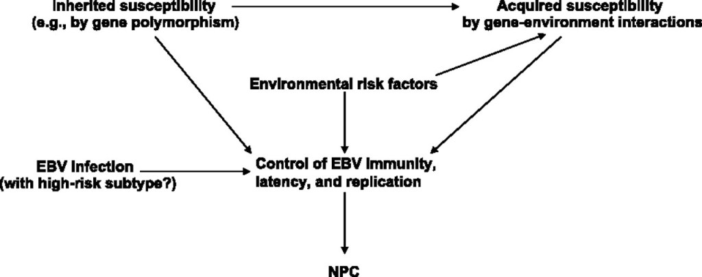
- Undifferentiated subtype of NPC:
- Is strongly associated with Epstein-Barr virus (EBV):
- EBV is also associated with earlier lesions:
- Such as carcinoma in situ
- EBV is also associated with earlier lesions:
- Is strongly associated with Epstein-Barr virus (EBV):
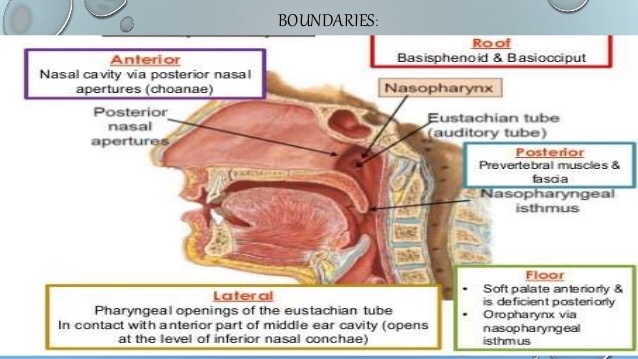
- The nasopharynx extends anteriorly from the posterior choana of the nasal cavity to the free border of the soft palate:
- It comprises a:
- Vault
- The lateral walls:
- Including the fossa of Rosenmüller and mucosa covering the torus tubarius
- A posterior wall
- The superior surface of the soft palate:
- Which is the floor
- The posterior lip of the opening of the Eustachian tube is the torus tubarius:
- Behind which is a mucosal fold:
- Called the fossa of Rosenmüller
- Behind which is a mucosal fold:
- It comprises a:
- The World Health Organization (WHO) classification for NPC encompasses:
- Keratinizing SCC
- Nonkeratinizing carcinomas:
- Well differentiated
- Undifferentiated
- Basaloid squamous cell carcinoma

- Keratinizing SCC (type 1):
- Is more common in North America:
- Not associated with EBV
- Is more common in North America:
- Nonkeratinizing carcinoma, undifferentiated type (type 2b):
- Is highly associated with EBV
- Accounts for 60% of all NPCs in adults
- The most frequent type in the pediatric population
- The first-echelon lymphatic drainage of NPC includes the :
- Retropharyngeal lymph nodes
- Superior jugular lymph nodes
- Posterior cervical chain nodes


- Lymph node metastasis from NPC is common:
- As many as 90% of patients:
- Have evidence of unilateral nodal involvement
- As many as 50% of patients:
- Have evidence bilateral nodal involvement
- As many as 90% of patients:
- The nasopharynx is the upper one-third of the pharynx and is separated from the oropharynx below by the soft palate:
- Anatomically:
- It is the space situated behind the nasal cavities
- Its mucosal lining starts immediately behind the posterior choana
- It is actually located in the center of the head:
- It is located more than 10 cm from the skin surface of the head in all directions
- The undersurface of the body of the sphenoid bone:
- Forms the roof (vault) of the nasopharynx:
- Which slants downwards to form the posterior wall of the nasopharynx:
- In front of the arch of the atlas and upper part of the body of the axis vertebrum
- Which slants downwards to form the posterior wall of the nasopharynx:
- Forms the roof (vault) of the nasopharynx:
- The floor of the nasopharynx:
- Is formed by the upper surface of the soft palate:
- Which separates the nasopharynx from the oropharynx below
- The lateral wall of the nasopharynx is formed by:
- The opening of the Eustachian tubes superiorly
- The upper part of the superior pharyngeal constrictor muscle inferiorly
- The orifice of the Eustachian (auditory tympanic) tube is delineated by:
- An incomplete cartilaginous ring:
- The deficient portion is in the inferolateral aspect
- The medial portion of the cartilaginous ring:
- Elevates the overlying mucosa to form the medial crusa:
- Also known as the Torus tubarus
- The slit-like space formed by this medial crusa and the posterior wall of the nasopharynx:
- Is the fossa of Rosenmüller
- Elevates the overlying mucosa to form the medial crusa:
- An incomplete cartilaginous ring:
- Is formed by the upper surface of the soft palate:
- Anatomically:

- The muscular wall of the nasopharynx is formed by the:
- Superior pharyngeal constrictors lying deep to the pharyngobasilar fascia:
- The fascial sheets join:
- To form a median raphe:
- Which extends from the skull base downwards along the entire posterior pharyngeal wall
- To form a median raphe:
- The fascial sheets join:
- Superior pharyngeal constrictors lying deep to the pharyngobasilar fascia:
- The lymph nodes that drain the nasopharynx:
- Lie in the retropharyngeal space:
- Outside the pharyngobasilar fascia
- In front of the prevertebral fascia
- Lie in the retropharyngeal space:
- The cranial nerves IX, X, XI and XII, the carotid sheath and the sympathetic trunk:
- Traverse the parapharyngeal space:
- Which is lateral to the superior pharyngeal constrictor
- Traverse the parapharyngeal space:
- The roof (vault) of the nasopharynx:
- Is lined by pseudostratified ciliated epithelium
- The posterior wall of the nasopharynx:
- Is lined with stratified squamous cells:
- The epithelium has a well-defined basement membrane and there is abundant lymphatic tissue in the lamina propria:
- This lymphoid tissue forms the pharyngeal tonsil or adenoid in children
- The epithelium has a well-defined basement membrane and there is abundant lymphatic tissue in the lamina propria:
- Is lined with stratified squamous cells:
- Branches of the internal maxillary artery:
- Supply the nasopharynx
- Venous drainage is to the:
- Pterygoid venous plexus:
- Then to the facial and internal jugular veins
- Pterygoid venous plexus:
- The sensory nerve supply of the region:
- Is from branches of the maxillary nerve (V2)
- The lymphatic supply of the nasopharynx drains into the retropharyngeal lymph nodes:
- Efferent lymphatics from these nodes and those that come directly from the nasopharynx:
- Drain to the deep cervical lymph nodes:
- The lymphatic drainage then passes down the neck nodes in an orderly fashion:
- From the high neck nodes to the lower ones
- The lymphatic drainage then passes down the neck nodes in an orderly fashion:
- Drain to the deep cervical lymph nodes:
- Efferent lymphatics from these nodes and those that come directly from the nasopharynx:
#Arrangoiz #HeadandNeckSurgeon #CancerSurgeon #SurgicalOncologist #NasopharyngealCancer #Teacher #Surgeon
