
- Ultrasound technology:
- Is becoming an integral part of diagnosing parathyroid adenomas
- Careful ultrasound evaluation with:
- B-mode
- Shear wave elastography
- Three-dimensional (3D) of parathyroid adenomas:
- May improve localization and outcome
- Introduction:
- A 60-year-old woman was referred for the evaluation of hyperparathyroidism.
- This patient gave her informed consent.
- She had a history of hypothyroidism and thyroid nodules.
- She was being treated with levothyroxine 50 mcg daily.
- Routine testing revealed hypercalcemia:
- The serum calcium was 11.2 (nL range 8.7–10.2 mg/dL), creatinine was 0.69 (nL range 0.57–1.00 mg/dL), intact parathyroid hormone (PTH) was 70 (nL range 15–65 pg/mL), phosphorus was 2.7 (nL range 2.5–4.5 mg/ dL), vitamin D was 38.7 (30–100 ng/mL), and 24 hours urine calcium was 362.9 (100–300 mg/24 hour).
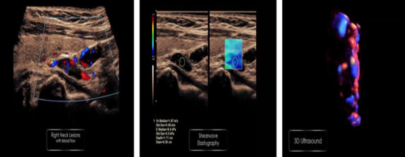
- The neck ultrasound showed:
- Two lesions one superior/posterior and the other in the inferior/posterior aspect of the right thyroid lobe measuring 11.6 · 4.4 · 9.7 mm and 14.6 · 5.0 · 10.0 mm, respectively.
- Both lesions resembled parathyroid adenomas.
- Shear wave velocity (SWV) measurements for the superior and inferior lesions were 1.67 and 1.77 m/second, respectively.
- For the adjacent thyroid tissue SWV was 2.3 m/second, significantly higher.
- 3D ultrasound examination demonstrated a polar artery in both lesions.
- A sestamibi scan showed a probable right parathyroid adenoma and she was referred for surgery.
- She was found to have two right parathyroid adenomas in the superior and inferior poles corresponding with the ultrasound finding.
- Intraoperative PTH level decreased from 139.9 to 17 pg/mL postresection.
- Six weeks after surgery, her calcium and PTH were normal.
- Materials and Methods:
- This patient was evaluated with ultrasound imaging, including:
- B- mode
- Shear wave elastography (SWE)
- 3D ultrasound
- This patient was evaluated with ultrasound imaging, including:
- Discussion:
- Most patients with primary hyperparathyroidism have a single parathyroid adenoma:
- Other causes include:
- Glandular hyperplasia
- Multiple adenomas
- Parathyroid carcinoma
- Other causes include:
- The role of ultrasound in diagnosing parathyroid adenomas:
- Is becoming more prominent because of:
- Improved technology
- Low cost
- Noninvasive nature
- Is becoming more prominent because of:
- SWE can be an added value to b-mode ultrasound in diagnosing parathyroid adenomas:
- Previous publications have reported that SWV measurement of parathyroid adenomas:
- May enhance other sonographic parameters to predict the diagnosis of parathyroid adenomas:
- Parathyroid adenomas appear to have:
- A more homogenous texture when compared with the thyroid gland
- Lower tissue stiffness when compared with the thyroid gland
- Parathyroid adenomas appear to have:
- May enhance other sonographic parameters to predict the diagnosis of parathyroid adenomas:
- Previous publications have reported that SWV measurement of parathyroid adenomas:
- It can be challenging to differentiate:
- Parathyroid adenomas from lymph nodes (LNs) and ectopic thyroid tissue at level VI, with b-mode ultrasound
- A combination of 3D ultrasound images with 3D color Doppler (CD):
- Might improve our ability to:
- Identify the polar artery and enhance differentiation from LN
- Might improve our ability to:
- 3D technology might improve the view:
- By adding coronal view to current b-mode that comprises of transverse and longitudinal views
- Most patients with primary hyperparathyroidism have a single parathyroid adenoma:
- Conclusion:
- Combining multiple image modalities, including:
- B-mode, shear wave elastography, and 3D technology:
- May improve our ability to identify parathyroid adenomas.
- B-mode, shear wave elastography, and 3D technology:
- Parathyroid adenomas have:
- A lower SWV compared with thyroid tissue
- 3D ultrasound technology may enhance view of the polar artery when adding 3D CD.
- Combining multiple image modalities, including:
#Arrangoiz
#ParathyroidExpert
#ParathyroidSurgeon
#Hiperparatiroidism #Hipercalcemia
#HeadandNeckSurgeon
#SociedadQuirurgica
#Hiperparatiroidismo
#ExpertoenParatiroides
