When a patient has a negative but close DCIS margin:
The margin width (distance between the edge of the DCIS and the inked margin) reflects the completeness of excision and is an important determinant of local recurrence in DCIS:
Particularly for patients considering omission of radiotherapy after breast-conserving surgery
In 2016, the Society of Surgical Oncology and American Society of Radiation Oncology developed consensus guidelines regarding margins for DCIS:
These guidelines were based on a meta-analysis of 22 trials enrolling 4,660 women treated with partial mastectomy and radiation therapy:
There was a 64% reduction in local recurrence risk in patients with negative margins compared to those with positive margins
Margin thresholds ≥ 2 mm were associated with fewer local recurrences
For patients with positive margins:
Either re-excision or mastectomy to achieve negative margins should be performed
For patients with close margins, multiple factors should be considered:
The volume / extent of DCIS
Its distribution throughout a specimen
The volume of the excision
The volume of DCIS deemed close to the margin (focal or extensive)
After review of pathology:
Re-excision and / or radiation boost should be performed
A post-excision mammogram:
May be considered to rule out residual suspicious calcifications in the partial mastectomy operative bed for targeting during re-excision:
Breast-conservation therapy may be re-attempted
If the close margins are extensive, mastectomy may be indicated
References
Morrow M, Van Zee KJ, Solin LJ, et al. Society of Surgical Oncology-American Society for Radiation Oncology-American Society of Clinical Oncology consensus guideline on margins for breast-conserving surgery with whole-breast irradiation in ductal carcinoma in situ. Ann Surg Oncol. 2016;23(12):3801-3810.
Van Zee KJ, Subhedar P, Olcese C, Patil S, Morrow M. Relationship between margin width and recurrence of ductal carcinoma in situ: analysis of 2996 women treated with breast-conserving surgery for 30 years. Ann Surg. 2015;262(4):623-631.
Dunne C, Burke JP, Morrow M, Kell MR. Effect of margin status on local recurrence after breast conservation and radiation therapy for ductal carcinoma in situ. J Clin Oncol. 2009;27(10):1615-1620.
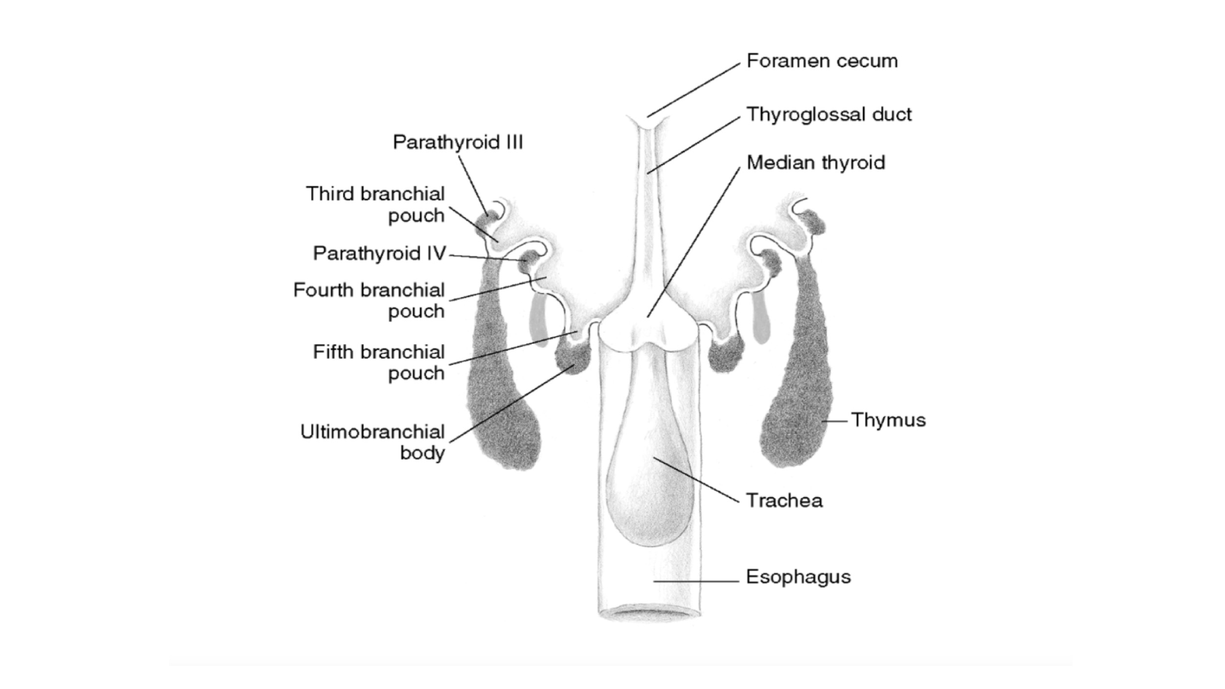
The development of the parathyroid glands in humans can be divided into five stages:
Preprimordial stage
Early primordial stage
Branchial complex stage
Isolation stage
Definitive form stage
The preprimordial stage:
Indicates the period between the formation of the pharynx and the earliest appearance of a recognizable parathyroid anlage:
During this stage, at 4 mm to 8 mm in length:
The third and fourth pharyngeal pouches show a slight dorsal extension.
The third pouch:
Which has the form of a tubelike lateral expansion of the primitive pharynx:
Makes contact with the ectoderm of the pharyngeal cleft and then continues its growth in a downward and ventral direction.
The early primordial stage:
When the embryo is about 9 mm in length:
The parathyroid tissue can be recognized
Proliferation and differentiation of large, clear cells occur in the third and fourth pouches:
Resulting in:
A thickening of the third and fourth pouches
Formation of a budlike nodule of the fourth pouch
The branchial complex stage:
The derivatives of the third and fourth pharyngeal pouches become separated from each other to reach independent positions.
During the early phase of this stage:
The pharyngeal pouches are still joined to the primitive pharynx by pharyngobranchial ducts:
These latter, subsequently, narrow and finally divide:
Which determines the definitive separation of the third and caudal pharyngeal complexes from the primitive pharynx.
At the beginning of this stage:
The primordial thymus and PIII are intimately joined:
Subsequently, the thymus begins a period of rapid ventral growth:
Until the lower pole comes in contact with the pericardium.
On the other hand, the growth of the PIII is not as rapid, and it remains a budlike projection from the superior end of the thymus cord.
Finally, it takes a sphere shape, intimately attaching to the upper pole of the thymus cord.
The position of the caudal pharyngeal complex in relation to the median anlage of the thyroid depends on:
Changes in form, size, and position of the rapidly growing lateral lobe of the median thyroid.
During this stage, the PIV rudiment is still attached to lateral thyroid body.
When the embryo is 13 mm to 14 mm long, the PIII and PIV migrate together with the thymus and ultimobranchial bodies, respectively.
Because of the extension of the cervical spine and the descent of the heart and great vessels:
The complex derived from the third branchial (parathymus) is drawn toward the superior mediastinum and, thus:
Migrates in a medial and caudal direction through the entire length of the embryonic neck to reach its final position, and separation of the PIII from the thymus begins.
The PIV follows the thyroid migration of the ultimobranchial bodies, which travel toward the lateral part of the main median thyroid rudiment:
Their descent in the neck is thus relatively limited.
They remain in contact with the posterior part of the middle third of the thyroid lobes.
The complex branchial stage ends when the embryo is approximately 18 to 20 mm in length.
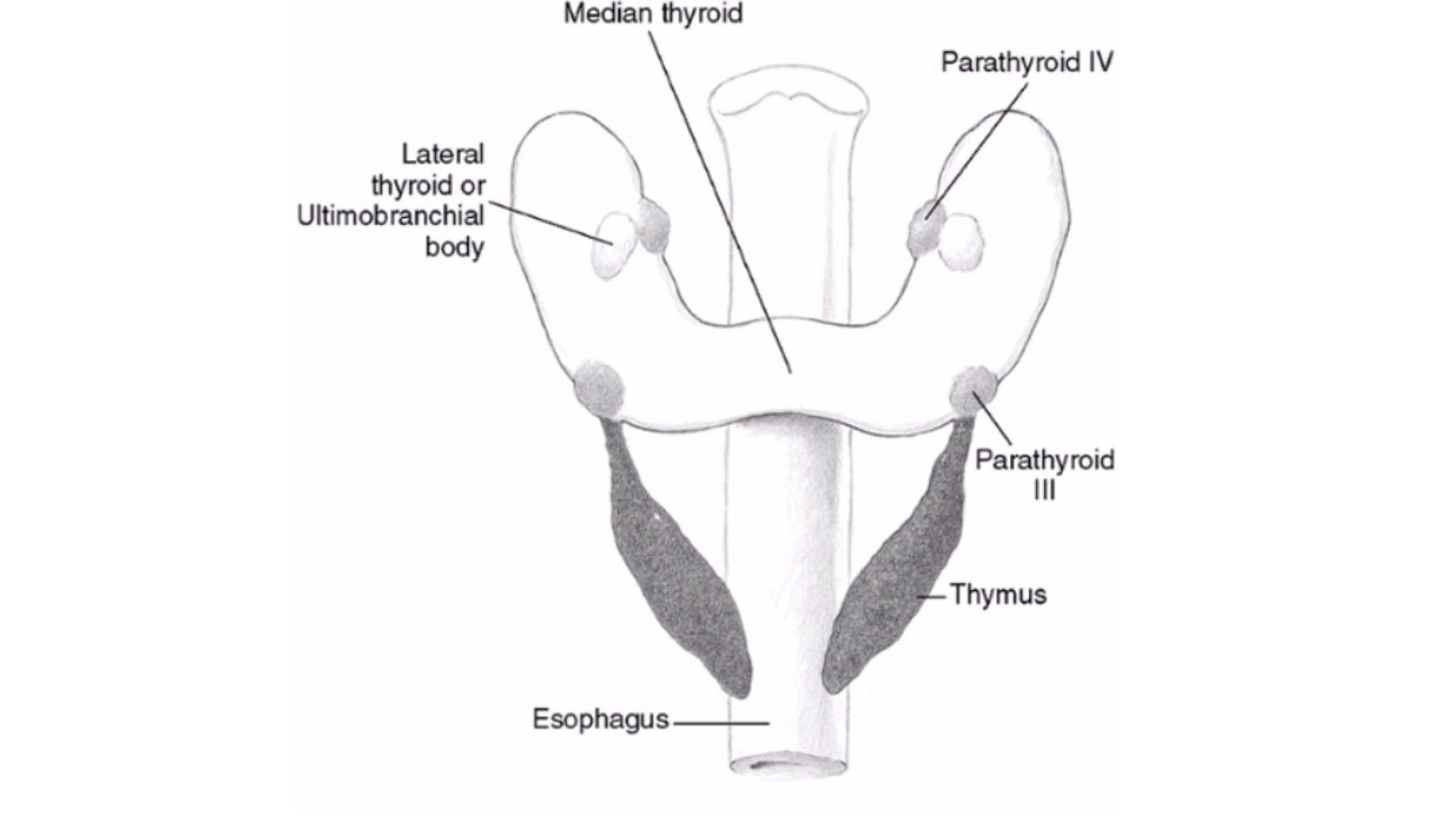
The isolation stage:
Is characterized by the separation of the parathyroid rudiments (PIII and PIV) from the other elements of the third (the thymus) and of the caudal pharyngeal complexes (the ultimobranchial bodies), respectively.
The isolation of the parathyroid glands is usually accomplished when the embryo is 20 mm in length.
After completing the descent through the neck:
The PIII increases in size and separation from the thymus occurs, because of cephalic regression of the last.
PIII is thus abandoned at the level of the anterior or posterolateral region of the inferior poles of the thyroid lobes, or at the level of the thyrothymic ligaments, vestigial structures indicative of their former connections.
The two elements of the caudal pharyngeal complex also grow separately and are conjoined by a connecting stalk:
The interruption of this stalk, determining the isolation of the PIV, occurs once the lateral and the median thyroid become incorporated.
The final position of the PIV in relation to the thyroid gland is determined by the place at which the inclusion of the ultimobranchial body (lateral thyroid element) occurs.
The definitive form stage:
Indicates the period from the end of the isolation to the time when the parathyroids assume their definitive form.
Rodrigo Arrangoiz MS, MD, FACS a head and neck surgeon / endocrine surgeon / surgical oncologist and is a member of Mount Sinai Medical Center:
He is an expert in the management parathyroid diseases.
Publication on parathyroid embryology and anatomy:
Parathyroid Embryology, Anatomy, and Pathophysiology of Primary Hyperparathyroidism:
Arrangoiz, R., Cordera, F., Caba, D., Juárez, M.M., More- no, E. and Luque, E. (2017) Parathyroid Em- bryology, Anatomy, and Pathophysiology of Primary Hyperparathyroidism. International Journal of Otolaryngology and Head & Neck Surgery, 6, 39-58.https://doi.org/10.4236/ijohns.2017.64007
Clinical hallmarks of Paget’s disease of the breast include:
Scaling
Erythema
Ulceration of the nipple:
Sometimes extending to the areola
Because the main differential diagnosis for this clinical presentation is eczema:
A short course of topical steroids was an appropriate initial step:
Failure to resolve should prompt tissue biopsy by punch or wedge technique and not additional steroid therapy
Pathology:
Revealing adenocarcinoma cells within the epidermis (Paget cells):
Confirms the diagnosis
HER2 amplification is found in 60% to 90% of cases of Paget’s disease of the breast:
But the patient should be fully evaluated prior to making decisions regarding the need for targeted therapy
Appropriate diagnostic imaging includes:
Mammography
Ultrasound
Breast MRI (when indicated)
As Paget’s disease is associated with an underlying malignancy 85% of the time
The appropriate surgical management of Paget’s disease is:
Breast conservation with central mastectomy (resection of the nipple-areolar complex) with resection of the primary tumor and irradiation or mastectomy, and not duct exploration
References
Chen CY, Sun LM, Anderson BO. Paget disease of the breast: changing patterns of incidence, clinical presentation, and treatment in the U.S. Cancer. 2006;107(7):1448-1458.
Killelea BK, Chagpar AB, Horowitz NR, Lannin DR. Characteristics and treatment of human epidermal growth factor receptor 2 positive breast cancer: 43,485 cases from the National Cancer Database treated in 2010 and 2011. Am J Surg. 2017;213(2):426-432.
Caliskan M, Gatti G, Sosnovskikh I, et al. Paget’s disease of the breast: the experience of the European Institute of Oncology and review of the literature. Breast Cancer Res Treat. 2008;112(3):513-521.
The Society of Surgical Oncology-American Society for Radiation Oncology (SSO-ASTRO) 2014 Consensus Guidelines:
Regarding margins of resection for invasive carcinoma of the breast:
Recommend the use of “no ink on tumor” as the standard of care:
For patients with invasive cancer:
Even with associated ductal carcinoma in situ (DCIS), are treated according to these guidelines
In a meta-analysis of 33 studies including 32,363 patients:
Odds of local recurrence were associated with margin status of positive vs. negative:
But not decreased with increasing margin distance for patients with invasive carcinoma
The study reported that rates of in-breast tumor recurrence are twice as high with positive margins:
Regardless of tumor biology, radiation boost, or endocrine therapy
There was no evidence that wide margins reduce recurrence:
Even in patients with extensive intraductal component
The American Society of Clinical Oncology (ASCO) guidelines:
Recommend consideration of post-excision mammography to document adequate resection in patients with microcalcifications
References
Moran MS, Schnitt SJ, Giuliano AE, et al. SSO-ASTRO consensus guideline on margins for breast-conserving surgery with whole breast irradiation in stage I and II invasive breast cancer. Int J Radiat Oncol Biol Phys. 2014;88(3):553-564.
Houssami N, Macaskill P, Marinovich ML, Morrow M. The association of surgical margins and local recurrence in women with early-stage invasive breast cancer treated with breast-conserving therapy: a meta-analysis. Ann Surg Oncol. 2014;21(3):717-730.
Buchholz TA, Somerfield MR, Griggs JJ, et al. Margins for breast-conserving surgery with whole-breast irradiation in stage I and II invasive breast cancer: American Society of Clinical Oncology endorsement of the Society of Surgical Oncology/American Society for Radiation Oncology consensus guideline. J Clin Oncol.2014;32(14):1502-1506.
After a median follow-up of 25.3 months continues to show a significant PFS benefit for the nivolumab / relatlimab combination over nivolumab alone in the first-line treatment of patients with unresectable or metastatic melanoma:
While no new safety findings were noted
Although investigators observed improvements in melanoma-specific survival and OS:
These did not meet the prespecified bar of significance
Analyses of outcomes with subsequent therapy suggested a continued benefit with nivolumab and relatlimab beyond initial treatment and first progression
The dual-checkpoint inhibitor combination of nivolumab and relatlimab continues to provide a significant efficacy benefit over nivolumab alone in patients with previously untreated unresectable or metastatic melanoma:
According to an update from the randomized, double-blind, phase 2/3 RELATIVITY-047 trial presented during the 2023 ASCO Annual Meeting
After a median follow-up of 25.3 months:
Nivolumab and relatlimab continued to demonstrate a consistent, significant improvement over nivolumab in progression-free survival (PFS; HR 0.81, 95% CI [0.67, 0.97]) and a trend toward improved overall survival (OS; HR 0.82, 95% CI [0.67, 1.02])
An exploratory analysis found a numerical improvement in melanoma-specific survival with nivolumab and relatlimab versus nivolumab (HR 0.77, 95% CI [0.61, 0.97])
This updated analysis confirms previous efficacy and safety data reported in the trial
Papillary thyroid carcinoma (PTC) is the most common subtype of thyroid cancer, and its genetics have been extensively studied. Here are some key genetic features and alterations associated with papillary thyroid carcinoma:
BRAF V600E mutation:
The BRAF V600E mutation is the most prevalent genetic alteration in PTC, occurring in approximately 40% to 60% of cases. This mutation leads to the activation of the MAPK signaling pathway, which plays a role in cell growth and proliferation. The presence of BRAF V600E mutation may be associated with a higher risk of disease recurrence and more aggressive tumor behavior.
RAS mutations:
RAS mutations, including NRAS and HRAS, are found in approximately 10% to 20% of PTC cases. These mutations also activate the MAPK signaling pathway, promoting cell growth and proliferation. RAS mutations are more common in older patients and are associated with a lower risk of disease recurrence compared to BRAF mutations.
RET/PTC rearrangements:
Rearrangements involving the RET gene, particularly RET/PTC1 and RET/PTC3, are found in a subset of PTC cases. These rearrangements result in the fusion of RET with other genes, leading to the constitutive activation of the RET tyrosine kinase. RET/PTC rearrangements are more commonly seen in radiation-induced PTC and are associated with a favorable prognosis.
Other genetic alterations:
Other less common genetic alterations found in PTC include NTRK rearrangements, EIF1AX mutations, and TERT promoter mutations. These alterations are present in a small proportion of PTC cases and may have implications for prognosis and targeted treatment strategies.
Adjuvant pembrolizumab continued to demonstrate significant improvements over placebo in DMFS and RFS:
After a median follow-up of 39.4 months
Whereas an earlier analysis suggested that there may be a lesser benefit with pembrolizumab in patients with stage IIC disease:
This was not observed with additional follow-up
The 3-year RFS rates were:
76.2% with pembrolizumab and 63.4% with placebo
In patients with stage IIB / IIC melanoma, adjuvant pembrolizumab provides a:
41% reduction in the risk of distant metastasis or death
38% reduction in the risk of recurrence or death:
According to an update from the KEYNOTE-716 trial presented at the 2023 ASCO Annual Meeting
KEYNOTE-716:
Was the first study to demonstrate a benefit for relapse-free survival [RFS] as well as distant metastasis-free survival [DMFS]:
Using anti–PD-1 immunotherapy (pembrolizumab) in patients with high-risk, non-nodal, stage IIB / IIC melanoma:
This updated analysis support[s] the durability of these findings over time
After a median follow-up of 39.4 months:
Adjuvant pembrolizumab continued to demonstrate significant improvements over placebo in DMFS (HR 0.59, 95% CI [0.44, 0.79]) and RFS (HR 0.62, 95% CI [0.49, 0.79]), consistent with prior reports
The investigators concluded that the results of this analysis support the use of pembrolizumab as adjuvant therapy in patients with resected stage IIB or IIC melanoma:
The effect of adjuvant anti–PD-1 antibodies is similar across stages of melanoma from stage IIB through stage IV resected disease
Having therapeutic options now validated in this setting is a big step forward for our patients and that the results of this final DMFS analysis confirm the important benefit observed in the previous DMFS report and further consolidate the role of pembrolizumab in this setting
Stage IIB / IIC disease is associated with a high risk of relapse
5-year melanoma-specific survival outcomes are similar to those for patients with stage IIIB disease:
Highlighting a need for additional treatment strategies for these patients
The randomized, phase 3 KEYNOTE-716 trial:
Was undertaken to evaluate the efficacy and safety of adjuvant pembrolizumab in patients with:
Newly diagnosed resected stage IIB / IIC melanoma with a negative sentinel lymph node biopsy
A total of 967 patients were randomly assigned to pembrolizumab or placebo:
Administered every 3 weeks for up to 17 cycles
In previous analyses, pembrolizumab demonstrated significant efficacy improvements in RFS and DMFS over placebo
In the last update, after a median follow-up of 27.4 months, median DMFS was not reached in either arm (HR 0.64, 95% CI [0.47, 0.88]; P = .0029)
In the current update:
After a median follow-up of 39.4 months, median DMFS was still not reached in either arm, and there was a consistent benefit with pembrolizumab (HR 0.59, 95% CI [0.44, 0.79];)
The RFS benefit with pembrolizumab was also maintained in the current follow-up:
The 3-year RFS rates were 76.2% with pembrolizumab and 63.4% with placebo, and median RFS was not reached in either arm (HR 0.62, 95% CI [0.49, 0.79])
The DMFS and RFS benefit with pembrolizumab was observed in both stage IIB / IIC melanoma:
An earlier analysis suggested that there may be a lesser benefit with pembrolizumab in patients with stage IIC disease:
This was no longer observed with additional follow-up
We now see that this discrepancy has been resolved with longer follow-up:
The overall impact of pembrolizumab as adjuvant therapy appears to be similar across stages
Safety outcomes were similar to those reported in the previous analyses
Grade 3/4 treatment-related adverse events (AEs) occurred in 17.2% of patients receiving pembrolizumab and 5.1% of those receiving placebo; grade 3/4 immune-related AEs and infusion reactions occurred in 11.0% and 1.2% of patients, respectively.
Treatment-related AEs led to discontinuation in 15.9% of patients receiving pembrolizumab and 2.5% receiving placebo
Investigators noted that the overall survival analyses are forthcoming
Next Steps
Looking ahead, one related area of discussion revolves around the role of sentinel lymph node biopsy for patients with melanoma:
If the patient has a deep primary melanoma, adjuvant pembrolizumab should be discussed with the patient whether the nodes are involved or not:
However, this remains an area of debate
Patient selection beyond the current AJCC classification will be key for the future:
To identify patients with a particularly favorable prognosis for whom treatment could be spared and to identify those patients with a poor prognosis who would benefit from additional therapy:
Biomarker studies are underway to guide us in this important next step
Another area of ongoing research is the potential use of combination immunotherapy regimens in the adjuvant setting:
Among the ongoing trials are:
KEYVIBE-010:
Evaluating pembrolizumab plus the anti-TIGIT antibody vibostolimab in patients with high-risk resected stage IIB, IIC, III, and IV melanoma
KEYNOTE-942:
Evaluating pembrolizumab plus a personalized neoantigen therapy (V940) for high-risk melanoma
References
Luke JJ, Rutkowski P, Queirolo P, et al. Pembrolizumab versus placebo as adjuvant therapy in completely resected stage IIB or IIC melanoma (KEYNOTE-716): a randomised, double-blind, phase 3 trial. Lancet. 2022;399(10336):1718-1729.
Long GV, Luke JJ, Khattak MA, et al. Pembrolizumab versus placebo as adjuvant therapy in resected stage IIB or IIC melanoma (KEYNOTE-716): distant metastasis-free survival results of a multicentre, double-blind, randomised, phase 3 trial. Lancet Oncol. 2022;23(11):1378-1388.
Gershenwald JE, Scolyer RA, Hess KR, et al. Melanoma staging: evidence-based changes in the American Joint Committee on Cancer eighth edition cancer staging manual. CA Cancer J Clin. 2017;67(6):472-492.
Egger ME, Bhutiani N, Farmer RW, et al. Prognostic factors in melanoma patients with tumor-negative sentinel lymph nodes. Surgery. 2016;159(5):1412-1421.
Patients diagnosed with estrogen receptor (ER)-positive breast cancer receive standard-of-care adjuvant treatment:
However, patients can experience disease recurrence:
With many recurrences occurring within the first 5 years
When disease recurrence does occur, it is often at a more advanced stage when the disease may be incurable:
Therefore, it is critical to prevent recurrences to better reach the original curative intent of treatment
Prior research demonstrated that ribociclib plus endocrine therapy:
Yielded significant improvements in overall and progression-free survival in patients with HR-positive / HER2-negative advanced breast cancer:
Based on that, NATALEE was designed to evaluate the combination in patients with early-stage disease
Another CDK4/6 inhibitor, abemaciclib, is U.S. Food and Drug Administration (FDA) approved for adjuvant treatment of adult patients with HR-positive / HER2-negative, node-positive early breast cancer at high risk of recurrence
What NATALEE now does:
If the FDA acknowledges the data for registration:
It enlarges the patient population who will have access to a CDK4/6 inhibitor