Blog

Galactoceles

  • Galactoceles:
    • Are milk retention cysts:
      • That result from a blocked milk duct
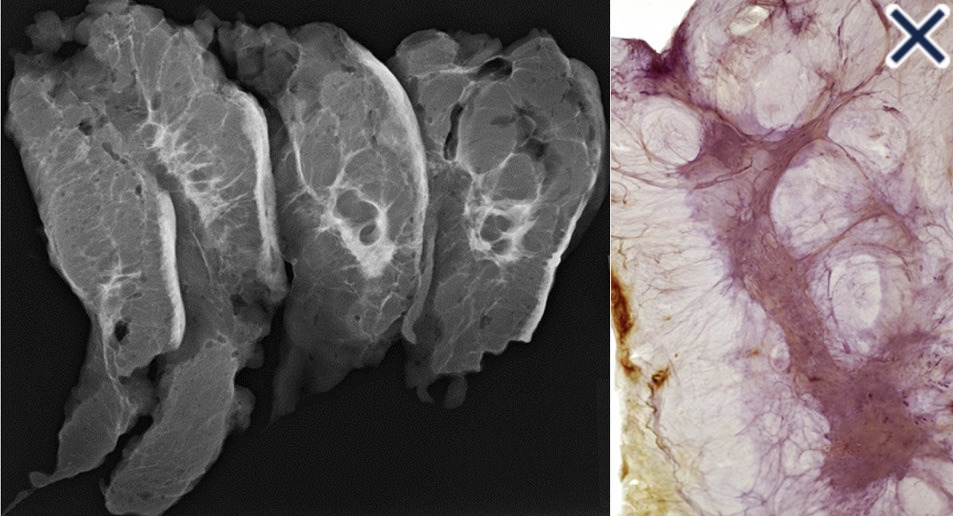
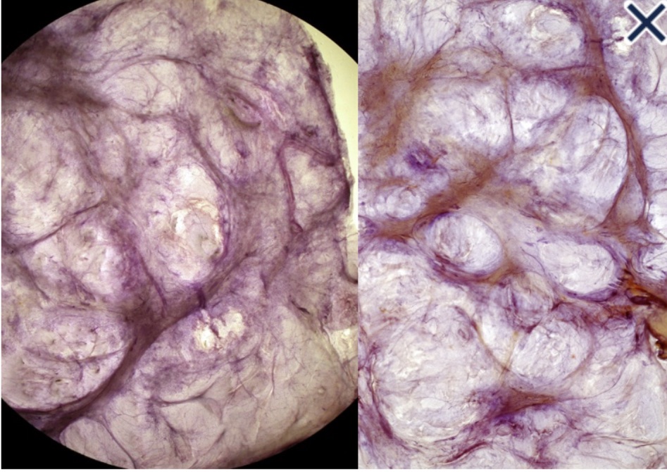
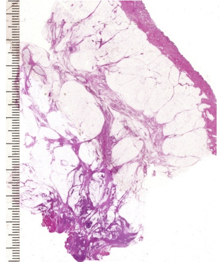
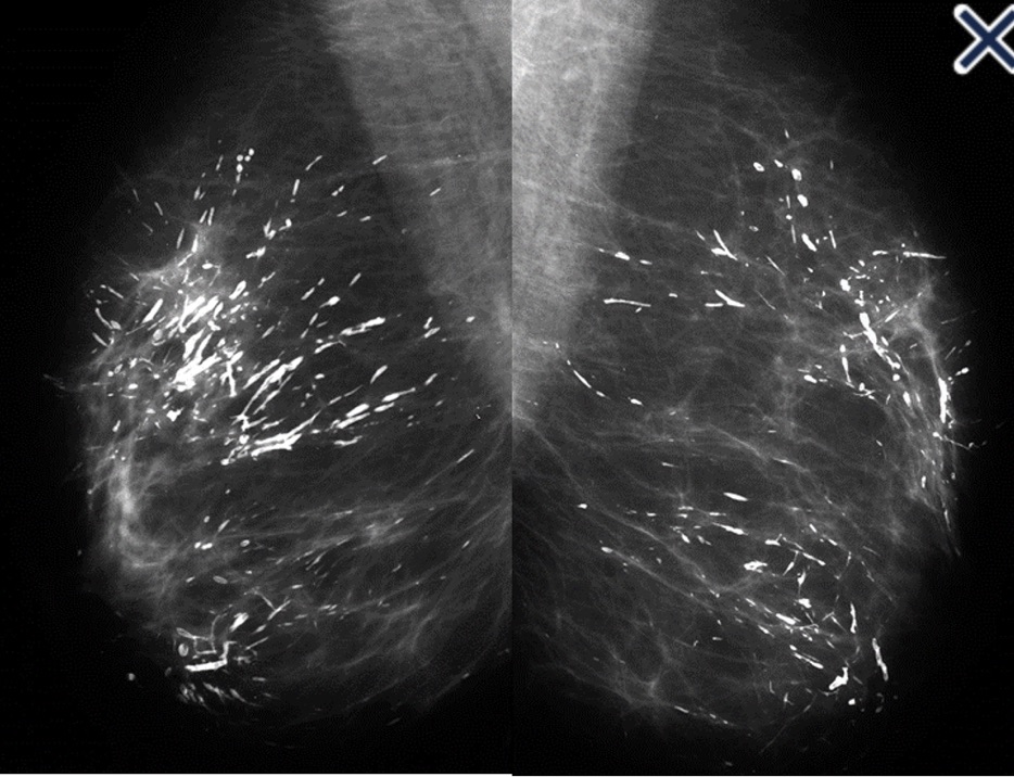
  • They present as cystic, sometimes very large masses:
    • During pregnancy, lactation, and after weaning
  • They are often painless unless they become infected
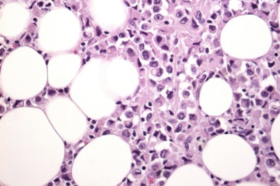
  • Initially, they contain milky fluid:
    • But over time, contents become thicker and more creamy or oily as the fluid is reabsorbed
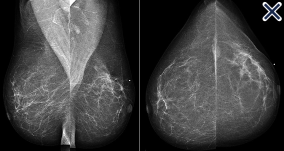
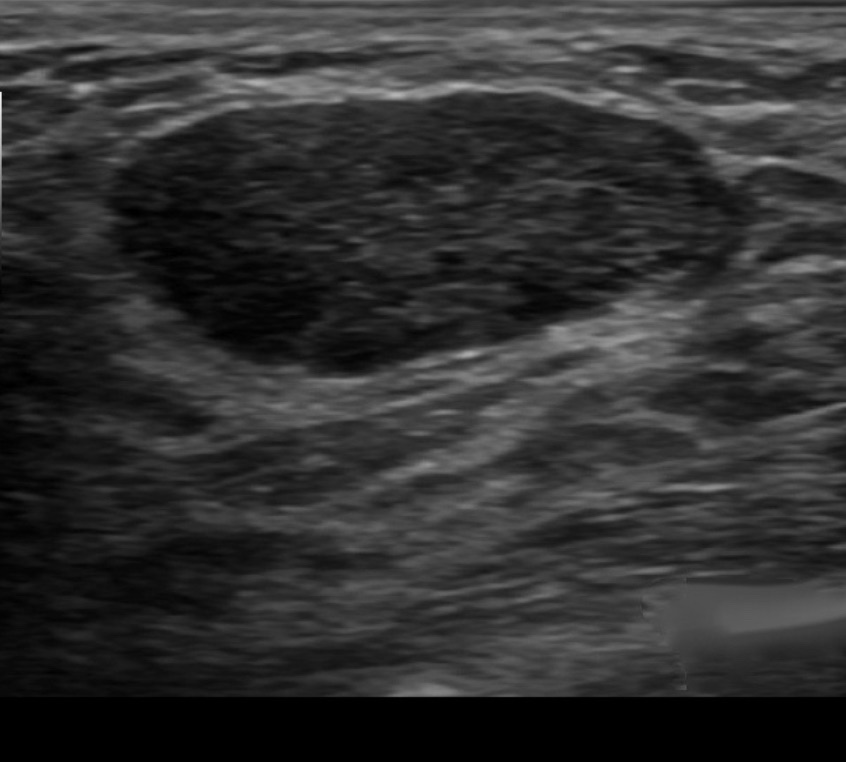
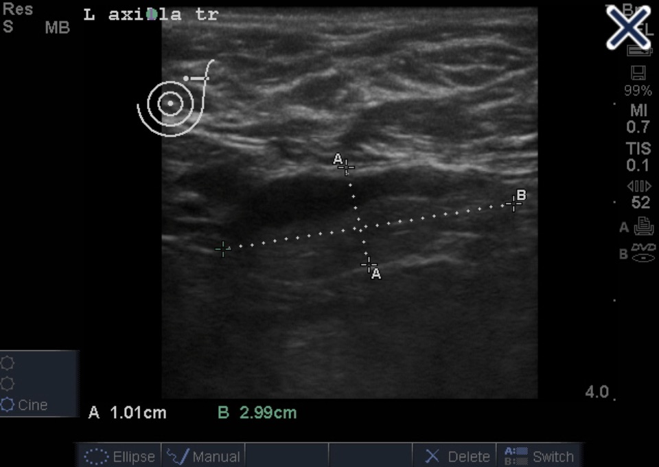
  • Ultrasound is the primary diagnostic method
    • Typical findings include a:
      • Well-defined lesion with thin echogenic walls
      • The internal appearance consists of either homogeneous contents or heterogeneous contents with fluid clefts and anechoic rims
  • Management consists of:
    • Needle aspiration demonstrating milky contents:
      • Which both confirms the diagnosis and excludes malignancy
  • Surgical resection is reserved for:
    • Cases refractory to conservative management
  • References
    • Sawhney S. Petkovska L, Ramadan S, Al-Muhtaseb S, Jain R, Sheikh M.Sonographic appearance of galactoceles. J Clin Ultrasound. 2002;30(1):18-22.
    • Sabate JM, Clotet M, Torrubia S, Gomez A, Guerrero R, de las Heras P. Radiologic evaluation of breast disorders related to pregnancy and lactation. Radiographics.2007;27(Suppl 1):S101-S124.

#Arrangoiz #BreastSurgeon #BreastCancer #Galactocele #CancerSurgeon

Less Frequent Mammography for Breast Cancer Survivors Has No Impact on Survival

  • Certain breast cancer survivors can safely reduce the frequency with which they undergo surveillance mammography 3 years after surgery, according to findings presented at the 2023 San Antonio Breast Cancer Symposium
  • The results are important, because currently in the US, breast cancer survivors are told to undergo an annual mammogram every year for an indefinite period of time after their surgery:
    • In the UK, survivors routinely undergo an annual mammogram for the first 5 years after surgery then de-escalate to once every 3 years afterward for those aged 50 and older
  • These findings presented at SABCS suggest that survivors can get screened less frequently than current guidelines allow.
  • In the phase 3 Mammo-50 trial:
    • Researchers randomly assigned women who underwent curative surgery for breast cancer to one of two groups.
    • One was assigned to undergo a mammogram every year, and another received mammograms less frequently: once every 2 years for those who had breast-conservation surgery and once every 3 years for those who had mastectomies
    • After a 5-year follow-up period, the groups had similar rates of both overall survival (OS) and breast-cancer specific survival
    • The annual group had a breast-cancer specific survival rate of 98.1% and an OS of 94.7%, whereas the less-frequent group had a breast-cancer specific survival rate of 98.3% and an OS of 94.5%
    • The two groups also had similar recurrence rates:
      • 5.9% for the annual group and 5.5% for those assigned to less frequent screening
    • Although the researchers acknowledged that the de-escalation arm had a lower compliance rate than the annual arm:
      • At 69% compared with 83%, they added that a sensitivity analysis revealed this difference in adherence did not impact their final results
  • In a concurrent substudy evaluating the participants’ quality of life using four different validated questionnaires, researchers reported no differences between the two groups in the domains of distress, mental well-being, and other areas
  • Researchers acknowledged that the study was limited by the fact that it focused only on women older than 50 who were already cancer-free for at least 3 years
  • De-escalation of mammographic surveillance reduces the burden on the health care system, decreases the inconvenience for women having to undergo these mammograms, and reduces the associated stress of waiting for results:
    • The trial demonstrated that the outcomes from undergoing less frequent mammograms were no worse than undergoing annual mammograms for this group of women

Younger Postmenopausal Patients May Skip Adjuvant Radiotherapy After Early Breast Cancer Surgery

  • Younger postmenopausal patients with low-risk, stage I HR-positive breast cancer and certain genetic markers may be able to safely forgo adjuvant radiotherapy:
    • According to study findings presented at the 2023 San Antonio Breast Cancer Symposium in Texas
  • In the IDEA (Individualized Decisions for Endocrine therapy Alone) trial:
    • 200 postmenopausal patients with stage I HR-positive, HER2-negative breast cancer:
      • All of whom had scores of 18 or less on the Oncotype DX recurrence assay:
        • Elected to skip adjuvant radiotherapy:
          • Provided they were willing to continue with endocrine therapy for 5 years
    • All participants were aged between 50 and 69 years and required to have a margin of 2 mm or more after breast-conserving surgery
    • The primary outcome:
      • Was the rate of disease recurrence at 5 years after surgery
    • Patients had a mean recurrence score of 11.2
    • Eighty-five patients had grade 1 tumors, whereas 109 had grade 2 tumors and 6 had grade 3 tumors
    • The tumors were a mean of 10 mm
    • The median follow-up was 5.21 years
    • Both the overall survival rate and breast-cancer survival rate were 100% at 5 years:
      • The researchers reported, with a 5-year freedom-from-recurrence rate of 99% (95% CI, 96-100):
        • However, 2 patients died after the 5-year follow-up period had expired
    • There were two breast cancer related events during the follow-up period:
      • One event, which occurred at the 21-month mark:
        • Was an isolated axillary recurrence:
          • This was treated with axillary dissection and breast irradiation as well as local irradiation
      • The other was an ipsilateral breast event:
        • Which occurred at 49 months and was treated with another breast-conserving surgery
    • Another 6 patients experienced recurrence after the 5-year follow-up period, the researchers reported:
      • Five of these were ipsilateral breast events, and one was an ipsilateral breast event with regional occurrence
    • For the entire follow-up period:
      • The crude rate of ipsilateral breast events was:
        • 3.3% (n = 2 of 60) for patients who were 50 to 59 years old
        • 3.6% (n = 5 of 140) for those who were 60 to 69 years old
      • The crude overall relapse rates in these age groups were 5% (n = 3) and 3.6%, respectively
  • These findings indicate that younger postmenopausal patients with stage I breast cancer who skip radiotherapy after breast-conserving surgery:
    • Have a very low risk of disease recurrence within 5 years:
      • However, 5 years is an early time point for this population, and longer-term follow-up of this study and others will be essential to determine whether this option can be safely offered to women in this age group
  • Reference:
    • Jagsi, R. Five-year outcomes of the IDEA trial of endocrine therapy without radiotherapy after breast-conserving surgery for postmenopausal patients age 50-69 with genomically-selected favorable stage I breast cancer. Abstract GS02-08. SABCS 2023.
#Arrangoiz @Doctor #Surgeon #CancerSurgeon #SurgicalOncologist #BreastSurgeon #BreastCancer #MSMC #MountSiniaMedicalCenter #Miami #Mexico

Radiation Changes in the Breast Seen on Mammogram

Right craniocaudad image.
Right mediolateral oblique image.
  • The mammogram:
    • Shows trabecular thickening and an increased thickness of the skin
  • Trabecular thickening:
    • Is an increased prominence of linear structures in the breast:
      • Primarily corresponding to Cooper’s ligaments
    • It is almost always seen in conjunction with skin thickening:
      • It indicates significant breast edema
    • It has a number of different causes, including:
      • Congestive heart failure
      • Renal failure
      • Venous or lymphatic obstruction
      • Diffusely infiltrating carcinoma
      • Radiation
    • When the cause is lymphatic obstruction:
      • The obstruction can be the result of surgery, radiation, or lymphatic invasion by cancer
  • Unilateral breast edema with increased trabecular and skin thickening:
    • Sometimes occurs with congestive heart failure
  • The pedal edema that develops during the daytime:
    • Decreases at night and the fluid accumulates in the dependent breast in patients who sleep on their side
  • References
    • Berg, WA, Birdwell RL, Kennedy A. Diagnostic Imaging: Breast. Salt Lake City, UT: Amirsys; 2006.
    • Verbelen H, Gebruers N, Beyers T, De Monie AC, Tjalma W. Breast edema in breast cancer patients following breast-conserving surgery and radiotherapy: a systematic review. Breast Cancer Res Treat. 2014;147(3):463-471.
    • Wratten CR, O’Brien PC, Hamilton CS, Bill D, Kilmurray J, Denham JW. Breast edema in patients undergoing breast-conserving treatment for breast cancer: assessment via high frequency ultrasound. Breast J. 2007;13(3):266-273.
    • Menta A, Fouad TM, Lucci A, Le-Petross H, Stauder MC, Woodward WA, et al. Inflammatory breast cancer: what to know about this unique, aggressive breast cancer. Surg Clin North Am. 2018;98(4):787-800.
#Arrangoiz #Doctor #Surgeon #CancerSurgeon #SurgicalOncologist #BreastSurgeon #MountSinaiMedicalCenter #MSMC #Miami Mexico #BreastCancer

Sonographic Appearance of Lymph Nodes

  • The sonographic appearance of a normal lymph node:
    • Is elliptical with a thin, hypoechoic cortex and an isoechoic to hyperechoic fatty hilum (Image)
Normal axillary lymph node
  • Metastatic carcinoma in a lymph node:
    • Would usually have an asymmetric thick cortex or have near-total or total obliteration of the hilum:
    • Resulting in a rounded, hypoechoic mass (Image)
Node with metastatic carcinoma
  • Axillary adenopathy can occur in association with rheumatoid arthritis:
    • But the sonographic findings would usually be a symmetrical, mild thickening of the cortex:
      • Usually with preservation of the hilum
  • Silicone granulomas:
    • Classically create a snowstorm appearance:
      • Which allows a definitive diagnosis by ultrasound alone
Silicone Granulomatous
  • The mass in the image has a rounded anterior border and “dirtyincoherent shadowing that obscures the posterior border of the lesion:
    • Nothing other than silicone can cause this sonographic appearance:
      • But it is difficult to distinguish free silicone that has migrated to the axilla from a node that has been replaced with silicone
  • In different stages of development, silicone extravasation can also mimic complex cysts or fibrotic nodules, depending on the amount of silicone extravasated and the amount of time from extravasation:
    • If a large amount of silicone is released into the tissue, the ultrasound pattern is that of a complex cyst
  • Silicone’s presence in tissues can cause a local inflammatory reaction:
    • Which may cause tissue fibrosis and a fibrotic nodule:
      • This is a late finding seen following extravasating
  • References:
    • Berg WA, Caskey CI, Hamper UM, Anderson ND, Chang BW, Sheth S, et al. Diagnosing breast implant rupture with MR imaging, US, and mammography. Radiographics. 1993;13(6):1323-1336.
    • Juanpere S, Perez E, Huc O, Motos N, Pont J, Pedraza S. Imaging of breast implants‒a pictorial review. Insights Imaging. 2011;2(6):653-670.

Mammographic Images in Diffusely Invasive Breast Cancer

  • Diffusely invasive carcinoma:
    • Has a mammographic appearance of:
      • Diffuse architectural distortion
    • Usually involving a large area:
      • Often larger than a lobe:
        • With no central tumor mass and no calcifications
    • It sometimes has the appearance of:
      • A “spider’s web” (Image 1)
    • The diffusely infiltrating cancer forms concave contours with the surrounding fat in a manner similar to normal fibroglandular tissue (Images 2 a-b)
Image 1: Mediolateral oblique and craniocaudal projections.
Mastectomy slice radiographs (a) and large format 3D histology image (b) showing concave contours similar to normal breast
  • The imaging findings of diffusely infiltrating breast cancer:
    • Are strikingly different from the imaging findings of breast cancers originating either from the terminal ductal lobular units (TDLUs) or the lactiferous ducts:
      • Suggesting that it may have a different site of origin
  • It has been recently proposed that diffusely infiltrating breast cancers may originate from:
    • Mesenchymal stem cells (progenitors):
      • Through a complex process of both epithelial-mesenchymal transformation and more frequently, mesenchymal-epithelial transformation
    • The clinical presentation is typically a:
      • Recently detected, extensive, firm lesion:
        • Often appearing as an interval cancer following a previous mammogram which was interpreted as normal
    • On clinical breast examination:
      • The cancer does not have a distinct tumor mass or focal skin retraction seen in other cancers:
        • But rather an indistinct “thickening” and eventually a shrinkage of the breast
    • In order to make the diagnosis before the development of a palpable mass and a decrease in size of the breast:
      • The radiologist and breast surgeon must have a high level of suspicion and a thorough knowledge of the underlying pathophysiology
    • The subgross (3D) histopathology images:
      • Show how growth of the mesenchymal tissue distorts the normal, harmonious connective tissue framework by causing nonuniform thickening of the fine sheets of connective tissue (Images 3a -b)
Large format subgross (3D) histology images of a diffusely infiltrating breast cancer
  • The predominance of mesenchyme in the diffusely infiltrating breast malignancy:
    • Allows it to be imaged with greater sensitivity by ultrasound than by mammography
  • The thin sheets or veils of tissue reflect the ultrasound waves, but are relatively easily penetrated by x-rays:
    • The structural / architectural distortion, while difficult to detect mammographically:
      • Is readily detectable on 2-mm thick coronal sections of automated breast ultrasound (Image 3c)
    • The hypoechoic changes can also usually be seen on hand-held ultrasound (Image 4).
3D automated ultrasound images. The 2-mm thick multi-slice series demonstrate the extensive architectural distortion, corresponding to the 3D histology
Hand-held ultrasound of diffusely infiltrating carcinoma
  • The growth pattern and cell type of diffusely invasive breast cancer:
    • Is very similar to that of diffuse gastric carcinoma (linitis plastica):
      • Both of these diseases can be associated with a deleterious mutation in the CDH1 gene:
        • Which is located on chromosome 16q22:
          • It codes for e-cadherin protein (Image 5a, Image 5b)
Large format histology slide of diffusely infiltrating breast cancer similar to growth pattern of linitis plastica
High-power histology of pleomorphic infiltrating breast cancer with cell type similar to linitis plastica.
  • References
    • Hansford S, Kaurah P, Li-Chang H, Woo M, Senz J, Pinheiro H, et al. Hereditary diffuse gastric cancer syndrome: CDH1 mutations and beyond. JAMA Oncol. 2015;1(1):23-32.
    • Tot T. The diffuse type of invasive lobular carcinoma of the breast: morphology and prognosis. Virchows Arch. 2003;443(6):718-724.
    • Tot T. Diffuse invasive breast carcinoma of no special type. Virchows Arch. 2016;468(2):199-206.
#Arrangoiz #Doctor #Surgeon #CancerSurgeon #SurgicalOncologist #Breast Surgeon #MountSinaiMedicalCenter #MSMC #Miami #Mexico #BreastCancer

Evaluation of a Breast Mass on Ultrasound

  • You first evaluate the lesion for any of the 10 malignant signs:
    • Shadowing
    • Hypoechoic echotexture
    • Spiculation
    • Angular margins
    • Thick echogenic halo
    • Microlobulation
    • Taller than wide
    • Duct extension
    • Branching pattern
    • Calcifications
  • Finding none, you move on to the second step in the evaluation process:
    • Specifically look for one of the 3 strictly defined benign signs:
      • If any of them are found, the lesion can be considered BIRADS 3
    • The 3 benign findings defined by Stavros are:
      • A purely hyperechoic lesion with no hypoechoic area larger than a normal duct or lobule
      • Elliptical, wider than tall, well-circumscribed and thin echogenic capsule
      • Gently lobulated, wider than tall, well-circumscribed and thin echogenic capsule
    • Combining the elliptical or gently lobulated shapes with the presence of a complete, thin echogenic capsule:
      • Is necessary because many circumscribed carcinomas and most ductal carcinoma in situ are encompassed in a thin, echogenic capsule:
        • However, the shape of circumscribed invasive carcinoma or pure ductal carcinoma in situ is rarely elliptical or gently lobulated
  • BIRADS 3:
    • A 6-month follow-up ultrasound would be appropriate unless the anxiety of the patient makes core biopsy a better option
  • References
    • Madjar H, Mendelson EB. The Practice of Breast Ultrasound. 2nd ed. Thieme; 2008;141-144.
    • Stavros AT. Breast Ultrasound. Philadelphia, PA: Lippincott Williams & Wilkins; 2004.
#Arrangoiz #Doctor #Surgeon #CancerSurgeon #SurgicalOncologist #BreastSurgeon #MountSinaiMedicalCenter #MSMC #Miami #Mexico #BreastCancer

Ultrasound Appearance of Axillary Lymph Nodes

Abnormal lymph node with eccentric cortical thickening
  • This axilla ultrasound shows a case of severe eccentric compression and displacement of the hilum to the edge of the node:
    • Favoring metastatic disease
  • Typical lymph node ultrasound appearances include:
    • Uniformly mildly thickened cortex
      • Typical of inflamed or reactive lymph node
    • Eccentric cortical thickening:
      • Favors metastatic disease (Image)
    • Convex indentations of the hilum:
      • Favors metastatic disease
    • Severe compression of the hilum to slit-like configuration:
      • Can occur in metastatic or severely inflamed lymph node
    • Severe eccentric compression and displacement of the hilum to the edge of the node:
      • Favoring metastatic disease
    • Complete obliteration of the hilum and rounding of the node:
      • Favoring metastatic disease:
        • Can also occur in severe necrotizing lymphadenitis
    • Perinodal invasion by metastasis:
      • The outer thin echogenic capsule cannot be identified:
        • Angular margins
Severe eccentric compression and displacement of the hilum to the edge of the node.
  • References
    • Stavros AT. Evaluation of regional lymph nodes in breast cancer patients. In: Stavros AT. Breast Ultrasound. Philadelphia, PA: Lippincott Williams & Wilkins; 2004:858-859.
    • AIUM curriculum for fundamentals of ultrasound physics and instrumentation. JUltrasound Med 2019;38(8):1933-1935. https://onlinelibrary.wiley.com/doi/epdf/10.1002/jum.15088. Accessed July 16, 2020.
#Arrangoiz #Doctor #Surgeon #CancerSurgeon #SurgicalOncologist #Breast Surgeon #MountSinaiMedicalCenter #MSMC #Miami #Mexico #BreastCancer

Types of Calcifications 1

  • Ductal calcifications:
    • Have a wide variety of presentations depending upon the underlying process that created them
  • When coarse rod-like ductal calcifications are diffuse, bilateral, and not confined to a single lobe:
    • They can be confidently assumed to result from plasma cell mastitis, and do not require further evaluation or biopsy (Image)
  • The process is called secretory disease:
    • Because there is a stagnant, viscous fluid that eventually petrifies and results in the smooth contoured calcifications
    • Some of them are branching and look like malignant casting type calcifications:
      • But the key distinguishing feature is the diffuse, multilobe, bilateral nature of the process:
        • Calcifications become much more worrisome when they are confined to a single lobe
  • The most frequent malignant, ductal “casting type” calcifications are:
    • Fragmented, linear, and branching, and they are the most reliable mammographic sign of malignancy (Image)
Casting type calcification.
  • The presence of fragmented and / or dotted casting type calcifications on the mammogram restricted to one lobe:
    • Is a pathognomonic sign of a diffuse, grade 3 breast cancer subtype that originates in the major ducts and usually has a solid or micropapillary pattern:
      • Traditionally, this subtype has been called “comedo carcinoma”
    • The cancer cells either produce a viscous, proteinaceous fluid, which gradually concentrates and eventually calcifies, or they undergo necrosis (apoptosis) followed by calcification:
      • In both instances, the intraluminal pressure increases, distending the ducts considerably
  • Dotted casting type calcifications:
    • Have been referred to as “snakeskin-like calcifications” and they accumulate in the fluid produced by either micropapillary or solid cancer cell growth patterns (Image)
    • The tips of the micropapillary growths may become detached and eventually calcify, contributing to the intraluminal calcifications (Image)
Snakeskin type calcifications.
The micropapillary growths break off and calcify in the lumen, resulting in the individual dots of calcification (the dark, almost black stained structures).
  • Occasionally, malignant ductal calcifications present in a manner that can be easily mistaken for a benign process:
    • It occurs when fluid production, rather than necrosis, dominates the picture
    • The intraductal carcinoma can be grades 1, 2, or 3 and a micropapillary and/or cribriform architecture is present

Dense Breast Screening

  • Debate on adjunct screening in women with dense breasts:
    • Has resulted from legislation mandating that women be informed if their mammograms show dense breast tissue, including informing them that other screening modalities are available
  • In addition to MRI and molecular imaging:
    • Both tomosynthesis and breast ultrasound are additional techniques for enhanced screening in patients with dense breasts
  • Multiple studies show significant benefits from the addition of tomosynthesis to conventional digital mammography alone in screening programs:
    • Ciatto et al:
      • Found an increase in detection rate of invasive breast cancer:
        • From 5.3/1000 to 8.1/1000
      • While also decreasing the recall rate by 17%
    • Skaane et al:
      • Found a 40% increase in the detection of invasive cancers with a 15% reduction in false negatives
    • Rose et al.3 and Haas et al:
      • Showed statistically significant relative reductions in recall rates of 37% and 30%, respectively
    • A recent retrospective review of 454,850 examinations in 13 screening centers in the United States:
      • Demonstrated a 41% increase in invasive cancer detection, a 15% reduction in call backs, and a 49% increase in the positive predictive value for recall
  • Implementation of tomosynthesis:
    • Did not lead to a significant reduction in biopsy rates as compared to digital mammography screening
  • As yet, there are no data that show a reduction in mortality with enhanced screening in dense breasts
  • A prospective multicenter study compared tomosynthesis with bilateral physician hand-held ultrasound screening in 3,231 asymptomatic patients with mammography-negative dense breasts:
    • In all, 24 additional cancers were detected, 23 of which were invasive
    • Tomosynthesis detected 13 cancers, and ultrasound detected 23
    • These data suggest that even though tomosysthesis significantly increases the number of cancers found in dense breasts, in the hands of a skilled breast radiologist, ultrasound may be even better
  • References
    • Ciatto S, Houssami N, Bernardi D, Caumo F, Pellegrino M, Brunelli S, et al. Integration of 3D digital mammography with tomosynthesis for population breast-cancer screening (STORM): a prospective comparison study. Lancet Oncol. 2013;14(7):583-589.
    • Skaane P, Bandos AI, Gullien R, Eben EB, Ekseth U, Haakenaasen U, et al. Comparison of digital mammography alone and digital mammography plus tomosynthesis in a population-based screening program. Radiology. 2013;267(1):47-56.
    • Haas BM, Kalra V, Geisel J, Raghu M, Durand M, Philpotts LE. Comparison of tomosynthesis plus digital mammography and digital mammography alone for breast cancer screening. Radiology. 2013;269(3):694-700.
    • Rose SL, Tidwell AL, Bujnoch LJ, Kushwaha AC, Nordmann AS, Sexton R Jr. Implementation of breast tomosynthesis in a routine screening practice: an observational study. AJR Am J Roentgenol. 2013;200(6):1401-1408.
    • Friedewald SM, Rafferty EA, Rose SL, Durand MA, Plecha DM, Greenberg JS, et al. Breast cancer screening using tomosynthesis in combination with digital mammography. JAMA. 2014;311(24):2499-2507.
    • Tagliafico AS, Calabrese M, Mariscotti G, Durando M, Tosto S, Monetti F, et al. Adjunct screening with tomosynthesis or ultrasound in women with mammography-negative dense breasts: interim report of a prospective comparative trial. J Clin Oncol. 2016;34(16):1882-1888.
#Arrangoiz #Doctor #Surgeon #CancerSurgeon #SurgicalOncologist #Breast Surgeon #MountSinaiMedicalCenter #MSMC #Miami #Mexico #BreastCancer