
Blog

Precancerous Lesions of the Oral Cavity

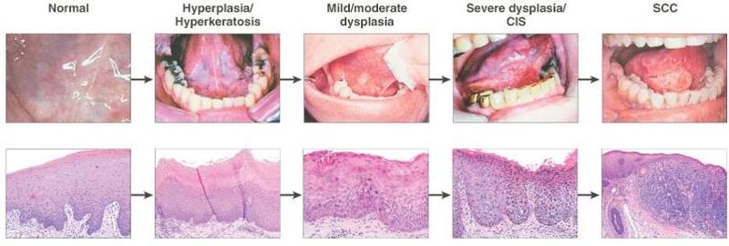
A. There is a sequence of disease progression from atypia / dysplasia through carcinoma in situ to frankly invasive cancer:

1. Leukoplakia and erythroplakia are terms applied to clinically identifiable lesions that may harbor invasive cancer or undergo malignant transformation.

B. Pathology:

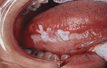
1. Leukoplakia results from chronic irritation of mucous membranes:
– This irritation stimulates the proliferation of white epithelial and connective tissue.

– Histopathologic examination reveals hyperkeratosis variably associated with underlying epithelial hyperplasia.

– In the absence of underlying dysplasia, leukoplakia rarely (less 5% of the cases) is associated with progression of disease to malignancy.
Philadelphia Illustration Dept./Elsevier


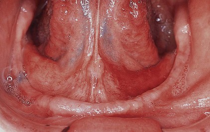
2. Erythroplakia is characterized by superficial, friable, red patches adjacent to normal mucosa.
Philadelphia Illustration Dept./Elsevier

– It is commonly associated with underlying epithelial dysplasia and has a much greater potential for malignancy than leukoplakia.

– Carcinoma is found in nearly 40% of erythroplakia cases.

3. Dysplasia is characterized by cellular atypia, loss of normal maturation, and loss of normal epithelial stratification.

– It is graded as mild, moderate, or severe, based on the degree of nuclear abnormality present.

– In the transition from mild to severe dysplasia, nuclear abnormalities become more marked, mitoses become more apparent, and these changes involve increasing depth of the epithelium.

– The likelihood of developing a carcinoma relates to the degree of dysplasia:

= In the case of severe dysplasia, as many as 24% of patients may develop invasive squamous cell cancer.

4. Carcinoma in situ is characterized by the presence of atypical changes throughout the epithelium, with complete loss of stratification.

– It is estimated that approximately 75% of invasive squamous cell carcinomas have an associated in situ component.

– Specific DNA mutations have also been identified in the sequence of disease progression from mild dysplasia to atypia to carcinoma in situ to invasive carcinoma.
_53022d86_13d85ffda7d__8000_00006167

Rodrigo Arrangoiz MS, MD, FACS
Cirugía General y Gastrointestinal
Michigan State University
Cirugía Oncológica
Fox Chase Cancer Center
Tumores de Cabeza y Cuello / Cirugía Endocrina
Fox Chase Cancer Center
Tumores de Cabeza y Cuello / Cirugía Endocrina
IFHNOS / Memorial Sloan Kettering Cancer Center
Maestría en Ciencias de Investigación
Drexel University
Certificado por el Colegio Americano de Cirugía
Fellow del Colegio Americano de Cirugía
Fellow de la Sociedad de Cirugia Oncológica 

Sociedad Quirúrgica S.C.
Hospital ABC Santa Fé
Av. Carlos Graef Fernández #154

Consultorio 515
Col. Tlaxala, Delg. Cuajimalpa
México, D.F. 05300
Tel: 1103 – 1600 Ext 4515 a la 4517
Fax:1664 – 7164
 

Cancer de la Cavidad Oral

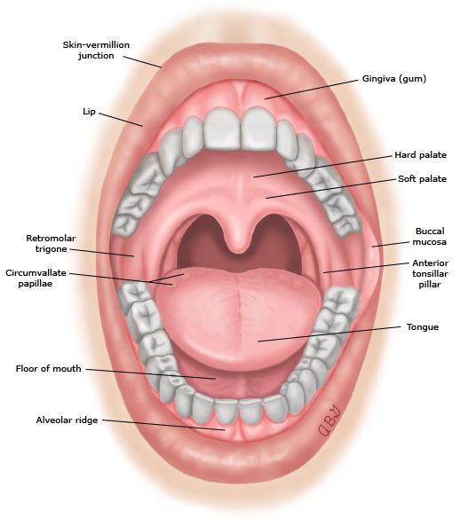
La cavidad oral representa la entrada al tracto aerodigestivo superior, que comienza en los labios y termina en la superficie anterior del arco facial.

Está cubierto por epitelio escamoso con glándulas salivales menores intercaladas.

La cavidad oral también contiene las estructuras dentoalveolares con la dentición superior e inferior.

La cavidad oral está continuamente expuesta a carcinógenos inhalados y consumidos, y por lo tanto es el sitio más común para el origen de neoplasmas epiteliales malignos en la región de la cabeza y el cuello.

Los carcinógenos conocidos en la cavidad oral incluyen aquellos presentes en el tabaco, el alcohol y las nueces de betel.

La asociación del virus del papiloma humano con el cáncer oral no está tan bien establecida como en los cánceres de la orofaringe.

Epidemiología

La incidencia estimada de cáncer de cavidad oral en los EE. UU. En 2018: 33 950 casos:

Lengua 17, 110 casos

Boca 13, 580 casos

Otros sitios de cavidad oral 3260 casos

La mortalidad estimada para el cáncer de cavidad oral en los EE. UU. En 2018 – 5160 muertes.

14699.myextj
Rodrigo Arrangoiz MS, MD, FACS
Cirugía General y Gastrointestinal
Michigan State University
Cirugía Oncológica
Fox Chase Cancer Center
Tumores de Cabeza y Cuello / Cirugía Endocrina
Fox Chase Cancer Center
Tumores de Cabeza y Cuello / Cirugía Endocrina
IFHNOS / Memorial Sloan Kettering Cancer Center
Maestría en Ciencias de Investigación
Drexel University
Certificado por el Colegio Americano de Cirugía
Fellow del Colegio Americano de Cirugía
Fellow de la Sociedad de Cirugia Oncológica 

Sociedad Quirúrgica S.C.
Hospital ABC Santa Fé
Av. Carlos Graef Fernández #154

Consultorio 515
Col. Tlaxala, Delg. Cuajimalpa
México, D.F. 05300
Tel: 1103 – 1600 Ext 4515 a la 4517
Fax:1664 – 7164
 

Oral Cavity Cancer

The oral cavity represents the entrance to the upper aerodigestive tract, which begins at the lips and ends at the anterior surface of the faucial arch.

  • It is lined by squamous epithelium with interspersed minor salivary glands.

The oral cavity also contains the dentoalveolar structures with the upper and lower dentition.

The oral cavity is continuously exposed to inhaled and consumed carcinogens, and thus it is the most common site for the origin of malignant epithelial neoplasms in the head and neck region.

Known carcinogens in the oral cavity include those present in tobacco, alcohol, and betel nuts.

  • The association of human papilloma virus with oral cancer is not as well established as in oropharyngeal cancers.

Epidemiology

  1. The estimated incidence of oral cavity cancer in the USA in 2018 – 33 950 cases:

    1. Tongue 17, 110 cases

    2. Mouth 13, 580 cases

    3. Other oral cavity sites 3260 cases

  2. The estimated mortality for oral cavity cancer in the USA in 2018 – 5160 deaths

14699.myextj
Rodrigo Arrangoiz MS, MD, FACS
Cirugía General y Gastrointestinal
Michigan State University
Cirugía Oncológica
Fox Chase Cancer Center
Tumores de Cabeza y Cuello / Cirugía Endocrina
Fox Chase Cancer Center
Tumores de Cabeza y Cuello / Cirugía Endocrina
IFHNOS / Memorial Sloan Kettering Cancer Center
Maestría en Ciencias de Investigación
Drexel University
Certificado por el Colegio Americano de Cirugía
Fellow del Colegio Americano de Cirugía
Fellow de la Sociedad de Cirugia Oncológica 

Sociedad Quirúrgica S.C.
Hospital ABC Santa Fé
Av. Carlos Graef Fernández #154

Consultorio 515
Col. Tlaxala, Delg. Cuajimalpa
México, D.F. 05300
Tel: 1103 – 1600 Ext 4515 a la 4517
Fax:1664 – 7164


Manejo de Pacientes con Alto Riesgo de Desarrollar Cancer de Mama

Según las pautas de NCCN, las mujeres con una predisposición genética al cáncer de seno deben estar familiarizadas con sus senos e informar cualquier cambio a su proveedor de atención médica.
Este conocimiento de los senos puede incluir un autoexamen periódico y consistente de los senos y debe comenzar a los 18 años.
Los exámenes clínicos de los senos por parte de un proveedor de servicios de salud se deben hacer cada 6 a 12 meses comenzando a los 20 a 25 años o 5 a 10 años antes del primer cáncer de mama conocido en la familia (lo que ocurra primero).
La vigilancia mejorada incluye imágenes de mama que comienzan entre 20 y 29 años en el síndrome de Li-Fraumeni, entre 25 y 29 años en el síndrome hereditario de cáncer de mama y ovario y entre 30 y 35 años en el síndrome de Cowden, o 5 a 10 años antes del síndrome de Cowden cáncer de mama en la familia (lo que ocurra primero).
La mamografía y la RM de mama generalmente se obtienen anualmente a intervalos de 6 meses.
La mamografía anual generalmente no se recomienda antes de los 30 años debido a la evidencia de que la exposición a la radiación de diagnóstico antes de los 30 años se asocia con un mayor riesgo de cáncer de seno.

Rodrigo Arrangoiz MS, MD, FACS
Cirugía General y Gastrointestinal
Michigan State University
Cirugía Oncológica
Fox Chase Cancer Center
Tumores de Cabeza y Cuello y Cirugia Endocrina
Fox Chase Cancer Center
Tumores de Cabeza y Cuello y Cirugía Endocrina
IFHNOS / Memorial Sloan Kettering Cancer Center
Maestría en Ciencias de Investigación
Drexel University
Certificado por el Colegio Americano de Cirugía
Fellow del Colegio Americano de Cirugía
Fellow de la Sociedad de Cirugia Oncológica

Sociedad Quirúrgica S.C.
Hospital ABC Santa Fé
Av. Carlos Graef Fernández #154
Consultorio 515
Col. Tlaxala, Delg. Cuajimalpa
México, D.F. 05300
Tel: 1103 – 1600 Ext 4515 a la 4517
Fax:1664 – 7164

rodrigo.arrangoiz@gmail.com
http://www.sociedadquirurgica.com
http://www.hiperparatiroidismo.info

Managing Breast Cancer Risk In High Risk Women

Per NCCN guidelines, women with a genetic predisposition to breast cancer should be familiar with their breasts and report any changes to their health care provider.

This breast awareness can include periodic, consistent breast self-examination and should begin at age 18.

Clinical breast examinations by a health care provider should be done every 6 to 12 months starting at age 20 to 25 years or 5 to 10 years before the earliest known breast cancer in the family (whichever comes first).

Enhanced surveillance includes breast imaging starting between ages 20 and 29 years in Li-Fraumeni syndrome, between 25 and 29 years in hereditary breast and ovarian cancer syndrome, and between 30 and 35 years in Cowden syndrome, or 5 to 10 years before the earliest known breast cancer in the family (whichever comes first).

Mammography and breast MRI are usually obtained yearly at 6-month intervals.

Annual mammography is generally not recommended before age 30 because of evidence that exposure to diagnostic radiation before age 30 is associated with an increased breast cancer risk.

Rodrigo Arrangoiz MS, MD, FACS
Cirugía General y Gastrointestinal
Michigan State University
Cirugía Oncológica
Fox Chase Cancer Center
Tumores de Cabeza y Cuello y Cirugia Endocrina
Fox Chase Cancer Center
Tumores de Cabeza y Cuello y Cirugía Endocrina
IFHNOS / Memorial Sloan Kettering Cancer Center
Maestría en Ciencias de Investigación
Drexel University
Certificado por el Colegio Americano de Cirugía
Fellow del Colegio Americano de Cirugía
Fellow de la Sociedad de Cirugia Oncológica

Sociedad Quirúrgica S.C.
Hospital ABC Santa Fé
Av. Carlos Graef Fernández #154
Consultorio 515
Col. Tlaxala, Delg. Cuajimalpa
México, D.F. 05300
Tel: 1103 – 1600 Ext 4515 a la 4517
Fax:1664 – 7164
rodrigo.arrangoiz@gmail.com
http://www.sociedadquirurgica.com
http://www.hiperparatiroidismo.info


Cowden Syndrome

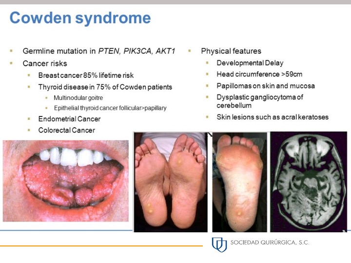
  • Cowden syndrome is caused by a mutation in the PTEN gene.

    • It is one of the few breast cancer predisposition syndromes with a clinically recognizable phenotype (Peutz-Jeghers syndrome is another).

  • Clinical features include extreme macrocephaly (head circumference >60 cm in a woman), multiple facial tricholemmomas (small skin-colored papules) and a family history of thyroid cancer before age 20 years or endometrial cancer before age 30 years.

  • It is a very rare syndrome, with only six cases identified among 2079 recently reported multigene panel tests.

  • It is clinically important, however, because the lifetime breast cancer risk may be as high as 85% and affected individuals have a very high risk of endometrial and thyroid cancer as well as a moderately increased risk of colorectal cancer and melanoma.

  • Current American Cancer Society guidelines support enhanced surveillance with annual mammogram and magnetic resonance imaging for women with PTEN mutations, though the timing of when this should begin is vague.

    • Data by Riegert-Johnson et al. suggest screening should commence around age 30 to 35 years as this is the time that the risk of breast cancer starts to increase.

    • Even after a cancer diagnosis, individuals with Cowden syndrome remain at increased risk of a variety of second primary cancers, especially breast, thyroid, and endometrial.

REFERENCES

  1. LaDuca H, Stuenkel AJ, Dolinsky JS, et al. Utilization of multigene panels in hereditary cancer predisposition testing: analysis of more than 2,000 patients. Genet Med. 2014;16:830-837.
  2. Ngeow J, Stanuch K, Mester JL, et al. Second malignant neoplasms in patients with Cowden syndrome with underlying germline PTEN mutations. J Clin Oncol. 2014;32:1818-1824.
  3. Riegert-Johnson DL, Gleeson FC, Roberts M, et al. Cancer and Lhermitte-Duclos disease are common in Cowden syndrome patients. Hered Cancer Clin Pract. 2010;8:6.
  4. Tan MH, Mester JL, Ngeow J, et al. Lifetime cancer risks in individuals with germline PTEN mutations. Clin Cancer Res. 2012;18:400-407.


Slide2

Rodrigo Arrangoiz MS, MD, FACS
Cirugía General y Gastrointestinal
Michigan State University
Cirugía Oncológica
Fox Chase Cancer Center
Tumores de Cabeza y Cuello / Cirugía Endocrina
Fox Chase Cancer Center
Tumores de Cabeza y Cuello / Cirugía Endocrina
IFHNOS / Memorial Sloan Kettering Cancer Center
Maestría en Ciencias de Investigación
Drexel University
Certificado por el Colegio Americano de Cirugía
Fellow del Colegio Americano de Cirugía
Fellow de la Sociedad de Cirugia Oncológica  

Sociedad Quirúrgica S.C.
Hospital ABC Santa Fé
Av. Carlos Graef Fernández #154

Consultorio 515
Col. Tlaxala, Delg. Cuajimalpa
México, D.F. 05300
Tel: 1103 – 1600 Ext 4515 a la 4517
Fax:1664 – 7164

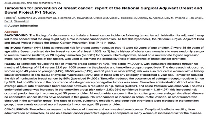
Chemoprevention

Slide2

Rodrigo Arrangoiz MS, MD, FACS

Cirugía General y Gastrointestinal
Michigan State University
Cirugía Oncológica
Fox Chase Cancer Center
Tumores de Cabeza y Cuello / Cirugía Endocrina
Fox Chase Cancer Center
Tumores de Cabeza y Cuello / Cirugía Endocrina
IFHNOS / Memorial Sloan Kettering Cancer Center
Maestría en Ciencias de Investigación
Drexel University
Certificado por el Colegio Americano de Cirugía
Fellow del Colegio Americano de Cirugía
Fellow de la Sociedad de Cirugia Oncológica
Sociedad Quirúrgica S.C.
Hospital ABC Santa Fé
Av. Carlos Graef Fernández #154

Consultorio 515
Col. Tlaxala, Delg. Cuajimalpa
México, D.F. 05300
Tel: 1103 – 1600 Ext 4515 a la 4517
Fax:1664 – 7164
 

Vigilancia con Resonancia Magnetica

La Sociedad Americana del Cáncer (American Cancer Society) respalda el examen con resonancia magnetica (RM) para cualquier persona con riesgo de cáncer de mama de por vida superior al 20%.

  • Esto incluiría a la mayoría de las personas con una mutación patógena en cualquiera de los genes de predisposición al cáncer de mama establecidos incluidos en los modernos paneles de pruebas genéticas.

Las directrices NCCN han respaldado específicamente la vigilancia mejorada con MRI para BRCA1, BRCA2, ATM, CDH1, CHEK2, NBN, NF1, PALB2, PTEN, STK11 y TP53.

Rodrigo Arrangoiz MS, MD, FACS

Cirugía General y Gastrointestinal
Michigan State University
Cirugía Oncológica
Fox Chase Cancer Center
Tumores de Cabeza y Cuello / Cirugía Endocrina
Fox Chase Cancer Center
Tumores de Cabeza y Cuello / Cirugía Endocrina
IFHNOS / Memorial Sloan Kettering Cancer Center
Maestría en Ciencias de Investigación
Drexel University
Certificado por el Colegio Americano de Cirugía
Fellow del Colegio Americano de Cirugía
Fellow de la Sociedad de Cirugia Oncológica
Sociedad Quirúrgica S.C.
Hospital ABC Santa Fé
Av. Carlos Graef Fernández #154

Consultorio 515
Col. Tlaxala, Delg. Cuajimalpa
México, D.F. 05300
Tel: 1103 – 1600 Ext 4515 a la 4517
Fax:1664 – 7164
 

ENHANCED SURVEILLANCE

The American Cancer Society supports screening MRI for anyone with lifetime breast cancer risk greater than 20%.

  • This would include all most anyone with a pathogenic mutation in any of the established breast cancer predisposition genes included in the modern genetic testing panels.

The NCCN guidelines have specifically endorsed enhanced surveillance with MRI for BRCA1, BRCA2, ATM, CDH1, CHEK2, NBN, NF1, PALB2, PTEN, STK11, and TP53.


Rodrigo Arrangoiz MS, MD, FACS

Cirugía General y Gastrointestinal
Michigan State University
Cirugía Oncológica
Fox Chase Cancer Center
Tumores de Cabeza y Cuello / Cirugía Endocrina
Fox Chase Cancer Center
Tumores de Cabeza y Cuello / Cirugía Endocrina
IFHNOS / Memorial Sloan Kettering Cancer Center
Maestría en Ciencias de Investigación
Drexel University
Certificado por el Colegio Americano de Cirugía
Fellow del Colegio Americano de Cirugía
Fellow de la Sociedad de Cirugia Oncológica
Sociedad Quirúrgica S.C.
Hospital ABC Santa Fé
Av. Carlos Graef Fernández #154

Consultorio 515
Col. Tlaxala, Delg. Cuajimalpa
México, D.F. 05300
Tel: 1103 – 1600 Ext 4515 a la 4517
Fax:1664 – 7164
 

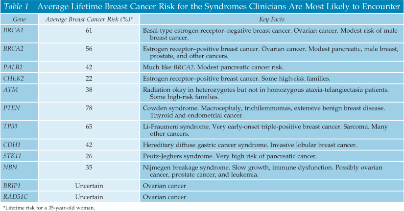
Estimating Breast Cancer Risk

The average probability of developing breast cancer by age 70 is estimated at 57% to 59% for BRCA1 gene mutation carriers and 49% to 51% for BRCA2 gene mutation carriers.

  • Emphasis is on the word “average” because these estimates are based on data from families with a lot of breast cancer cases as well as families with very few breast cancers.
    • Previous studies that had only included families with many breast cancer cases had concluded that lifetime breast cancer risk was more in the range of 80% to 85%.

This illustrates an important point:

  • The risk conferred by a pathologic mutation in any breast cancer predisposition gene will vary widely by family:
    • This is most likely due to gene-gene interactions, which are the modifying effects of small differences (e.g., polymorphisms) in many different genes.

Average Lifetime Brest Cancer Risk for the Syndromes

Rodrigo Arrangoiz MS, MD, FACS
Cirugía General y Gastrointestinal
Michigan State University
Cirugía Oncológica
Fox Chase Cancer Center
Tumores de Cabeza y Cuello / Cirugía Endocrina
Fox Chase Cancer Center
Tumores de Cabeza y Cuello / Cirugía Endocrina
IFHNOS / Memorial Sloan Kettering Cancer Center
Maestría en Ciencias de Investigación
Drexel University
Certificado por el Colegio Americano de Cirugía
Fellow del Colegio Americano de Cirugía
Fellow de la Sociedad de Cirugia Oncológica  

Sociedad Quirúrgica S.C.
Hospital ABC Santa Fé
Av. Carlos Graef Fernández #154

Consultorio 515
Col. Tlaxala, Delg. Cuajimalpa
México, D.F. 05300
Tel: 1103 – 1600 Ext 4515 a la 4517
Fax:1664 – 7164