Blog

Sociedad Quirúrgica S.C.

Sociedad Quirúrgica S.C. es el único grupo quirúrgico en México que todos sus socios han tenido entrenamiento en los mejores centros académicos de los estados unidos.

Cuatro de sus socios realizaron todo su entrenamiento como cirujanos y sub especialista en los  Estados Unidos, lo que implica que tienen los consejos de sus especialidades en México y en los Estados Unidos de America.

Rodrigo Arrangoiz MS, MD, FACS

Cirugia General – Michigan State University

(https://surgery.msu.edu/faculty/index.php)

images

Surgical Oncology / Head and Neck Surgery / Endocrine Surgery – Fox Chase Cancer Center (https://www.foxchase.org)

Unknown

Head and Neck Surgery and Oncology / Endocrine Surgery – IFHNOS/Memorial Sloan Kettering Cancer Center

(http://www.ifhnos.net/global_online_fellowship.html)

Master in Science (Clinical research for health care professionals) – Drexel University

Unknown

Fernando Cordera MD, FACS

Cirugia General – Mayo Clinic (http://www.mayo.edu)

images

Surgical Oncology / Head and Neck Surgery / Endocrine Surgery – Fox Chase Cancer Center (https://www.foxchase.org)

Unknown

Head and Neck Surgery and Oncology  – IFHNOS/Memorial Sloan Kettering Cancer Center

(http://www.ifhnos.net/global_online_fellowship.html)

David Caba MS, MD, MPH

Cirugia General –  Dartmouth (http://gme.dartmouth-hitchcock.org/general_surgery.html)

images-1

Surgical Oncology – University of Chicago

(https://surgery.uchicago.edu/specialties/general/csp/oncology)

Unknown

Manuel Muñoz MD, FACS, FACRS

Cirugia General – Mayo Clinic (http://www.mayo.edu)

images

Colorectal surgery – Mayo Clinic (http://www.mayo.edu)

images

Visiten nuestra pagina:

http://www.sociedadquirurgica.com


Unknown

Pleomorphic Adenoma

Pleomorphic adenoma is a benign salivary gland tumor that exhibits wide cytomorphologic and architectural diversity.

The tumor has the following three components:

  • An epithelial cell component

  • A myoepithelial cell component

  • A stromal (mesenchymal) component

Unknown

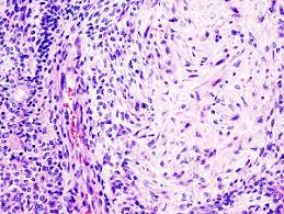
Identification of these three components, which may vary quantitatively from one tumor to another, is essential to the recognition of pleomorphic adenoma.

This tumor is also referred to as a benign mixed tumor.

Epidemiology

Pleomorphic adenoma is the most common salivary gland tumor in both children and adults.

In most series in the literature, it represents approximately 45% to 75% of all salivary gland neoplasms.

images-2

The annual incidence is approximately 2.0 to 3.5 cases per 100,000 population.

Pleomorphic adenoma occurs in individuals of all ages. However, it is most common in the third to sixth decades of life.

The average age at presentation is between 43 and 46 years.

Pleomorphic adenoma is seen more often in females than in males (2:1 ratio).

Etiology

Although the etiology of pleomorphic adenoma is unknown, the incidence of this tumor has been found to increase 15 to 20 years after exposure to radiation.

One study suggests that the simian virus (SV40) may play a causative role in the development of pleomorphic adenoma.

Location

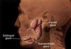
Among the major salivary glands (parotid gland, submandibular gland, and sublingual gland), the tail of the superficial lobe of the parotid salivary gland is the most common site of occurrence for pleomorphic adenoma (70% to 80% of cases), although this lesion can occur in any parotid location.

The tumor is less commonly seen in the submandibular salivary gland (10%) and is seldom encountered in the sublingual gland (1%).

With regard to the minor salivary glands (5% to 10% of cases):

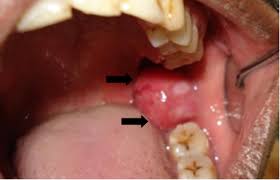
  • The palate (specifically, the junction of the soft and hard palates) and the lip are the most common sites for pleomorphic adenoma.
  • Other sites of minor salivary gland involvement include the nose, the paranasal sinuses, and the larynx.

Rare or unusual sites of occurrence include ectopic salivary gland tissues (eg, in the mandible, neck lymph nodes, or axilla). A case has been reported of a pleomorphic adenoma presenting as a midline nodule in the isthmus of the thyroid in a 66-year-old man.

Multiple tumors are unusual (1:40,000), but metachronous and synchronous tumors do occur.

Synchronous occurrence of pleomorphic adenoma and Warthin tumor (the second most common benign salivary gland tumor) has been reported

Clinical Features 

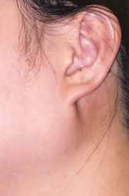
Pleomorphic adenoma usually presents as a slow-growing, painless mass, which may be present for many years. Symptoms and signs depend on the location.

When the tumor occurs in the parotid gland, signs of facial nerve weakness are seldom encountered; however, if the tumor is large and has been neglected, facial nerve weakness is likely to arise as the result of malignant change.

Pleomorphic adenoma in the deep lobe of the parotid gland may present as an oral retrotonsillar mass or parapharyngeal space tumor; indeed, tumors arising at this site are the source of the most common parapharyngeal space tumors.

images

Rapid enlargement of a tumor nodule should raise concern about the development of malignant change.

Patients with minor salivary gland tumors may present with a variety of symptoms, depending on the site of the tumor; such symptoms include dysphagia, dyspnea, hoarseness, difficulty in chewing, and epistaxis.

Macroscopic Characteristics

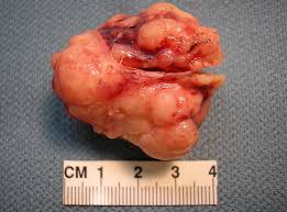
On gross examination, a pleomorphic adenoma is a single firm, mobile, well-circumscribed mass.

Its color may vary from whitish-tan to gray to bluish, and its size may range from a few millimeters to quite large or even giant.

Pleomorphic adenomas are irregularly shaped and have a bosselated surface.

 images-4.jpeg

Degenerative and cystic changes may be seen on sectioning.

It is not unusual to observe evidence of focal or massive infarction.

Recurrent tumors characteristically tend to present as multiple nodules of variable size.

Rodrigo Arrangoiz MS, MD, FACS

Cirugía General y Gastrointestinal
Michigan State University
Cirugía Oncológica
Fox Chase Cancer Center
Tumores de Cabeza y Cuello / Cirugía Endocrina
Fox Chase Cancer Center
Tumores de Cabeza y Cuello / Cirugía Endocrina
IFHNOS / Memorial Sloan Kettering Cancer Center
Maestría en Ciencias de Investigación
Drexel University
Certificado por el Colegio Americano de Cirugía
Fellow del Colegio Americano de Cirugía
Fellow de la Sociedad de Cirugia Oncológica
Sociedad Quirúrgica S.C.
Hospital ABC Santa Fé
Av. Carlos Graef Fernández #154
Consultorio 515
Col. Tlaxala, Delg. Cuajimalpa
México, D.F. 05300
Tel: 1103 – 1600 Ext 4515 a la 4517
Fax:1664 – 7164
 

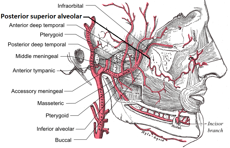
Anatomy of the Maxillary Artery

A. The maxillary artery supplies the deep structures of the face.

1. It branches from the external carotid artery just deep to the neck of the mandible.

B. Structure:

1. The maxillary artery, the larger of the two terminal branches of the external carotid artery:
– Arises behind the neck of the mandible, and is at first imbedded in the substance of the parotid gland.

2. It passes forward between the ramus of the mandible and the sphenomandibular ligament, and then runs, either superficial or deep to the lateral pterygoid muscle, to the pterygopalatine fossa.

3. It supplies the deep structures of the face.

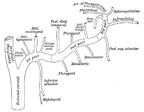
4. May be divided into:
– Mandibular portion (first part / bone portion)
– Pterygoid portion (second part / muscular portion)
– Pterygopalatine portions (third part).

C. First portion:

1. The first or mandibular portion (or bony portion) passes horizontally forward, between the neck of the mandible and the sphenomandibular ligament:
– Where it lies parallel to and a little below the auriculotemporal nerve.

2. It crosses the inferior alveolar nerve, and runs along the lower border of the lateral pterygoid muscle.

3. Branches include:
– Deep auricular artery
– Anterior tympanic artery
– Middle meningeal artery
– Inferior alveolar artery: which gives off its mylohyoid branch just prior to entering the mandibular foramen
– Accessory meningeal artery

D. Second Portion:

1. The second or pterygoid portion (or muscular portion) runs obliquely forward and upward under cover of the ramus of the mandible and insertion of the temporalis muscle:
– On the superficial (very infrequently on the deep) surface of the lateral pterygoid muscle.
– It then passes between the two heads of origin of this muscle and enters the pterygopalatine fossa.

2. Branches include:
– Masseteric artery
– Pterygoid branches
– Deep temporal arteries
= Anterior and posterior
– Buccal (buccinator) artery

E. Third Portion:

1. The third or pterygopalatine portion lies in the pterygopalatine fossa in relation with the pterygopalatine ganglion.

2. This is considered the terminal branch of the maxillary artery.

3. Branches include:
– Sphenopalatine artery:  also known as the nasopalatine artery which is the terminal branch of the maxillary artery
– Descending palatine artery:
= Greater palatine artery
= Lesser palatine artery
– Infraorbital artery
– Posterior superior alveolar artery
– Artery of pterygoid canal
-Pharyngeal artery
– Middle superior alveolar artery (could be a branch of the infraorbital artery)
– Anterior superior alveolar arteries (could be a branch of the infraorbital artery)
Posterior_superior_alveolar_arteryGray511


Rodrigo Arrangoiz MS, MD, FACS
Cirugía General y Gastrointestinal
Michigan State University
Cirugía Oncológica
Fox Chase Cancer Center
Tumores de Cabeza y Cuello / Cirugía Endocrina
Fox Chase Cancer Center
Tumores de Cabeza y Cuello / Cirugía Endocrina
IFHNOS / Memorial Sloan Kettering Cancer Center
Maestría en Ciencias de Investigación
Drexel University
Certificado por el Colegio Americano de Cirugía
Fellow del Colegio Americano de Cirugía
Fellow de la Sociedad de Cirugia Oncológica 

Sociedad Quirúrgica S.C.
Hospital ABC Santa Fé
Av. Carlos Graef Fernández #154

Consultorio 515
Col. Tlaxala, Delg. Cuajimalpa
México, D.F. 05300
Tel: 1103 – 1600 Ext 4515 a la 4517
Fax:1664 – 7164

 

Role of Axillary Radiation without Axillary Dissection (AMAROS Study)

The international After Mapping of the Axilla: Radiotherapy Or Surgery (AMAROS) study randomized 1,425 sentinel lymph node–positive patients to completion ALND or nodal field irradiation without ALND.

  • Nodal radiotherapy included axillary, supraclavicular, and low-neck fields.
  • There was no statistically significant difference in:
    • Disease-free or overall survival between the treatment arms.
  • Axillary radiotherapy was shown to have lower morbidity than ALND, with lower complication rates:
    • Specifically lower rates of lymphedema.
  • The 10-year overall survival with extensive nodal radiotherapy was 83% compared with 82% with ALND, and this difference was not statistically significant.
  • Axillary recurrence was observed in four of the 744 patients (< 1%) in the ALND group and seven of the 681 patients (1%) in the radiotherapy group, which was also not statistically significantly different.

There have been no randomized trials comparing radiotherapy with SLNB alone.

  • The axillary recurrence rate was 1% at 6 years in patients with positive sentinel nodes who received axillary radiation in the AMAROS trial.
    • This is comparable to the axillary recurrence rate of less than 2% at 10 years in the patients randomized to the sentinel lymph node–only arm of ACOSOG Z0011.
    • If the patients and axillary recurrence rates were similar with and without radiation, the utility of nodal radiation for patients with one to two positive sentinel lymph nodes is questionable.


Rodrigo Arrangoiz MS, MD, FACS
Cirugía General y Gastrointestinal
Michigan State University
Cirugía Oncológica
Fox Chase Cancer Center
Tumores de Cabeza y Cuello / Cirugía Endocrina
Fox Chase Cancer Center
Tumores de Cabeza y Cuello / Cirugía Endocrina
IFHNOS / Memorial Sloan Kettering Cancer Center
Maestría en Ciencias de Investigación
Drexel University
Certificado por el Colegio Americano de Cirugía
Fellow del Colegio Americano de Cirugía
Fellow de la Sociedad de Cirugia Oncológica  

Sociedad Quirúrgica S.C.
Hospital ABC Santa Fé
Av. Carlos Graef Fernández #154

Consultorio 515
Col. Tlaxala, Delg. Cuajimalpa
México, D.F. 05300
Tel: 1103 – 1600 Ext 4515 a la 4517
Fax:1664 – 7164


Salivary Gland Tumors

Current thinking on malignant salivary gland neoplasms

Rodrigo Arrangoiz*, Pavlos Papavasiliuo, David Sarcu, Thomas J. Galloway, John A. Ridge, Miriam Lango

Temple University, Fox Chase Cancer Center, 333 Cottman Avenue, Philadelphia USA 19111

Email address:

rodrigo.arrangoiz@gmail.com(R. Arrangoiz), pavlos.papavasiliuo@fccc.edu(P. Papavasiliuo), david.sarcu@tuhs.temple.edu(D. Sarcu), thomas.galloway@fccc.edu(T. J. Galloway), john.ridge@fccc.edu(J. A. Ridge), miriam.lango@fccc.edu(M. Lango)

To cite this article:

Rodrigo Arrangoiz, Pavlos Papavasiliuo, David Sarcu, Thomas J. Galloway, John A. Ridge, Miriam Lango. Current Thinking on Malignant Salivary Gland Neoplasms. Journal of Cancer Treatment and Research. Vol. 1, No. 1, 2013, pp. 8-24.
doi: 10.11648/j.jctr.20130101.12


Click to access a8c039c6e049d0c696bb936662de04c16bf1.pdf

Abstract:Malignant salivary gland neoplasms are rare, representing approximately 3% to 7% of all head and neck cancers. Contrasting from the more common mucosal head and neck cancers, which, in general, are ascribed to excessive tobacco, alcohol use, and more recently to viral infection, specific carcinogenic factors for malignant salivary gland growths have not been as clearly identified. Histologically, they represent a heterogeneous group of tumors. Forty histologic types of epithelial tumors of the salivary glands have been reported; some are exceedingly rare and may be the topic of only a few case reports. Salivary tumors can arise in the major salivary glands or in one of the minor salivary glands (predominantly mucus secreting glands), which are distributed throughout the upper aerodigestive. Most patients who develop malignant salivary gland tumors are in the sixth or seventh decade of life. FNA should be considered as part of the diagnostic evaluation but due to its varying sensitivities and specificities imaging modalities such as ultrasound, CT scans, and MRI should also be used as diagnostic adjuncts. Surgery is the primary modality for management of these tumors, nontraditional surgical approaches and instrumentation, as well as facial nerve monitoring, can be selectively utilized to try and decrease the morbidity associated with these surgical procedures. Adjuvant treatment is primarily achieved with radiation therapy. Chemotherapy continues to have a palliative role in the management of salivary gland tumors; however, research in this field is trying to identify a therapeutic role for chemotherapy in order to improve overall survival.

Keywords: Salivary Gland Tumors, Epidemiology of Salivary Gland Tumors, Types of Salivary Gland Tumors, Diagnosis and Treatment of Salivary Gland Tumors


Book Chapter

Malignant Salivary Gland Neoplasms Literature Review

Rodrigo Arrangoiz*, Fernando Cordera, David Caba, Luis Fernando Negrete, Manuel Muñoz Juarez, Eduardo Moreno Paquentin and Enrique Luque de León

Sociedad Quirúrgica S.C. at the American British Cowdray Medical Center, Mexico

*Corresponding Author: Rodrigo Arrangoiz, Av. Carlos Graef Fernandez # 154 – 515, Colonia Tlaxala, Delegación Cuajimalpa, Mexico City 05300, Mexico, Tel: (5255) 16647200; Email: rodrigo.arrangoiz@gmail.com

First Published February 28, 2018


Abstract

Malignant salivary gland neoplasms represent approximately 3% to 7% of all head and neck cancers making them extremely rare tu- mors. Contrasting from the more common mucosal head and neck cancers, which, in general, are attributed to excessive tobacco, alcohol use, and more recently to viral infection, specific carcinogenic factors for malignant salivary gland growths have not been as clearly identi- fied. Histologically, they represent a heterogeneous group of tumors. Salivary tumors can arise in the major salivary glands or in one of the minor salivary glands (predominantly mucus secreting glands), which are distributed throughout the upper aerodigestive. Most pa- tients who develop malignant salivary gland tumors are in the sixth or seventh decade of life. FNA should be considered as part of the di- agnostic evaluation but due to its varying sensitivities and specificities imaging modalities such as ultrasound, CT scans, and MRI should also be used as diagnostic adjuncts. Surgery is the primary modality for management of these tumors, nontraditional surgical approaches and instrumentation, as well as facial nerve monitoring, can be selectively utilized to try and decrease the morbidity associated with these surgical procedures. Adjuvant treatment is primarily achieved with radiation therapy. Chemotherapy continues to have a palliative role in the management of salivary gland tumors; however, research in this field is trying to identify a therapeutic role for chemotherapy in order to improve overall survival.


Click to access malignant-salivary-gland-neoplasms-literature-review.pdf

Rodrigo Arrangoiz MS, MD, FACS
Cirugía General y Gastrointestinal
Michigan State University
Cirugía Oncológica
Fox Chase Cancer Center
Tumores de Cabeza y Cuello / Cirugía Endocrina
Fox Chase Cancer Center
Tumores de Cabeza y Cuello / Cirugía Endocrina
IFHNOS / Memorial Sloan Kettering Cancer Center
Maestría en Ciencias de Investigación
Drexel University
Certificado por el Colegio Americano de Cirugía
Fellow del Colegio Americano de Cirugía
Fellow de la Sociedad de Cirugia Oncológica 

Sociedad Quirúrgica S.C.
Hospital ABC Santa Fé
Av. Carlos Graef Fernández #154

Consultorio 515
Col. Tlaxala, Delg. Cuajimalpa
México, D.F. 05300
Tel: 1103 – 1600 Ext 4515 a la 4517
Fax:1664 – 7164

 

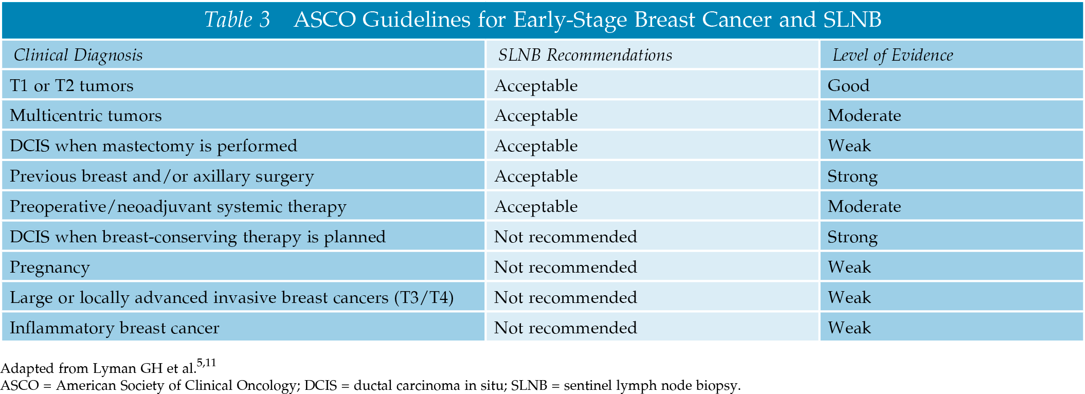
The Current ASCO Guidelines for SLNB


Rodrigo Arrangoiz MS, MD, FACS
Cirugía General y Gastrointestinal
Michigan State University
Cirugía Oncológica
Fox Chase Cancer Center
Tumores de Cabeza y Cuello / Cirugía Endocrina
Fox Chase Cancer Center
Tumores de Cabeza y Cuello / Cirugía Endocrina
IFHNOS / Memorial Sloan Kettering Cancer Center
Maestría en Ciencias de Investigación
Drexel University
Certificado por el Colegio Americano de Cirugía
Fellow del Colegio Americano de Cirugía
Fellow de la Sociedad de Cirugia Oncológica 

Sociedad Quirúrgica S.C.
Hospital ABC Santa Fé
Av. Carlos Graef Fernández #154

Consultorio 515
Col. Tlaxala, Delg. Cuajimalpa
México, D.F. 05300
Tel: 1103 – 1600 Ext 4515 a la 4517
Fax:1664 – 7164
 

Segmental Mandibulectomy

The current indications for a segmental mandibulectomy include:

(1) Gross invasion by oral cancer;

(2) Invasion of inferior alveolar nerve or inferior alveolar canal by a tumor;

(3) Massive soft tissue disease adjacent to the mandible;

(4) A primary malignant tumor of the mandible;

(5) A tumor that has metastasized to the mandible.


Rodrigo Arrangoiz MS, MD, FACS
Cirugía General y Gastrointestinal
Michigan State University
Cirugía Oncológica
Fox Chase Cancer Center
Tumores de Cabeza y Cuello / Cirugía Endocrina
Fox Chase Cancer Center
Tumores de Cabeza y Cuello / Cirugía Endocrina
IFHNOS / Memorial Sloan Kettering Cancer Center
Maestría en Ciencias de Investigación
Drexel University
Certificado por el Colegio Americano de Cirugía
Fellow del Colegio Americano de Cirugía
Fellow de la Sociedad de Cirugia Oncológica  

Sociedad Quirúrgica S.C.
Hospital ABC Santa Fé
Av. Carlos Graef Fernández #154

Consultorio 515
Col. Tlaxala, Delg. Cuajimalpa
México, D.F. 05300
Tel: 1103 – 1600 Ext 4515 a la 4517
Fax:1664 – 7164

Càncer de la Cavidad Oral

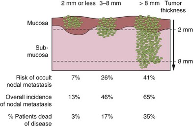
La característica histológica más importante del tumor primario que afecta la selección del tratamiento y el pronóstico final es su profundidad de infiltración.

  • Las lesiones delgadas y superficialmente invasivas tienen un riesgo menor de metástasis a los ganglios linfáticos regionales, son altamente curables y ofrecen un excelente pronóstico.

Unknown

  • Por otro lado, las lesiones más gruesas que infiltran profundamente los tejidos blandos subyacentes tienen una incidencia significativamente mayor de metástasis en los ganglios linfáticos regionales y un impacto adverso en el pronóstico.

Unknown

El riesgo de metástasis ganglionares y tasas de supervivencia en relación con el espesor de la lesión primaria para los carcinomas escamosos T1 y T2 de la lengua y el piso de la boca se muestran en la siguiente figura:

B9780323055895000081_f008-039-9780323055895

Aunque sería ideal conocer el espesor exacto de la lesión antes de la intervención quirúrgica, en muchos casos tener esa información antes de la extirpación quirúrgica del tumor primario es clínicamente poco práctico.

  • En general, sin embargo, el grosor de la lesión apreciado por la palpación es un indicador razonablemente bueno de lesiones profundamente invasivas versus lesiones superficiales para estimar el grado de resección de tejido blando y / o hueso para la lesión primaria y para decidir sobre la necesidad de disección electiva de los ganglios linfáticos regionales en riesgo en un cuello con hallazgos clínicos negativos.

Rodrigo Arrangoiz MS, MD, FACS
Cirugía General y Gastrointestinal
Michigan State University
Cirugía Oncológica
Fox Chase Cancer Center
Tumores de Cabeza y Cuello / Cirugía Endocrina
Fox Chase Cancer Center
Tumores de Cabeza y Cuello / Cirugía Endocrina
IFHNOS / Memorial Sloan Kettering Cancer Center
Maestría en Ciencias de Investigación
Drexel University
Certificado por el Colegio Americano de Cirugía
Fellow del Colegio Americano de Cirugía
Fellow de la Sociedad de Cirugia Oncológica 

Sociedad Quirúrgica S.C.
Hospital ABC Santa Fé
Av. Carlos Graef Fernández #154

Consultorio 515
Col. Tlaxala, Delg. Cuajimalpa
México, D.F. 05300
Tel: 1103 – 1600 Ext 4515 a la 4517
Fax:1664 – 7164

 

Oral Cavity Cancer

The most important histological feature of the primary tumor that affects selection of treatment and eventual prognosis is its depth of infiltration.

  • Thin and superficially invasive lesions have a lower risk of regional lymph node metastasis, are highly curable, and offer an excellent prognosis.

Unknown

  • On the other hand, thicker lesions that deeply infiltrate the underlying soft tissues have a significantly increased incidence of regional lymph node metastasis and an adverse impact on prognosis.

Unknown

The risk of lymph node metastasis and survival rates in relation to the thickness of the primary lesion for T1 and T2 squamous carcinomas of the oral tongue and floor of mouth are shown in the following figure:

B9780323055895000081_f008-039-9780323055895

Although it would be ideal to know the exact thickness of the lesion before surgical intervention, in many instances having that information before surgical excision of the primary tumor is clinically impractical.

  • In general, however, thickness of the lesion as appreciated by palpation is a reasonably good indicator of deeply invasive lesions versus superficial lesions to estimate the extent of soft tissue and/or bone resection for the primary lesion and to decide on the need for elective dissection of the regional lymph nodes at risk in a neck with negative clinical findings.

Rodrigo Arrangoiz MS, MD, FACS
Cirugía General y Gastrointestinal
Michigan State University
Cirugía Oncológica
Fox Chase Cancer Center
Tumores de Cabeza y Cuello / Cirugía Endocrina
Fox Chase Cancer Center
Tumores de Cabeza y Cuello / Cirugía Endocrina
IFHNOS / Memorial Sloan Kettering Cancer Center
Maestría en Ciencias de Investigación
Drexel University
Certificado por el Colegio Americano de Cirugía
Fellow del Colegio Americano de Cirugía
Fellow de la Sociedad de Cirugia Oncológica 

Sociedad Quirúrgica S.C.
Hospital ABC Santa Fé
Av. Carlos Graef Fernández #154

Consultorio 515
Col. Tlaxala, Delg. Cuajimalpa
México, D.F. 05300
Tel: 1103 – 1600 Ext 4515 a la 4517
Fax:1664 – 7164



¿Quien debe estar tratando el cancer de tiroides?

Les dejo la respuesta de Ashok R. Shaha, MD, FACS (profesor de dos de los cirujanos oncologos de Sociedad Quirùrgica S.C.)
 
 
Sociedad Quirùrgica tiene un cirujano oncologo con sub especialidad en tumores de cabeza y cuello / cirujano endocrino:
 
Rodrigo Arrangoiz MS, MD, FACS
Cirugía Oncológica
Fox Chase Cancer Center
 
Tumores de Cabeza y Cuello / Cirugía Endocrina
Fox Chase Cancer Center
 
Tumores de Cabeza y Cuello / Cirugía Endocrina
IFHNOS / Memorial Sloan Kettering Cancer Center
 
Maestría en Ciencias de Investigación
Drexel University
 
Certificado por el Colegio Americano de Cirugía
Fellow del Colegio Americano de Cirugía
Fellow de la Sociedad de Cirugia Oncológica
 
Sociedad Quirúrgica S.C.
Hospital ABC Santa Fé
Av. Carlos Graef Fernández #154
Consultorio 515
Col. Tlaxala, Delg. Cuajimalpa
México, D.F. 05300
Tel: 1103 – 1600 Ext 4515 a la 4517
Fax:1664 – 7164

 
rodrigo.arrangoiz@gmail.com