Blog

Radiofrequency Ablation for Papillary Thyroid Microcarcinoma Is Safe and Effective in Long-Term Follow-up

  • Clin Thyroidol 2021;33:121–123.
  • Background
    • Low-risk papillary thyroid microcarcinomas (PTMCs) have been shown to have an:
      • Indolent course
    • This observation has led to alternative management approaches such as:
      • Active surveillance (AS) rather than standard treatment with surgery
    • Studies examining AS have shown promising results, with a low incidence of disease progression such as:
      • Tumor enlargement (5.3%)
      • Lymph node involvement (1.6%)
      • No evidence of distant metastasis
    • However, delayed surgeries during AS do occur:
      • Likely driven by patient and physician:
        • Anxiety
    • Therefore ultrasound-guided thermal ablation, particularly radiofrequency ablation (RFA):
      • Has been proposed as an alternative therapy in this setting
    • A recent meta-analysis of 11 cohorts:
      • Has shown RFA to be safe and effective in the management of PTMC over a relatively short follow-up period of up to two years
    • As clinicians consider the management of PTMC:
      • Longer-term data on the durability of RFA outcomes are needed
    • This study was a single-institution report of the authors’ experience with RFA in a cohort of patients with PTMC
  • Methods
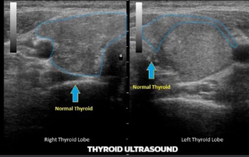
    • This was a retrospective study of 84 PTMCs in 74 patients at a single center in South Korea
    • The study population was derived from a previously reported cohort of 133 patients who underwent RFA after September 2008 and had a follow-up of longer than 5 years
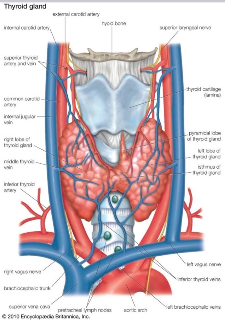
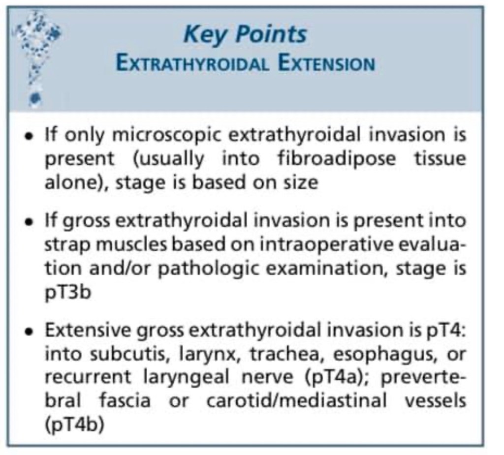
    • Participants had PTMC, with exclusion of aggressive subtypes on cytology, while imaging was used to exclude gross extrathyroidal extension, cervical adenopathy and distant metastases
    • All participants had either a contraindication to surgery or refused it
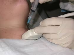
    • The RFA procedure was performed under local anesthesia by a single radiologist, and the approach was trans-isthmic, using the moving-shot technique
    • After RFA, patients were followed using ultrasound, interpreted by the same radiologist who performed the procedure, as well as clinical evaluations at 1, 6, and 12 months and annually thereafter for a minimum of 5 years
    • The outcomes of interest included PTMC volume changes, newly developed PTMCs, lymph node involvement, distant metastasis, delayed surgery during the follow-up period, and RFA safety, defined by immediate and long-term complications
  • Results
    • The study cohort consisted of 74 adults with a mean age of 46 years
    • There were 84 PTMCs treated with RFA, with 62% of them measuring < 5 mm (range, 3 to 10 mm maximum diameter)
    • Only 13 lesions required a second RFA session because of incomplete ablation during the first RFA session (average, 1.2 RFA sessions / patient)
    • Over a mean (±SD) follow-up period of 72±18 months:
      • There were no cases of tumor progression, lymph node or distant metastases, or delayed surgery
    • Complete tumor disappearance was noted by 60 months in all PTMCs:
      • With 98.8% disappearance by 36 months
    • Four new PTMCs developed in three participants’ remaining thyroid glands:
      • These were also treated effectively with RFA
    • Minor complications occurred in 4.1% of participants and included:
      • Hematomas and first-degree burns
    • Major complications occurred in 1.4% and included:
      • Voice changes, with recovery by 2 months
  • Conclusions
    • In adults with low-risk PTMCs, RFA has been shown to be effective and safe in over 5 years of follow-up
    • This approach has led to complete disappearance of all treated lesions and prevention of local tumor progression, metastasis, or delayed surgery, all with minimal risk of complication
  • This study is the first to report on long-term follow-up of a sizable cohort of PTMC patients treated with RFA and found excellent efficacy and safety
  • Previous reports were limited by a short duration of follow-up—a significant limitation considering the indolent course of PTMC
  • However, there are some notable considerations:
    • First, these data reflect experience at a single center with a single and highly experienced ultrasound and RFA operator, thus limiting external validity to centers with similar expertise:
      • Experience with RFA is paramount given the reported doubling in ablated lesion volume early-on post ablation
        • Due to ablation of parenchyma surrounding PTMCs:
          • Which needs to be separated from residual tumor presence with need for repeat RFA
          • Thus, without adequate expertise regarding interpretation of post-RFA imaging, unnecessary procedures might be performed
    • Second, the favorable outcomes with regard to tumor recurrence and LN metastasis are in contrast with some previous reports:
      • This is likely related to selection of less aggressive tumors:
        • As evidenced by cytologic criteria and small tumor size (greater than 62% of participants had a tumor size less than 0.5 cm)
      • Since thyroid cancer screening is no longer pursued, it is likely that the median size of RFA-treated PTMCs will increase, hence the need for revisiting the efficacy of the procedure periodically
  • Overall, these results are certainly promising, but their application to all PTMCs remains limited
  • In our practice, we do not routinely biopsy thyroid nodules less than 10 mm, in line with current American Thyroid Association (ATA) guidelines
  • For greater adoption of RFA, this approach would need to be reconsidered
  • At the same time, as RFA is being considered as an alternative to AS and surgery, patient-centered outcomes such as anxiety, pain, quality of life, and cost will be essential in future studies
  • Currently, without direct comparison between RFA, AS, and surgery:
    • RFA should be individualized to patient-specific situations:
      • Small low-risk PTMCs
      • Poor candidates for surgery or refusing surgery
      • Anxiety about AS
    • It should be considered only after careful discussion of standard treatment options
    • For patients interest in RFA, special emphasis should be placed on the required expertise for this procedure, in order to replicate the excellent results described here

#Arrangoiz #CancerSurgeon #HeadandNeckSurgeon #SurgicalOncologist #ThyroidSurgeon #ThyroidExpert #CASO #ThyroidNodules #CenterforAdvancedSurgicalOncology

Thyroid Nodules

👉Thyroid nodules are a major health problem worldwide.

👉The prevalence of palpable thyroid nodules in the general population is approximately 5% in women and 1% in men living in parts of the world with sufficient iodine.

👉High resolution neck and thyroid ultrasound can detect thyroid nodules in a signif- icant proportion of randomly selected individuals, with higher frequencies in women and the elderly population.

👉The importance of thyroid nodules lies in the need to rule out cancer. The majority of thyroid nodules are benign, clinically irrelevant, and can be safely managed with a good surveillance program.

👉The detection and diagnosis of differentiated thyroid cancer have evolved over the years with increased use of high resolution cervical and thyroid ultrasound, fine needle aspiration biopsy (FNAB), molecular testing, and thyroglobulin as a serum tumor marker.

👉An algorithm that utilizes high resolution ultrasound and, when indicated, FNAB, and molecular testing for the diagnosis of thyroid nodules, facilitates a personalized, risk-based protocol that promotes high-quality care and minimizes cost and unnecessary testing.

Click to access 63f85b77cd57d842f1dd19ec8e4abf06e99e.pdf

#Arrangoiz

#ThyroidSurgeon

Parathyroid Awareness

👉The majority of patients with both hyperparathyroidism and osteoporosis who undergo parathyroidectomy benefit from surgery with improvement in their bone mineral density.

👉https://pubmed.ncbi.nlm.nih.gov/23040710/

#CheckYourCalcium #Arrangoiz #ParathyroidSurgeon #ParathyroidExpert #HeadandNeckSurgeon

Parathyroid Gland Identification During Thyroid Surgery

#Arrangoiz #ParathyroidSurgeon #ParathyroidExpert #Hyperparathyroidism #CASO #CenterforAdvancedSurgicalOncology

Combination Pembrolizumab Plus Lenvatinib May Be Option in Anaplastic and Poorly Differentiated Thyroid Cancers

  • Clin Thyroidol 2021;33:131–133.
  • Background
    • Anaplastic thyroid carcinoma (ATC) and metas‐ tasized poorly differentiated thyroid carcinoma (PDTC):
      • Are rare aggressive malignancies with low overall survival despite the availability of extensive multimodal therapies
    • Tumors are highly proliferative:
      • With frequently increased tumor mutational burden as compared with differentiated thyroid carcinomas and elevated programmed cell death ligand 1 (PD-L1) levels
  • Methods
    • In a retrospective study, the authors analyzed six patients with metastasized ATC and two with PDTC:
      • Who received a combination therapy of lenvatinib and pembrolizumab
    • Lenvatinib was started at 14 to 24 mg daily and combined with pembrolizumab at a fixed dose of 200 mg every 3 weeks
    • Maximum treatment duration with this combination:
      • Was 40 months:
        • Three of the six patients with ATC were still on therapy
    • Patients’ tumors were characterized by whole-exome sequencing and PD-L1 expression levels (tumor proportion score [TPS], 1–90%)
  • Results
    • The best overall response within ATCs was:
      • 66% (4 of 6) with complete remission
      • 16% (1 of 6) with stable disease
      • 16% (1 of 6) with progressive disease
    • The best overall response in PDTCs was:
      • Partial remission (in 2 of 2)
    • Median progression-free survival was:
      • 17.75 months for all patients
      • 16.5 months for ATCs:
        • With treatment durations of 1, 4, 11, 15, 19, 25, 27, and 40 months
    • Grade III of IV toxicities developed in 4 of 8 patients and required dose reduction or discontinuation of lenvatinib
    • The median overall survival was 18.5 months:
      • With three ATC patients still alive without relapse (at 40, 27, and 19 months) despite metastatic disease at the start of treatment (International Union against Cancer [UICC] stage 4C)
    • All patients with long-term (greater than 2 years) or complete responses had either:
      • An increased tumor mutational burden or a PD-L1 TPS greater than 50%
  • Conclusions
    • These results suggest that the combination of lenvatinib and pembrolizumab might be safe and effective in patients with ATC or PDTC, leading to complete and long-term remissions
    • The combination treatment is now being systematically examined in a phase two clinical trial (Anaplastic Thyroid Carcinoma Lenvatinib Pembrolizumab – ATLEP; NCT04171622) in patients with ATC and PDTC
  • This small retrospective study demonstrates a rather provocative and high response rate in patients with ATC using the:
    • Multi–tyrosine kinase inhibitor lenvatinib combined with the immune checkpoint inhibitor pembrolizumab
  • While there is little doubt that pembrolizumab is active in ATC with activity that is likely comparable to that of spartalizumab (a similar PD-1 inhibitor that was tested in a robust phase two study of ATC and demonstrated a radiographic response rate of 19%), questions remain about the use of lenvatinib in this disease
  • Following initial reports from Japan with similarly remarkable response rates and a subsequent phase two study that seemed to confirm some efficacy, an international follow-up study failed to reproduce these results and was terminated after finding only one positive response in 33 patients (3) (NCT02657369)
  • Why there was such a discrepancy between those results?
    • At least one possible explanation is that in the Japanese study not all cases might have been anaplastic thyroid cancer, but could have been a mix between anaplastic and poorly differentiated thyroid cancer due to a lack of central pathology review
    • In PDTC, the high response rate to lenvatinib has been well described
    • One could argue that the combination of a checkpoint inhibitor plus lenvatinib could result in synergy:
      • With one drug enhancing the other’s activity perhaps through exposure of a broader range of neoantigens to the immune system
    • This hypothesis was the basis for a recently presented phase two study in radioiodine-refractory differentiated thyroid cancer conducted by the International Thyroid Oncology Group, which also combined pembrolizumab plus lenvatinib and which used the rate of complete responses as the primary end point
    • Unfortunately, there was no signal of synergistic activity, and the results were very much in line with those observed with lenvatinib alone
    • With that in mind, it is somewhat difficult to anticipate that the current data will be reproducible in the planned phase two study, but given the generally bad prognosis in this disease, every possible lead should be explored

#Arrangoiz #ThyroidSurgeon #ThyroidExpert #CancerSurgeon #ThyroidCancer #HeadandNeckSurgeon #CASO #CenterforAdvancedSurgicalOncology

Parathyroid Hormone Physiology

Parathyroid hormone (PTH) acts to raise blood calcium levels via its action on 3 organs. 1. The kidneys by Increased calcium reabsorption, Decreased phosphate reabsorption, Increased conversion of vitamin D to its final form – 1,25(OH) vitamin D; 2) Bones (increased resorption); 3) Intestines (increased absorption largely due to increased vitamin D in its final form).

Find out more at https://www.scirp.org/pdf/IJOHNS_2017072615211601.pdf

#Arrangoiz #ParathyroidSurgery #PararhyroidSurgeon #CASO #CenterforAdvancedSurgicalOncology

The Usefulness of Contrast-Enhanced Ultrasound to Evaluate Small Solid Thyroid Nodules Compared to TI-RADS

  • Clin Thyroidol 2021;33:114–116.
  • Background
    • Thyroid nodules are very common and frequently incidentally detected with the increasing use of imaging
    • The primary concern is how to identify those with suspicious features in order to avoid unnecessary diagnostic exams like fine-needle aspiration biopsy (FNAB) and overtreatment
    • Even though the Thyroid Imaging Reporting and Data System (TI-RADS):
      • Created by the American College of Radiologists:
        • Is widely accepted as an easy and useful tool to avoid unnecessary FNAB and close follow-up it still suffers from some biases, including experience and heterogeneity among sonographers
    • Contrast-enhanced ultrasound (CEUS) is a novel technology:
      • That can help differentiate between benign and malignant thyroid nodules
    • TI-RADS does not recommend FNAB for nodules up to 1 cm:
      • Even though such nodules have mild, moderate, or high (TI-RADS 3, 4, and 5, respectively) sonographic risk for malignancy
    • However, CEUS represents a new tool in the evaluation of thyroid nodules
    • The aim of this study was to compare the use of CEUS versus conventional ultrasound (as TI-RADS) in the risk stratification of thyroid nodules
  • Methods
    • In this study, 185 solid thyroid nodules between 0.5 and 1 cm in 154 patients were evaluated by conventional ultrasound and CEUS at the same time and by the same investigator
    • In conventional ultrasound, each target nodule was scored based on the components of:
      • Echogenicity, shape, edge, and strong echogenic focus, according to the 2017 edition of ACR TI-RADS in order to ascertain a TI-RADS classification
    • Qualitative indicators of CEUS analysis in the targeted thyroid nodules included:
      • Enhancement intensity, patterns of enhancement, internal homogeneity, the presence of perfusion defect, clearness of boundary, morphology, and size at enhance‐ ment peak
    • The CEUS characteristics of benign and malignant thyroid nodules with the significant differential diagnosis were scored, and the total scores of each thyroid nodule were calculated
  • Results
    • Of 185 thyroid nodules, the diagnosis was confirmed in 133 by surgical pathology, with 90 nodules (67.67%) showing papillary thyroid cancer (PTC) and 43 (32.33%) having benign results
    • In 52 thyroid nodules, cytology from the FNAB showed PTC in 11 nodules (21.15%) and nonmalignancy in 41 (78.85%)
    • Overall, there were 101 malignant thyroid nodules
    • When comparing the risk assessment scores:
      • CEUS outperformed TI-RADS using conventional ultrasound for benign diseases, but this was not the case in malignant nodules
    • Comparing the diagnostic performance between TI-RADS TR5 nodules and CEUS nodules that scored 5 points:
      • Sensitivity was 90.10% versus 86.13%, specificity 55.95% versus 89.29%, accuracy 74.59% versus 87.57%, positive predictive value 72.22% versus 90.63%, negative predictive value 82.46% versus 84.27%, and the area under the receiver-operating-characteristic curve 0.738 (95% CI, 0.663–0813) versus 0.916 (95% CI, 0.871– 0.961).
  • Conclusions
    • This study found that CEUS:
      • When evaluating small solid thyroid nodules, had higher diagnostic performance and a higher specificity than TI-RADS using conventional ultrasound
      • CEUS may be a valuable imaging tool to select patients with thyroid nodules for FNAB or surgery
    • Conventional ultrasound is currently the initial diagnostic tool for the risk stratification of thyroid nodules
    • To increase the diagnostic confidence and avoid unnecessary FNABs, many societies have proposed systems to stratify malignancy risks by ultrasound and guide physicians in when to perform FNAB:
      • One of the most popular and practical methods is TI-RADS
    • However, how to accurately differentiate malignant from benign nodules remains a significant challenge in many situations:
      • Since at least a half of all biopsied nodules are benign and up one third of FNABs are inconclusive
    • The new imaging method of CEUS can show tumor perfusion and vascular distribution after intravenous injection of a microbubble contrast agent:
      • Since vascular structures in the nodule differ from those of normal tissues
  • The specific characteristics of CEUS, like enhancement intensity, patterns of enhancement, and internal homogeneity, seem to be valuable in determining which nodules should undergo an FNAB and which may be kept under observation
  • In this study evaluating small nodules:
    • CEUS had significantly higher performance than TI-RADS in differentiating malignant from benign nodules:
      • Suggesting that CEUS qualitative analysis could be more effective in excluding malignant nodules and avoiding unnecessary biopsies
    • The use of CEUS in clinical practice has some crucial limitations, including the:
      • Cost of the exam, especially if the patient has more than one nodule to be analyzed:
        • Each nodule requires injection of a contrast agent
      • In addition, the small numbers of radiologists trained to perform this imaging study, as well as the absence of well-estab‐ lished criteria and consensus regarding the patterns of enhancement and classification of thyroid nodules, make its broad use in clinical practice controversial at present

#Arrangoiz #CancerSurgeon #ThyroidSurgeon #ThyroidExpert #HeadandNeckSurgeon #SurgicalOncologist #EndocrineSurgery #CASO #CenterforAdvancedSurgicalOncology

Risk of Clinically Significant Thyroid Cancer Is Low During Long-Term Population-Based Follow-up of Thyroid Nodules

  • Background
    • Fine-needle aspiration (FNA) using the recently revised Bethesda System for Reporting Thyroid Cytopathology accurately discriminates benign from malignant thyroid nodules:
      • However, the risk of malignancy in each Bethesda category has been studied using inconsistent methods, and most studies focus on nodules with corresponding surgical histology, which introduces selection bias
    • The present study sought to determine the real-world risk of malignancy in each Bethesda category using data from a large retrospective cohort of thyroid nodules with a mixture of surgical and long-term population-based clinical follow-up
  • Methods
    • All thyroid FNAs from the University of California, San Francisco (UCSF) Pathology database (collected between January 1, 1997, and December 31, 2004) were reviewed and recoded using the 2017 Bethesda system reporting standard
    • If multiple nodules were biopsied and had different results, the highest-grade Bethesda category was used
    • Corresponding patients were matched to the UCSF cancer registry, along with the California Cancer Registry (CCR; a statewide, population-based registry)
    • Patients who were not detected in either registry were considered to be free of malignancy
    • Exclusion criteria included a prior diagnosis of thyroid cancer, prior thyroid surgery, or development of a cancer in the lobe con- tralateral to the biopsy site
    • The date of the original FNA was the time origin, and the interval time to repeat FNA, surgery, or final follow-up on July 10, 2015 (date of matching to the CCR), was recorded
    • Kaplan–Meier survival curves and Cox proportional hazards models were used to estimate incidence rates of malignancy for each category and the instantaneous risk of malignancy, respectively
  • Results
    • A total of 2233 patients with 2758 FNA reports were available:
      • 26 were excluded, for a final count of 2207
    • The median age was 48 years (range, 7 to 92) and 1880 (85.2%) were female
    • Of the 2207 test results:
      • 236 (10.7%) were determined to be nondi- agnostic
      • 1575 (71.4%) benign
      • 57 (2.58%) atypia of undetermined significance (AUS)
      • 78 (3.53%) follicular lesion of undetermined significance (FLUS)
      • 107 (4.85%) follicular neoplasm or Hürthle-cell neoplasm
      • 20 (0.9%) suspicious for malignancy
      • 134 (6.07%) malignant
    • Median follow-up after the initial FNA was 13.9 years (range, 10.5 to 18.4):
      • 279 (12.6%) patients were diagnosed with thyroid malignancy during that period
    • Compared to the benign reference group, hazard ratios were:
      • 2.09 (95% CI, 1.2–3.7) for a nondiagnostic read
      • 8.8 (95% CI, 5.7–13.6) for AUS / FLUS
      • 10.9 (95% CI, 7.0–17.0) for follicular neoplasm
      • 49.1 (95% CI, 27.1–88.9) for suspicious for malignancy
      • 201 (95% CI, 138–293) for a malignant read
    • When AUS and FLUS were split into separate categories:
      • AUS had a higher hazard ratio (13.0; 95% CI, 7.7–22.0)
    • Malignancy rates per 1000 person-years were:
      • 4.82 (95% CI, 3.0–7.9) for a nondiagnostic read
      • 2.42 (95% CI, 1.9–3.2) for a benign read
      • 22.4 (95% CI, 16.0–31.6) for AUS / FLUS
      • 29.1 (95% CI, 20.4–41.3) for follicular neoplasm
      • 183 (95% CI, 108–310) for suspicious for malignancy, and 980 (CI) for a malignant read
    • A total of 52 (3.2%) of 1575 were false-negative results:
      • 29 (1.7%) of which were papillary thyroid microcarcinomas
    • A total of 15 patients died from thyroid cancer:
      • None of them had an initial benign FNA
  • Conclusions
    • FNA and the Bethesda System for Reporting Thyroid Cytopathology are highly accurate in detecting thyroid malignancy
    • Long-term combined clinical and histopathologic follow-up reveal a low false-negative rate, low rates of malignancy in nondiagnostic specimens, and extremely low rates of mortality, especially in benign and nondiagnostic categories
  • Thyroid nodules are common:
    • 5% to 15% of them are malignant:
      • The majority of which are well-differentiated thyroid cancers with a good prognosis
  • The Bethesda system has standardized reporting of thyroid nodule cytopathology:
    • Thereby facilitating efficient and precise communication, research, and articulation of evidence-based management guidelines:
      • However, even the most robust classification systems meet with variable adoption and implementation in the real-world clinical setting, and generalizability depends on validation in diverse popula- tions of patients and care systems
  • The present study sought to address two limitations in estimating risk of malignancy in validation studies:
    • Use of surgical histopathology as the gold standard:
      • Which leads to a higher estimated risk of malignancy in lower-risk nodules since they are less likely to undergo surgery
      • Inconsistent statistical handling of “indeterminate” categories:
        • Which have a wide range of preva- lence across institutions
  • Strengths of the study include:
    • A long duration of follow-up of a robust number of patients and use of an institutional and comprehensive population-based cancer registry to detect and provide a narrative summary of malignant cases and patient mortality
  • Limitations of the study include:
    • Its retrospective, single-institution design (which improves standardization at the cost of generalizability) and the assumption that patients without registry data did not develop malignancy, which misses patients who moved away or did not follow up, thereby underestimating the risk of malignancy
    • It is worth noting that even a median follow-up of 13.9 years is relatively short when dealing with an indolent disease, especially in light of long-term data showing minimal growth of biopsy-proven small papillary thyroid carcinomas
  • Given the marked indolence of most thyroid cancers, coupled with a high and rising incidence of small and incidental cancers:
    • Perhaps the most important task for clinicians is to avoid missing patients harboring clinically significant malignancy
  • The most notable aspect of this study is:
    • The confirmation that the false-negative rate for benign FNA was low, at 3.2%:
      • More than half of which were innocent papillary microcarcinomas), and that no patients with a false-negative FNA died of thyroid cancer
    • While risk of malignancy of nondiagnostic specimens was roughly double that of benign ones:
      • It was still relatively low, and the long-term outcomes for patients were similar
    • It is not surprising that thyroid cancer– related mortality was low (0.7%) across all categories during the follow-up period
    • Lastly, it was interesting that the AUS category had a higher risk of malignancy than FLUS:
      • When they were separated on the basis of nuclear (AUS) and architectural (FLUS) atypia:
        • While the Bethesda system designates AUS/ FLUS as a single category:
          • The current study corroborates others showing that subcategorization, while controversial, is worthy of discussion
          • Additional studies with large data sets and long-term follow-up are needed
    • Ultimately, while thoughtful management and longitudinal surveillance of patients with thyroid nodules requires integration of cytopathology with clinical and sonographic risk assessment:
      • It is reassuring to see confirmation that most patients with thyroid cancer do well, and that our standard-of- care approach of using FNA and Bethesda system reporting standards is accurate and rarely misses malignancy

#Arrangoiz #CancerSurgeon #ThyroidSurgeon #ThyroidExpert #HeadandNeckSurgeon #ThyroidCancer #SurgicalOncologist #CASO #CenterforAdvancedSurgicalOncology #Miami

Interpreting Microscopic Positive Margins in Differentiated Thyroid Cancer

  • Clin Thyroidol 2021;33:184–186.
  • Background
    • Disease-specific survival for differentiated thyroid cancer (DTC) is lengthy and most profoundly impacted by:
      • Patient age (55 years)
      • Distant metastases
      • Extent of gross extrathyroidal extension (ETE)
      • Nodal status
    • Microscopic margin positivity (R1 margin):
      • Has shown no impact on local recurrence, but it has not been studied as:
        • A marker of completeness of resection
    • This National Cancer Database (NCDB) study evaluated the impact of R1 margin on overall survival in DTC to determine the presence of modifiable factors such as institutional annual thyroidectomy volume and hospital setting of thyroidectomy
  • Methods
    • The NCDB was queried for adults (ages 18 to 90) who underwent surgery from 2004 to 2016 for treatment of well- and moderately-differentiated thyroid carcinoma with a primary tumor size of 1 to 4 cm
    • Patients with histologically aggressive variants were excluded
    • Patients with gross ETE into the strap muscles (T3b); subcutaneous soft tissues, larynx, trachea, esophagus, or recurrent laryngeal nerve (T4a); or invading prevertebral fascia, encasing carotid or mediastinal vessels (T4b), or nodal disease were included if primary tumor size was less than 4 cm
    • Binary logistic-regression analyses evaluated factors affecting margin positivity after adjusting for confounders including age, gender, comorbidity score (modified Charlson–Deyo), extent of thyroid operation (lobectomy or total), margin status, lymphovascular invasion, multifocality, institutional annual case volume for thyroidectomy, and institu- tion type (community, comprehensive community, academic, or integrated network
  • Results
    • This study included 14,471 patients with DTC (median age, 47.52 years [range, 18–90]; 74.7% female)
    • After correcting for confounders, disease-specific (nonmodifiable) factors impacting survival included:
      • ETE:
        • 25.2% of patients; OR, 1.47; P = 0.001), lateral neck nodal disease (12.6%; OR, 1.73; P = 0.001
      • Lymphovascular invasion:
        • 15.7%; OR, 1.32; P = 0.044
      • R1 margin:
        • 15.3%; OR, 1.46; P = 0.038
    • Modifiable factors impacting survival included:
      • Institutional case volume and type of facility:
        • Overall, institutions had a mean case volume of 30.3 thyroidectomies per year (range, 4 to 485):
          • When evaluating R1 margin status as a modifiable risk factor for survival:
            • Treatment in academic / research facilities (OR, 0.623; 95% CI, 0.527–0.738; P<0.001), integrated networks (OR, 0.782; 95% CI, 0.654–0.934; P = 0.009), or facilities with higher case volumes (OR, 0.979; 95% CI, 0.978–0.982; P = 0.004):
              • Was associated with lower odds of R1 margin
    • This study was sufficiently powered to detect a small impact of R1 margin on 8-year survival:
      • OR, 1.464; 95% CI, 1.039–2.121; P = 0.038
  • Conclusions
    • In this large database study:
      • R1 margin in DTC impacted overall survival
      • Modifiable risk factors associated with decreased risk of R1 margin were:
        • Treatment in an academic / research facility or integrated network and higher institutional annual case volume
  • As the thyroid gland has an incomplete capsule:
    • Interpreting an R1 margin as a prognostic indicator in DTC has been the source of some confusion
  • A meta-analysis of patients with DTC:
    • Found no association between R1 margin and local recurrence
  • This study aimed to evaluate the impact of negative versus R1 margins in a more homogeneous population of patients with smaller DTCs
  • The major modifiable factor in this study was care setting:
    • Higher-volume thyroid surgeons are more likely to operate in academic settings
    • Worldwide, higher individual surgeon volume for thyroidectomy (30 to 100 cases / year) is protective against complications and yields better oncologic outcomes
    • A similar NCDB study with a more heteroge- neous thyroid cancer population confirmed that treatment in an academic center was associated with a lower probability of R1 margin and higher overall survival
  • Rather than contradicting prior studies about the seeming irrelevance of R1 margin in DTC:
    • This study highlights how R1 margin may be a marker of incomplete resection when thyroidectomy is performed in low-volume centers
    • When I receive pathologic “positive margins,” assuming no ETE was present intraoperatively:
      • I generally reassure the patient that all tumor was appropriately resected:
        • However, this study raises important questions about how to interpret pathology reports documenting R1 margin from lower-volume centers
  • In contrast to this study’s findings of improvement in mean overall survival with negative margins:
    • Prior retrospective studies have not found that R1 margin impacts overall survival
  • Although this study corrected for confounders:
    • ETE was the variable most strongly correlated with an R1 margin (OR, 2.8):
      • ETE decreases survival in thyroid cancer, and it makes sense that ETE would be a natural continuation of R1 margins
        • However, further research is needed to determine the prognostic significance of thyroid cancers with positive inked margins without a finding of gross ETE

#Arrangoiz #CancerSurgeon #SurgicalOncologist #HeandandNeckSurgeon #ThyroidSurgeon #EndocrineSurgery #ThyroidExpert #ThyroidCancer #CASO #CenterforAdvancedSurgicalOncology

Withholding Radioactive Iodine for Lower-Risk Papillary Thyroid Cancers Is Safe, but Delays the Certainty of Treatment Response

  • Clin Thyroidol 2021;33:117–120.
  • Background
    • The past 10 to 15 years have seen two related paradigm shifts in the care of patients with papillary thyroid cancer:
      • Risk-stratification systems:
        • Have been developed and validated, which have allowed clinicians to recognize patients with an excellent prognosis
      • Treatment guidelines:
        • Have also undergone rapid change, particularly for patients with lower-risk cancers
      • These changes, which have led to a trend of less aggressive treatment of low-risk patients:
        • Have been controversial
    • One area of extreme controversy has been when to use postoperative radioactive iodine ablation
    • There are important reasons why the conser‐ vative use of radioactive iodine is ideal:
      • In the face of an excellent prognosis, salivary and lacrimal treatment-related adverse events, or even the potential of an increase in the rate of second cancers, become important:
    • However, proponents for the increased use of radioactive iodine point to:
      • Its usefulness in completing risk stratification
    • This study was a retrospective analysis of patient outcomes from a single Italian center (Sapienza University of Rome) before and after a policy change for the use of postoperative radioactive iodine
    • Prior to 2011 at this institution, most patients with papillary thyroid cancer had received radioactive iodine following total thyroidectomy as a standard procedure
    • In 2011, the default position changed for patients in whom the estimated risk of recur‐ rence was ≤ 8% and decision-making was deferred for approximately 12 months:
      • In this later group, delayed radioactive iodine ablation was recommended if:
        • The serum thyroglobulin concentration was ≥ 1 ng/mL
        • There is imaging evidence of persistent disease or
        • At the patients request
  • Methods
    • Patients initially treated between 2005 and June 2011 were managed in the more aggressive radioactive iodine era (cohort 1; 116 patients)
    • Cohort 2 included 156 patients initially treated from July 2011 to December 2018
    • Follow-up was performed 3 months after surgery and then at least yearly, with analysis performed at 12 months, 3 years after surgery, and at last contact
    • Outcomes were classified according to dynamic risk-stratification criteria validated for patients undergoing total thyroidectomy followed by radioactive iodine, as stratified by:
      • Excellent response:
        • No clinical, biochemical, or structural evidence of disease with a very sensitive cutpoint for serum thyroglobulin of ≤ 0.2 ng/ml)
      • Indeterminate response:
        • Detectable but low serum thyroglobulin levels, positive thyroglobulin antibodies, or abnormal but nonspecific imaging findings
      • Biochemical incomplete response:
        • Abnormal serum thyroglobulin levels or rising thyroglobulin antibodies
      • Structural incomplete response:
        • Identifiable locoregional or distant metastatic disease
    • Analyses were performed by comparing numbers of patients with structural incomplete response with those with other outcomes, and “gray-zone” responses (indeterminate or biochemical incomplete response) with “black-or-white” responses (excellent or structural incomplete responses)
    • Modeling approaches attempted to account for potential confounding
  • Results
    • In cohort 1, of the 116 patients, 90 (plus one additional patient during a median follow-up of 8 years) received postoperative radioactive iodine ablation
    • In cohort 2, of the 156 patients, 10 (plus three additional patients during a median follow-up of 4 years) received postoperative radioactive iodine
    • Apart from follow-up time, the only key tumor feature that differed between cohorts was a smaller median tumor size in the group from the later period (10 mm vs. 7 mm; range, 1–45 and 1–60 respectively)
    • Structurally incomplete responses were very rare in both cohorts and at all time points analyzed (1% to 3%, P=not significant)
    • Significantly higher proportion of gray-zone responses was apparent at 1 year of follow-up in cohort 2, driven mostly by the presence of more indeterminate responses due to serum thyroglobulin antibody positivity in that group (8.6% in cohort 1 vs. 18.6% in cohort 2)
    • While these statistically significant differences did not persist at final follow-up, gray-zone responses were very common and remained numerically higher in cohort 2 (30% in cohort 1 and 44% in cohort 2, from a combination of detectable serum thyroglobulin levels, persistent positive serum thyroglobulin antibodies, and non‐specific imaging findings
  • Conclusions
    • In this longitudinal cohort study, structural incomplete responses were rare in lower-risk papillary thyroid cancer patients, whether or not they received radioactive iodine ablation:
      • However, less use of postoperative radioactive iodine does lead to a higher rate of “uncertain” disease response status, at least initially
  • This study supports the success of modern risk-stratification systems in identifying patients with very low risk of structurally persistent or recurrent differentiated thyroid cancer:
    • Whether or not postoperative radioactive iodine ablation is given
  • It builds on previous work showing low rates of recurrence in appropriately selected patients treated with surgery alone and should not surprise those familiar with the literature
  • Clinicians can be reassured that withholding radioactive iodine in lower-risk patients rarely results in the development of clinically significant recurrent disease:
    • Acknowledging that most patients treated in this study had small, localized tumors and that follow-up was relatively short
  • The other key message of the study is the trade-offs that occur between a more aggressive and a more conservative approach to radioactive iodine pre‐ scription:
    • At least in the short term, not administering postoperative radioactive iodine led to a lower chance of “certainty” in the success of the treatment response:
      • This gray-zone outcome was due to a combination of:
        • Detectable serum thyroglobulin (at extremely low levels), persistently positive serum thyroglobulin antibodies, and
        • Nonspecific imaging findings being more common in patients not receiving radioactive iodine
  • Without a considered response, higher rates of gray-zone outcomes could lead to increased patient (and physician) anxiety and / or an increase in diagnostic procedures associated with hunting for low-volume structural recurrence
  • Limiting these possibilities (and the side effects of overtreatment with radioactive iodine) requires attention to the biology of each patient’s thyroid cancer, a care team working together with consistent communication, careful discus‐ sion with patients about how common gray-zone responses are and relating that the majority do not represent recurrent disease, and assessment of the trends in each patient’s biochemical and imaging response over time
  • Consultations take longer, and depending on a clinic’s imaging protocols, may result in an increase in ultrasound use to follow up on nonspecific imaging findings
  • Flexibility is also required, particularly in recognizing that some patients’ anxiety can be reduced only by radioactive ablation and that the rare requirement for delayed radioactive iodine administration is not necessarily a treatment failure
  • This study, therefore, adds a valuable contribution to the literature, in highlighting the issues of certainty trade-offs that occur with current trends toward less aggressive treatment of lower-risk differentiated thyroid cancers

#Arrangoiz #CancerSurgeon #ThyroidSurgeon #ThryoidExpert #ThyroidCancer #HeadandNeckSurgoen #SurgicalOncologist #CASO #CenterforAdvancedSurgicalOncology #EndocrineSurgery