

The treatment landscape for HER2-positive early breast cancer (EBC) is evolving rapidly — and trastuzumab deruxtecan (T-DXd) is emerging as a potential new standard in both the neoadjuvant and adjuvant settings.
🔹 Neoadjuvant Setting
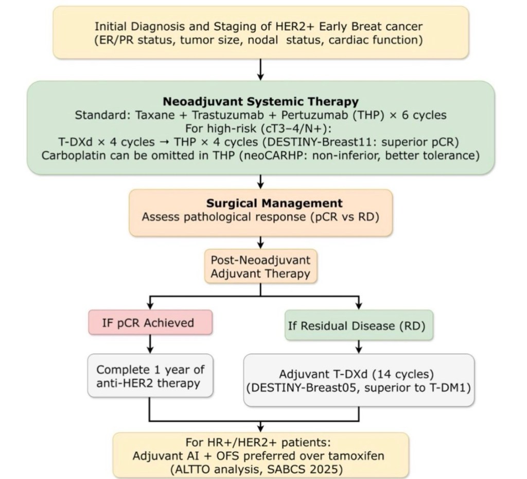
DESTINY-Breast11
T-DXd followed by THP (docetaxel + trastuzumab + pertuzumab) demonstrated:
Significantly higher pathologic complete response (pCR) rates compared with standard anthracycline-based regimens A chemotherapy-sparing strategy with reduced anthracycline exposure Favorable tolerability profile consistent with prior T-DXd data
📊 Early reports show pCR rates approaching ~65–70%, exceeding historical benchmarks for standard neoadjuvant regimens (typically ~55–60%).
Clinical Implication:
We may be entering an era of antibody–drug conjugate (ADC)-based neoadjuvant intensification, potentially redefining the backbone of HER2-directed therapy.
Reference:
Hurvitz SA et al. DESTINY-Breast11. Presented at ESMO 2024 / SABCS 2024 (late-breaking data).
🔹 Adjuvant Setting
DESTINY-Breast05
For patients with residual invasive disease after neoadjuvant therapy, T-DXd demonstrated:
53% reduction in risk of invasive disease–free survival (iDFS) events compared with T-DM1 Superior invasive disease–free survival Manageable toxicity, with ILD rates consistent with prior experience
This builds upon the paradigm established by KATHERINE, where T-DM1 replaced trastuzumab in patients with residual disease.
Now, T-DXd appears poised to replace T-DM1 in this high-risk population.
Reference:
DESTINY-Breast05. Presented at ASCO 2025.
von Minckwitz G et al. KATHERINE trial. NEJM. 2019;380:617–628.
🔬 Why This Matters
We are witnessing:
A shift from monoclonal antibodies → ADC-based escalation Earlier deployment of highly potent HER2-directed agents Refinement of risk-adapted therapy based on response
If adopted into guidelines (NCCN, ASCO, ESMO), this could:
Redefine the management of residual disease Potentially reduce recurrence risk further in high-risk HER2+ EBC Change neoadjuvant sequencing strategies
⚠️ Considerations
ILD/pneumonitis risk requires vigilance Cost-effectiveness and long-term survival data pending Optimal sequencing with pertuzumab still being clarified
📌 Bottom Line
T-DXd is no longer just a metastatic drug.
It is rapidly reshaping the curative-intent HER2+ early breast cancer algorithm.
Choledochal Cysts – Types and Management
Choledochal cysts are congenital cystic dilatations of the biliary tree. They are associated with an abnormal pancreaticobiliary junction and carry a significant lifetime risk of malignancy (especially cholangiocarcinoma).
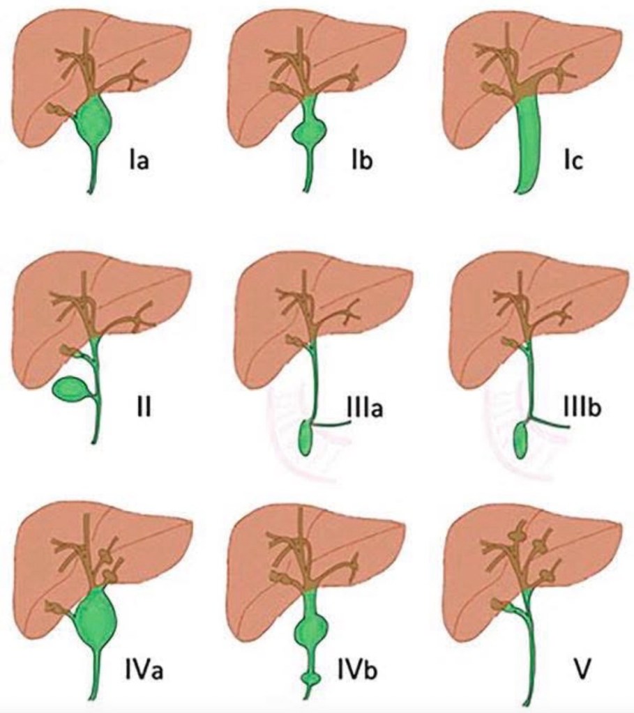
Classification (Todani Classification)
The most widely used system is the Todani classification, which divides choledochal cysts into five main types:
Type I – Extrahepatic bile duct dilatation (most common, 50–80%)
• Ia – Diffuse cystic dilatation of CBD
• Ib – Focal segmental dilatation
• Ic – Fusiform dilatation of CBD
Management:
→ Complete excision of extrahepatic bile duct + Roux-en-Y hepaticojejunostomy
Type II – True diverticulum of CBD
• Saccular outpouching from extrahepatic bile duct
Management:
→ Diverticulectomy ± primary closure of CBD
Type III – Choledochocele
• Intraduodenal dilatation of distal CBD (within ampulla)
Management:
→ Endoscopic sphincterotomy (often sufficient)
→ Surgical excision if large/symptomatic
Type IV – Multiple cysts
• IVa – Both intrahepatic and extrahepatic involvement
• IVb – Multiple extrahepatic cysts only
Management:
→ Excision of extrahepatic bile duct + Roux-en-Y hepaticojejunostomy
→ Liver resection if localized intrahepatic disease
→ Liver transplant if diffuse severe intrahepatic disease
Type V – Caroli Disease
• Multiple intrahepatic cystic dilatations only
Associated with congenital hepatic fibrosis.
Management:
→ Segmental liver resection (localized)
→ Liver transplantation (diffuse disease)
Clinical Presentation
• Children: classic triad (rarely complete)
• Abdominal pain
• Jaundice
• Palpable mass
• Adults:
• Recurrent cholangitis
• Pancreatitis
• Biliary colic
• Incidental finding
Investigations
• Ultrasound – initial test
• MRCP – investigation of choice
• CT if malignancy suspected
• LFTs
ERCP mainly therapeutic (type III).
Complications
• Cholangitis
• Pancreatitis
• Stones
• Strictures
• Rupture (rare)
• Cholangiocarcinoma (10–30% lifetime risk if untreated)
Principles of Management (Important for Practice)
Surgical Standard Operation
Cyst excision + Roux-en-Y hepaticojejunostomy
→ Gold standard for Type I and IV
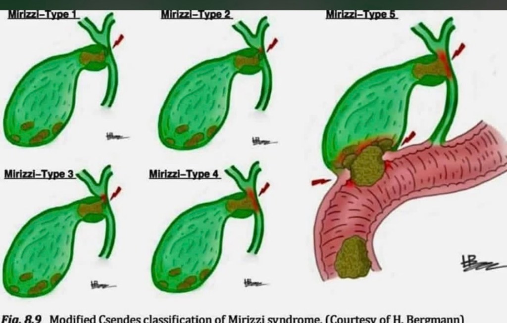
Mirizzi Syndrome: The rare but challenging complication where an impacted gallstone in the cystic duct or Hartmann’s pouch causes external compression or fistulization into the common bile duct. The modified Csendes classification grades severity from Type 1 (external compression only) through Type 5 (cholecystobiliary fistula with gallstone ileus). Type 1 shows simple compression without fistula formation. Type 2 involves erosion affecting less than one-third of the bile duct circumference. Type 3 extends to involve one-third to two-thirds of the duct. Type 4 shows complete destruction of the bile duct wall. Type 5 adds the complication of cholecystoenteric fistula with gallstone ileus. Recognition is critical during cholecystectomy as misidentification can lead to bile duct injury. Higher types require bile duct reconstruction
New 5-Year Evidence Supporting Radiofrequency Ablation (RFA) in Early-Stage Breast Cancer
I’m pleased to share results from the RAFAELO Phase 3 multicenter trial — published online in Annals of Surgical Oncology (Feb 18, 2026) — assessing radiofrequency ablation (RFA) as a minimally invasive alternative to partial mastectomy in early-stage breast cancer.
🔍 Study Overview
• Design: Multicenter, single-arm, Phase 3 clinical study.
• Population: 370 women with solitary Tis–T1 (≤1.5 cm), N0M0 breast carcinomas.
• Intervention: Percutaneous RFA followed by whole-breast radiation (45–60 Gy).
• Primary Endpoint: 5-year ipsilateral breast tumor recurrence-free survival (IBTRFS).
📈 Key Findings
✔ At 5 years, IBTRFS was 98.6% (90% CI 97.1–99.3%), exceeding the pre-specified noninferiority margin of 90%.
✔ Only 2 ipsilateral recurrences were observed at 5 years.
✔ Grade ≥3 skin ulceration was rare (1/370 patients), underscoring a favorable safety profile.
✔ These results suggest that RFA with adjuvant radiation may be comparable to partial mastectomy in appropriately selected early-stage patients.
🏷 Clinical Significance
This large prospective trial provides the most robust long-term evidence to date that RFA — a less invasive approach — may be a viable local-control strategy in small, node-negative breast cancers. These findings reinforce ongoing interest in expanding treatment options that balance oncologic safety with patient-centred care (e.g., cosmesis, procedural morbidity).
Optional Add-Ons for Engagement
🔹 Thanks to the RAFAELO Study Group and contributing centers for advancing patient-centred oncology.
🔹 Looking forward to longer follow-up, quality-of-life data, and comparative trials against standard surgery
Paper summary (Eur Arch Otorhinolaryngol, 2026) — “The impact of drains on surgical outcomes in thyroid surgery”
This is a meta-analysis of randomized controlled trials comparing drain vs no drain after adult thyroid surgery (search Jan 1995–Aug 2025). It included 10 RCTs (n=1,078) and assessed haematoma/seroma (primary) plus SSI, return-to-theatre, pain, and length of stay.
Key findings
No significant difference with drains for: Haematoma (p=0.15) Seroma (p=0.64) Return-to-theatre (p=0.22) Drains were associated with worse outcomes: Higher SSI (4.2% vs 0.5%, p=0.01) Longer LOS (≈ +1.2 days, p<0.0001) More pain (MD ≈ +2.2, p=0.001)
Conclusion of the authors: routine drains don’t reduce clinically important collections/bleeding outcomes and should be selective/patient-specific.
Additional high-yield evidence on the same question
Systematic reviews
2017 meta-analysis (14 studies, n=1,927): drains increased infection and length of stay, with no significant differences in haematoma/seroma or RLN palsy/hypoparathyroidism. Cochrane review: highlights the key limitation of drains—they can block with clot and do not replace meticulous haemostasis / re-exploration when bleeding occurs; overall evidence did not support routine use.
Randomized trials (examples)
2013 RCT (Uganda, n=68): no-drain group had shorter LOS and less pain, with no signal that drains prevented important complications. 2023 RCT (lobectomy + central neck dissection, n=104): no routine drain needed; no-drain group had shorter LOS and better comfort metrics.
Evidence-based recommendation (practical)
1) Default position
For uncomplicated thyroidectomy/hemithyroidectomy, the best available RCT/meta-analysis evidence supports NO routine drain because it does not reduce haematoma/seroma and does increase SSI, pain, and LOS.
2) When a drain may be reasonable (selective use)
Consider a drain selectively when you believe a drain will meaningfully manage expected ongoing output or permit monitoring in a high-risk scenario, e.g.:
Extensive dissection / large dead space (e.g., combined procedures, broad flap elevation) Significant intraoperative oozing despite optimization (coagulopathy, difficult hemostasis) Reoperative thyroid surgery Very large goiter/substernal component (case-dependent) Neck dissection / lateral compartment work (many surgeons drain these by default; note: classic drain trials often exclude lateral neck dissections)
(Even in these settings, it’s worth emphasizing: drains don’t “prevent” a dangerous post-thyroidectomy hematoma—rapid recognition and evacuation remain key, and drains may clot off.)
3) What to do instead of routine drains (high-impact steps)
Meticulous hemostasis + Valsalva before closure Layered closure / dead-space minimization Standardized post-op neck checks and early warning protocol (swelling, tightness, voice change, stridor) Clear hematoma pathway (immediate bedside opening vs OR depending on severity/resources)

A cutting-edge study published in npj Breast Cancer today reports validation of the HER2DX genomic test as a robust prognostic tool in first-line advanced HER2-positive breast cancer treated with trastuzumab, pertuzumab, and a taxane (THP).
🔬 What was done: Researchers combined real-world data from 215 patients across Spanish and Polish cohorts. They assessed baseline tumor tissue using the HER2DX assay to derive genomic scores linked to outcomes in patients receiving standard first-line HER2-targeted therapy (THP).
📊 Key findings:
• A high ERBB2 mRNA score was associated with significantly longer progression-free and overall survival, as well as higher objective response rates — independent of traditional clinical variables.
• The team developed a HER2DX metastatic prognostic score that outperformed ERBB2 mRNA levels alone in predicting outcomes, suggesting genomic profiling can further refine risk stratification in advanced disease.
📈 Implications: This study supports the clinical utility of HER2DX in identifying patients with HER2-positive advanced breast cancer who might derive the greatest benefit from existing first-line therapies — and highlights the growing role of genomic tools in precision oncology.
Rodrigo Arrangoiz, MD
Surgical Oncologist, Mount Sinai Medical Center (MSMC)
Head & Neck and Breast Cancer Specialist
https://www.nature.com/articles/s41523-026-00909-0
