My name is Rodrigo Arrangoiz I am a breast surgeon/ thyroid surgeon / parathyroid surgeon / head and neck surgeon / surgical oncologist that works at Center for Advanced Surgical Oncology in Miami, Florida.
I was trained as a surgeon at Michigan State University from (2005 to 2010) where I was a chief resident in 2010. My surgical oncology and head and neck training was performed at the Fox Chase Cancer Center in Philadelphia from 2010 to 2012. At the same time I underwent a masters in science (Clinical research for health professionals) at the University of Drexel. Through the International Federation of Head and Neck Societies / Memorial Sloan Kettering Cancer Center I performed a two year head and neck surgery and oncology / endocrine fellowship that ended in 2016.
Mi nombre es Rodrigo Arrangoiz, soy cirujano oncólogo / cirujano de tumores de cabeza y cuello / cirujano endocrino que trabaja Center for Advanced Surgical Oncology en Miami, Florida.
Fui entrenado como cirujano en Michigan State University (2005 a 2010 ) donde fui jefe de residentes en 2010. Mi formación en oncología quirúrgica y e n tumores de cabeza y cuello se realizó en el Fox Chase Cancer Center en Filadelfia de 2010 a 2012. Al mismo tiempo, me sometí a una maestría en ciencias (investigación clínica para profesionales de la salud) en la Universidad de Drexel. A través de la Federación Internacional de Sociedades de Cabeza y Cuello / Memorial Sloan Kettering Cancer Center realicé una sub especialidad en cirugía de cabeza y cuello / cirugia endocrina de dos años que terminó en 2016.
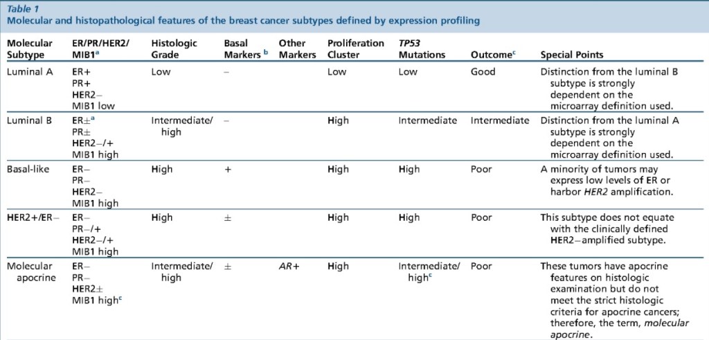
Following the description of the main intrinsic breast cancer molecular subtypes based on gene expression analysis:
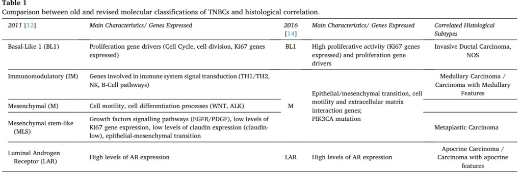
Lehmann et al. demonstrated the complex and heterogeneous nature of TNBCs, and that their definition simply based on interpretation of histopathological features might be of limited efficacy in understanding prognostic behaviour and therapeutic implications
They described six subtypes of TNBCbased on gene expression analysis of 21 breast cancer data sets:
Basal-like type 1 (BL1):
Characterized by high proliferative activity demonstrated by elevated Ki67 mRNA expression
Basal-like type2 (BL2):
Showing basal-myoepithelial phenotype
Immunomodulatory (IM) subtype:
That includes gene ontologies related to immune cell processes involved in immune signal transduction (such as TH1/TH2, NK and B-cell receptor pathways), in absence of significant correlation with stromal inflammatory cell infiltrate
Mesenchymal (M) subtype:
That displays genetic patterns responsible for cell motility and cell differentiation processes (i.e. Wnt pathway, ALK pathway)
Mesenchymal stem-like (MLS) subtype:
Shows genetic profiles associated with growth factors signalling pathways (i.e. EGFR, PDGF) and, particularly, low rates of proliferation genes and low expression of claudins’ family proteins (lately described as claudin-low cancer subtype)
LAR subtypes (luminal androgen receptor subtype):
Express high levels of androgen receptor hormones (AR) and correlates also with tumors showing apocrine differentiation on histologic examination
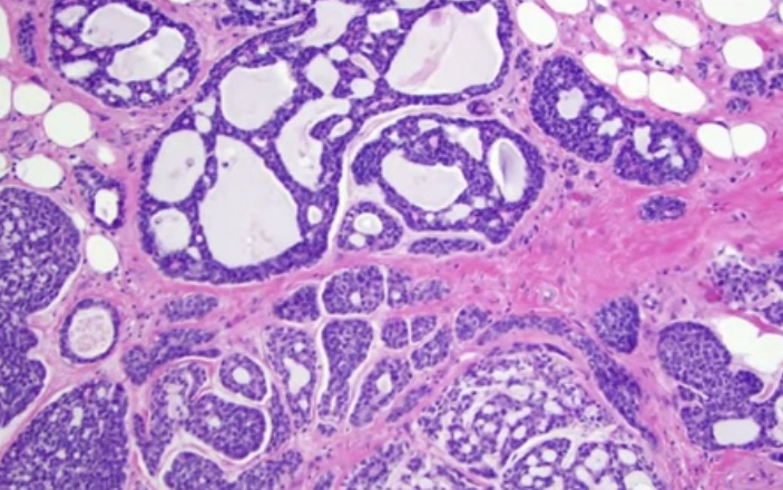
Exhibit tubular, trabecular, cribriform, and / or solid patterns
Cribriform is the classic pattern
Characterized by MYB-NFIB t(6;9)(q22-23;p23-24)
Adenoid Cystic Carcinoma of the BreastTumor Infiltrating Lymphocytes (TIL)
Tumor infiltrating lymphocytes (TIL):
Recommendations for assessing TILS in breast cancer:
Evaluated for the stromal component (% of stromal TIL)
Evaluated with the borders of the invasive tumor
Exclude TILs outside of the tumor border, around DCIS and normal lobules
Lymphocytes and plasma cells, exclude neutrophils
Full sections are preferred over biopsies:
Cores can be used in the pre therapeutic neoadjuvant setting
Average TILs in the tumor area (do not focus on hotspots)
The number of TILS correlate with complete pathologic response in the neoadjuvant setting
No formal recommendations for a clinically relevant TILS threshold(s) can be given at this stage
PD-L1 and Breast Cancer:
The PD-L1 on tumor cells, when combined with its PD-1 on immune cells:
Causes an inhibition of immune response mediated by CD8+ T cells
Breast tumor that have PD-L1 tend to have high number of TILs, and the majority are of the triple negative type
Tumors arising in BRCA 1 carriers:
BRCA 1 is involved in:
DNA repair
Cell cycle regulation
Transcriptional regulation
Chromatin remodeling
Loss of BRCA 1 leads to:
Deficiency in repair of DNA doble-strand breaks
75% of all tumors developing in BRCA 1 germ line mutation carriers are TNBC:
High histologic grade
High proliferation rate
Residual cancer burden after neoadjuvant chemotherapy (NAC):
Parameters required to calculate residual cancer burden (RCB):
Submission of the entire area of the tumor bed
Tumor dimensions (at least in two dimensions)
Percentage invasive carcinoma in the tumor bed
Percentage of the in situ carcinoma in the tumor bed
The number of positive lymph nodes
The largest diameter of nodal metastasis
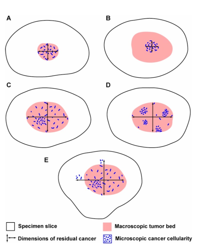
In these diagrams, the macroscopic tumor bed dimensions in examples A, C, D also define the final dimensions of the residual tumor bed after microscopic review. However, the macroscopic tumor bed dimensions in example B overestimate the extent of residual cancer, and so the dimensions of the residual tumor bed (d1 and d2) would be revised after microscopic evaluation of the extent of residual cancer in the corresponding slides from the gross tumor bed. In a different example (E), microscopic residual cancer extends beyond the confines of the macroscopic tumor bed. Again, the dimensions of the residual tumor bed (d1 and d2) would be revised after microscopic evaluation of the recognizable extent of residual cancer beyond the macroscopic tumor bed. This approach accounts for differences in the concentration and distribution of residual cancer within a tumor bed. In the illustration above, the estimated % CA in example A would be high (in a small area), whereas the estimated % CA for examples C and D would be lower (in a larger area). In examples C and D, the estimated % CA would likely be similar, even though the distribution of cancer within the residual tumor bed is different in those two examples.A practical way to estimate % CA in a slide is to encircle with ink dots the tumor bed on each slide from the grossly defined residual tumor bed (e.g., slides A1-A5 in the example above). Then use the microscope to estimate the cellularity in each microscopic field across the area of tumor bed. In each microscopic field, % CA can be estimated by comparing the proportion of residual tumor bed area containing cancer (invasive or in situ). Estimate an average of the readings for % CA in the cross-sectional area. The same can be done for in situ component (% CIS). Estimates are to the nearest 10%, but include 0%, 1%, and 5% for areas with low cellularity. The average cellularity within the tumor bed from each slide across the tumor bed can then be estimated (illustrated above).
It is recommended to repeat ER, PR, and HER2 on invasive TNBC after neoadjuvant therapy
Distant metastasis in patient with residual disease after NAC:
Factors associated with increased distant metastatic rate:
Triple negative breast cancer (TNBC) is a subtype of breast tumor lacking hormone receptors expression and HER2 gene amplification:
Represents 24 % of newly diagnosed breast neoplasms
TNBC is usually characterized by poor prognosis and lack of wide choice therapeutic agents due to the absence of targetable hormone receptors and HER2 expression:
Therefore is considered a very interesting and challenging topic for breast cancer research
TNBC is a functional term that defines a wide spectrum of entities:
With different biology and clinical behavior, with marked genetic, transcriptional, histologic and clinical differences
The definition of a new classification for breast cancer based on its gene expression pattern divided breast tumors into four “intrinsic subtypes”:
Luminal subtype:
Divided in Luminal A and Luminal B:
Characterized by estrogen receptor gene expression
The HER2 subtype:
Characterized by HER2 gene amplification
The so-called basal-like subtype:
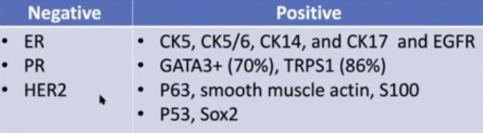
A particular breast cancer showing positivity for basal and myoepithelial markers and lack of hormone receptors and HER2 gene amplification
“Normal breast-like” subtype:
With triple-negative phenotype but cellular derivation typical of normal breast epithelium
Notably, both the basal-like and normal breast-like subtypes were already recognized as triple negative
All breast cancers arise in the terminal duct lobular units (the functional unit of the breast) of the collecting duct
The histological and molecular characteristics:
Have important implications for therapy:
Several classifications on the basis of molecular and histological characteristics have been developed
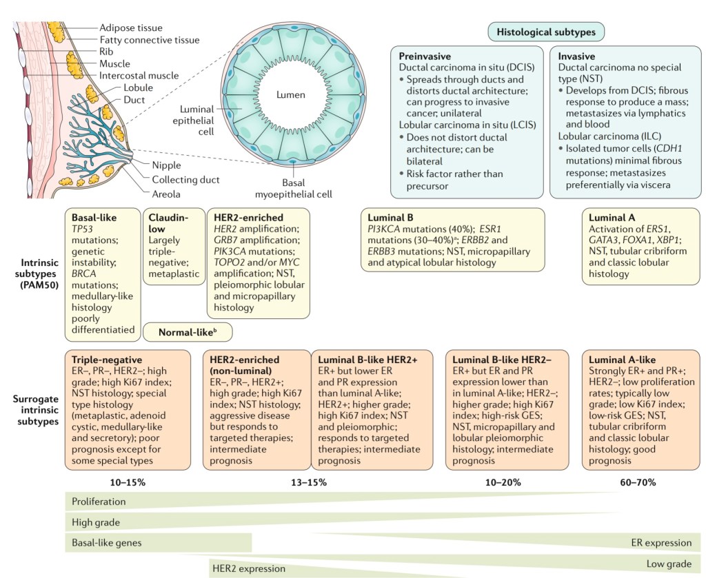
The histological subtypes described here (Figure) are the most frequent subtypes of breast cancer:
Ductal carcinoma (now referred to as ‘no special type’ (NST)) and lobular carcinoma:
Are the invasive lesions
Their pre-invasive counterparts are:
Ductal carcinoma in situ and lobular carcinoma in situ (or lobular neoplasia), respectively
The intrinsic subtypes of Perou and Sorlie:
Are based on a 50-gene expression signature (PAM50)
The surrogate intrinsic subtypes are typically used clinically and are based on histology and immunohistochemistry expression of key proteins:
Estrogen receptor (ER), progesterone receptor (PR), human epidermal growth factor receptor 2 (HER2) and the proliferation marker Ki-67
Tumors expressing ER and / or PR are termed ‘hormone receptor-positive’
Tumors not expressing ER, PR and HER2 are called ‘triple-negative’
The normal breast terminal duct lobular unit:
It is formed by two layers:
Inner or luminal layer (epithelial cell layer)
Outer or basal layer (myoepithelial cell layer)
Cancer can arise from both of this layers
Molecular subtypes that are triple negative:
Basal-like:
TP53 mutations
Genetic Instability
BRCA mutations
Medullary-like histology
Poorly differentiated
Claudin-low:
Largely triple negative
Metaplastic
Normal breast-like
Molecular apocrine
Interferon rich
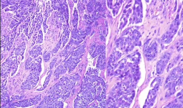
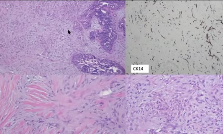
We can classify TNBC according to the grade of the tumor. Representative micrographs of low-grade and high- grade variants of TNBC.Histologic Types of TNBCs and their key genetic features/potential therapeutic targets.
TNBC can be classified into:
Low-grade and high-grade histologic types
Several histologic types of low-grade TNBC, including:
Salivary gland-like tumors of the breast and solid papillary carcinoma with reverse polarity:
Are underpinned by specific / pathognomonic genetic alterations
In contrast, acinic cell carcinoma and high-grade variants of TNBC have somatic genomic landscape similar to those of conventional TNBC
Low- grade variants of metaplastic breast carcinomas (MBCs) are unlikely to be underpinned by specific genetic alterations; however, the genetic analyses performed to date included only a few or single cases
Progression to high-grade TNBC has been described in most low-grade forms of TNBC:
However, it occurs at a different rate
Whereas fairly common in acinic cell carcinoma, it is a rare event in the salivary gland-like tumors of the breast and solid papillary carcinoma with reverse polarity
It should be noted that evidence for the presence of PRKD1 E710D mutations or PRKD1/2/3 rearrangements in polymorphous carcinoma of the breast remains to be documented
Substantially reduces the risk of developing invasive breast cancer in patients with lobular carcinoma in situ (LCIS) and should be offered
Negative margins are generally not required for classic LCIS:
Whereas they are recommended for:
Pleomorphic LCIS
The patient’s future risk of developing breast cancer is:
Approximately 1% annually
Bilateral prophylactic mastectomy:
Is generally considered to be more invasive than is required in this setting and, although it could be discussed as an option, should not be routinely recommended
References:
Page DL, Kidd TE Jr, Dupont WD, Simpson JF, Rogers LW. Lobular neoplasia of the breast: higher risk for subsequent invasive cancer predicted by more extensive disease. Hum Pathol. 1991;22(12):1232-1239.
Andersen JA. Lobular carcinoma in situ of the breast. An approach to rational treatment. Cancer. 1977;39(6):2597-2602.
Akashi-Tanaka S, Fukutomi T, Nanasawa T, Matsuo K, Hasegawa T, Tsuda H. Treatment of noninvasive carcinoma: fifteen-year results at the National Cancer Center Hospital in Tokyo. Breast Cancer. 2000;7(4):341-344.
Walt AJ, Simon M, Swanson GM. The continuing dilemma of lobular carcinoma in situ. Arch Surg. 1992;127(8):904-907.
Haagensen CD, Bodian C, Haagensen DE. Neoplasia (lobular carcinoma in situ). In Breast Carcinoma: Risk and Detection. Philadelphia, PA: WB Saunders, 1981.
Wong SM, King T, Boileau JF, Barry WT, Golshan M. Population-based analysis of breast cancer incidence and survival outcomes in women diagnosed with lobular carcinoma in situ. Ann Surg Oncol. 2017;24(9):2509-2517.
Fisher ER, Land SR, Fisher B, Mamounas E, Gilarski L, Wolmark N. Pathologic findings from the National Surgical Adjuvant Breast and Bowel Project: twelve-year observations concerning lobular carcinoma in situ. Cancer. 2004;100(2):238-234.
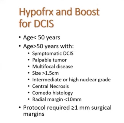
Hypofractionation whole breast irradiation (WBI) may be used as an alternative to conventional fractionation (CF) CF-WBE in patients with DCIS
Recommendation strength: conditional
Quality of evidence: Moderate
Consensus: 86%
Age, grade, and margins for DCIS:
Statement KQ2D:
A tumor boost may be used for patients with DCIS who meet any of the following criteria:
Age =/< 50 years
High grade
Close (< 2 mm) or positive margins
Recommendation strength: conditional
Quality of evidence: Moderate
Consensus: 92%
This two statements from ASTRO rely of data from two randomized trials:
The DBCG Hypo Trial:
Entry criteria:
> 40 years of age
BCS for node-negative breast cancer
DCIS (13% of the cohort)
Primary endpoint:
Grade 2 to 3 breast induration assuming no inferiority regarding locoregional recurrence
Median follow-up of 7.26 years
The local control were the same between hypofractionation vs conventional fractionation.Grade 2 to 3 induration rates were similar between hypofractionation vs conventional fractionation.BIG 3-07 / TROG 07.01 Trial
BIG 3-07 / TROG 07.01:
Background:
Whole breast irradiation (WBI) after conservative surgery for ductal carcinoma in situ (DCIS) reduces local recurrence.
They investigated whether a tumor bed boost after WBI improved outcomes, and examined radiation dose fractionation sensitivity for non-low-risk DCIS.
Methods:
The study was an international, randomized, unmasked, phase 3 trial involving 136 participating centres of six clinical trials organisations in 11 countries (Australia, New Zealand, Singapore, Canada, the Netherlands, Belgium, France, Switzerland, Italy, Ireland, and the UK).
Eligible patients were women aged 18 years or older with unilateral, histologically proven, non-low-risk DCIS treated by breast-conserving surgery with at least 1 mm of clear radial resection margins.
They were assigned to one of four groups (1:1:1:1) of no tumour bed boost versus boost after conventional versus hypofractionated WBI, or randomly assigned to one of two groups (1:1) of no boost versus boost after each center prespecified conventional or hypofractionated WBI.
The conventional WBI used was 50 Gy in 25 fractions, and hypofractionated WBI was 42.5 Gy in 16 fractions. A boost dose of 16 Gy in eight fractions, if allocated, was delivered after WBI.
Patients and clinicians were not masked to treatment allocation. The primary endpoint was time to local recurrence.
Findings:
Between June 25, 2007, and June 30, 2014, 1608 patients were randomly assigned to have no boost (805 patients) or boost (803 patients).
Conventional WBI was given to 831 patients, and hypofractionated WBI was given to 777 patients.
Median follow-up was 6.6 years.
The 5-year free-from-local-recurrence rates were 92.7% (95% CI 90·6-94·4%) in the no-boost group and 97.1% (95·6-98·1%) in the boost group (hazard ratio 0·47; 0·31-0·72; p<0·001).
The boost group had higher rates of grade 2 or higher breast pain (10% [8-12%] vs 14% [12-17%], p=0·003) and induration (6% [5-8%] vs 14% [11-16%], p<0·001).
Interpretation:
In patients with resected non-low-risk DCIS, a tumor bed boost after WBI reduced local recurrence with an increase in grade 2 or greater toxicity.
The results provide the first randomised trial data to support the use of boost radiation after postoperative WBI in these patients to improve local control.
The international scale of the study supports the generalizability of the results.
Radiation Techniques for DCIS:
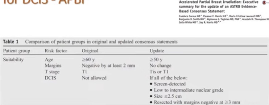
Accelerated partial breast irradiation:
ASTRO Evidence Based Consensus Statement
Summary:
Moderately hypo-fractionation WBI is a standard treatment
Consider boost for:
High grade DCIS
> 2 cm tumors
Positive or < 2 mm margins
Pre-menopausal patients
Patients less than 50 years of age
APBI in DCIS is safe and effective option in ASTRO “suitable”candidates
Not all techniques (just validated with external beam radiation)
Consider hormone therapy in aggressively minded patients or those wishing to decrease risk of contralateral breast cancer
Consider genomic assay assistance to aid in radiation decisions in select patients:
Postmenopausal patients with otherwise low-risk disease
Started to think if there is a sub group of patients that we can omit radiation therapy
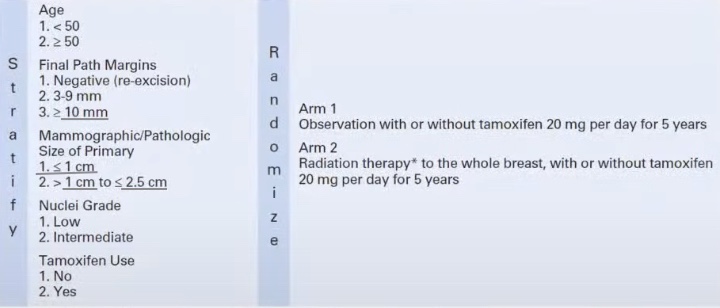
This trials looked into high risk factors for local regional recurrence (LRR) in DCIS:
Prognostic factors associated with increased locoregional recurrence:
Age less than 50 years
Size > than 2 cm
Grade 3
Positive of close < margins
Additional risk factors:
Symptomatic:
Palpable / bloody discharge
Comedo, solid types of DCIS
Black race
ER and / or PR negative
What is the optimal surgical margin in DCIS?
The use of a 2-mm margin as the standard for an adequate margin in DCIS treated with whole-breast irra- diation is associated with lower rates of IBTR and has the potential to decrease re-excision rates, improve cosmetic outcomes, and decrease health care costs:
Clinical judg- ment should be used in determining the need for further surgery in patients with negative margins narrower than 2 mm.
The second generation DCIS trials:
RTOG 9804:
They included:
Mammographically detected DCIS
Low to intermediate-grade DCIS
Less than 2.5 cm
Margins =/> than 3 mm
This study has two different perspectives:
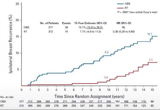
Although the results are significant:
Local recurrence (LR) rates without RT are less than 10%
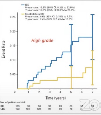
Longer follow-up of the RTOG 9804 trial: LR rates were as high as 15.1% in omitted radiation arm
The second generation DCIS trials:
ECOG E5194:
From 1997 to 2022:
Has only one arm
Inclusion criteria:
Non-palpable DCIS
Cohort 1:
Low to intermediate grade DCIS < than 2 cm
Cohort 2:
High grade DCIS < than 1 cm
> 3 mm margins / no residual calcifications on postoperative mammogram
Lumpectomy alone:
NO radiation
Starting in the year 2000:
Patients could take tamoxifen
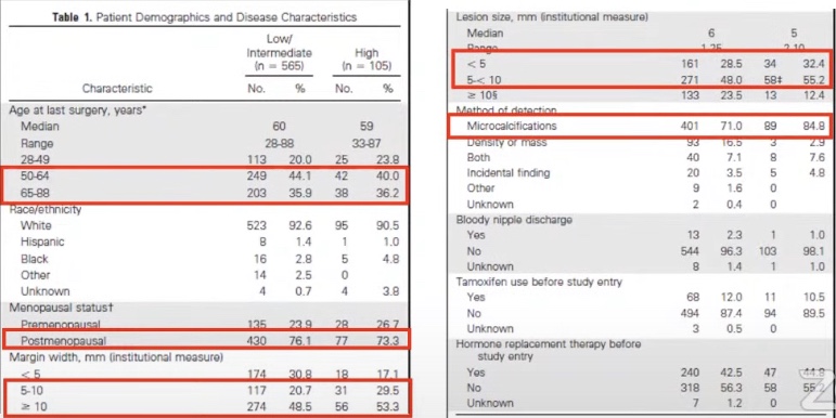
Like in the RTOG 9804 trial patients in the ECOG E5194 trial tended to be older, postmenopausal, have wider margins of resection, and had smaller tumors (detected on mammogram).With long-term follow-up in the ECOG E5194 the LR rates in high risk DCIS were almost 25% and in lower risk DCIS was 14.4%, but local control rates were still high and these data should be taken into account in share decision making.
What can we use to help us determine risk of recurrence in DCIS?
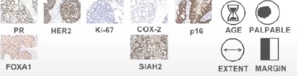
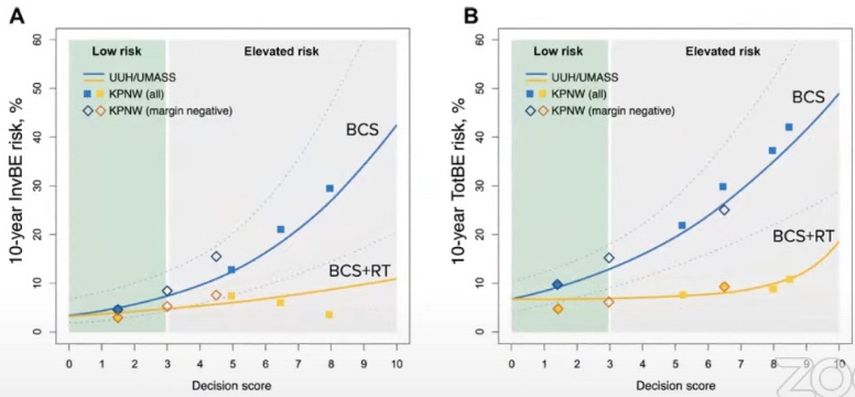
Genomic Assays – DCISion RT (Prelude DX):
Seven gene biological risk signature developed from three cohorts:
UCSF (n=324)
Uppsala Univeristy Hospital, Sweden (n=458)
University of Massachusetts (n=300)
Decision score 0 to 10:
Low risk 0 to 3
High risk score > 3 to 10
This biological signature was validated in a retrospective cohort
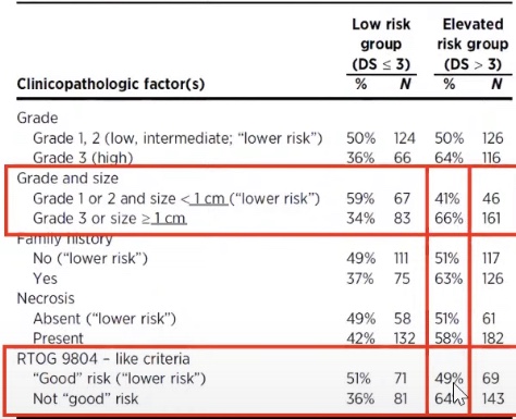
The patients that were defined as low risk by clinical and pathological factors were could actually be higher risk patients based on genetic analysis in 41% to 49% of the cases. 34% to 36% of high risk DCIS patients were found to be low risk by DCISion RT.
This tool is available but it has not be validated in a prospective clinical trial
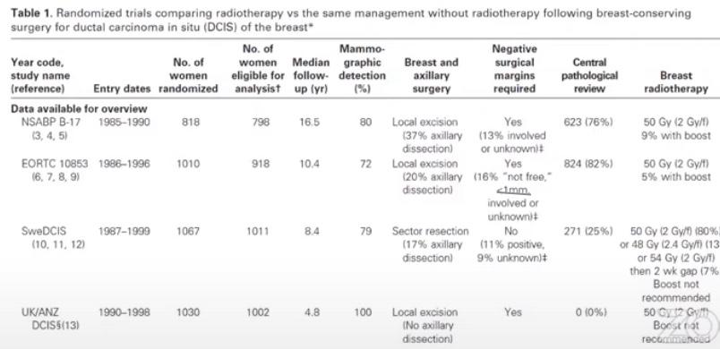
Seminal first generation trials of radiation therapy after breast conserving surgery (BCS) for DCIS:
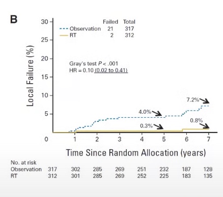
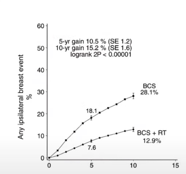
Meta-Analysis of DCIS Trials. These trials showed that radiation therapy in DICS significantly decreased local recurrence rates of both DCIS and IBC.
Radiation therapy is associated with a roughly 50% relative risk reduction in local recurrence and absolute risk reduction of 15%:
In these studies most patients did not receive tamoxifen
The first generation seminal trials:
Were conducted in the 1980’s and 1990’s
Many patients had symptomatic DCIS:
With palpable tumors of bloody nipple discharge:
Screening mammogram was uncommon
10% to 20% of patients had a positive or close margins
Greater than 1/3 of the patients had high grade DCIS
The DCIS patients that we see in our clinics today are:
Smaller, low grade, and are diagnosed earlier:
Because of the screening mammograms
The effect of tamoxifen on local control:
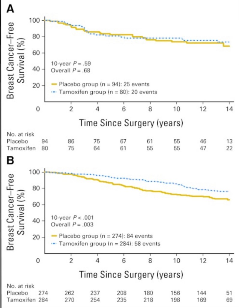
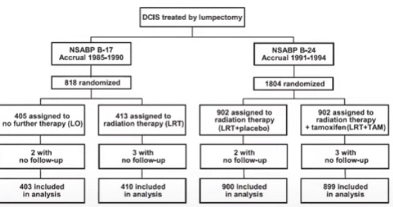
NSABP B 24. Lumpectomy + RT vs. Lumpectomy + RT + Tamoxifen. A. Estrogen receptor negative patients B. Estrogen receptor positive patients
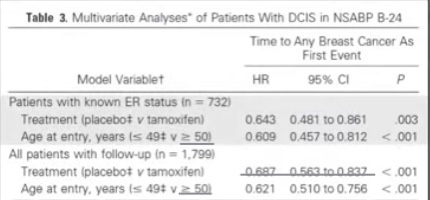
In the NSABP B 24 trial in multivariable analysis:
Young age and no tamoxifen were adverse prognostic factors
Long-term outcomes of invasive ipsilateral breast cancer recurrences after lumpectomy in the NSABP B-17 and B-24 randomized trials for DCIS:
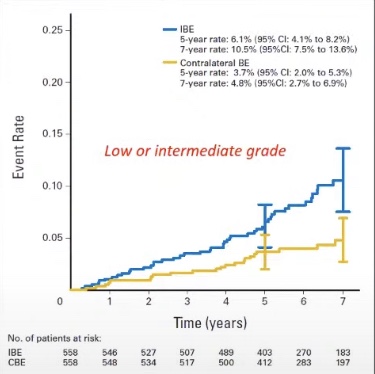
The cumulative analysis of these trials showed that radiation therapy has a relatively greater impact on preventing local recurrence than tamoxifen. Tamoxifen plays a role in significantly decreasing the risk of contralateral breast cancer.
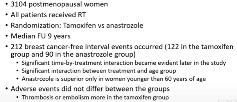
Anastrozole vs Tamoxifen in DCIS (IBIS-II DCIS Trial):
2980 postmenopausal women
71% of the patients received radiation therapy
Randomization:
Anastrozole vs tamoxifen
Median follow-up:
7.2 years
144 breast cancer recurrences
The non-inferiority of anastrozole was established:
But its superiority to tamoxifen was not
Adverse effects:
Anastrozole:
More fractures, musculoskeletal events, hypercholesterolemia, and strokes
Tamoxifen:
More muscle spasms, gynecological cancers and symptoms, vasomotor symptoms, and deep vein thromboses
NSABP B-35 Trial
Summary of radiation therapy in DCIS:
If prevention of ipsilateral breast tumor recurrence (IBTR) is the primary goal:
Tamoxifen cannot replace the impact of radiation therapy on local control after BCS
Tamoxifen may further decrease local regional recurrence risk after BCS therapy with radiation and significantly reduce contralateral breast cancer
In postmenopausal women less than 60 years of age:
Instead of trying to identify patients with underlying invasion:
Let’s find a way to mark the sentinel lymph node (SLN) in advance
Senti nel Lymph Node Dissection in DCIS and how to
Not to do it
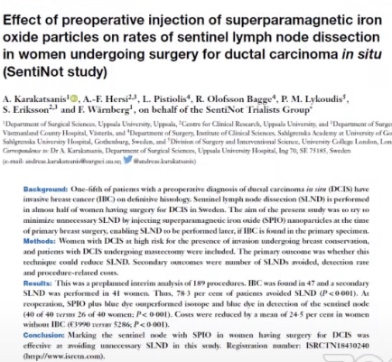
The SentiNot Trial
Concept:
Inject supraparamagnetic iron oxide particles (SPIO) at the primary operation:
Mark the SLN but do not remove it
If pathology report shows invasion:
Then we can go back and perform the SLNB
A multicenter cohort study:
Has been completed
Multicenter randomized trial:
Is ongoing
Inclusion criteria: Grade 2, > 20 mm, any grade 3, mass forming DCIS, Andy DCIS planned for mastectomyThe detection rate in the WLE were not different but in the setting of OPBCT and mastectomy the detection rate was inferior to standard technique The procedure was not accurate in any setting others than WLE
Nipple blebs are caused by trauma from shallow infant latch
Science:
Blebs appear as:
Small white, yellow, or red blisterlike lesions on the surface of a nipple
They are inflammatory lesions that may occlude a nipple orifice:
They reflect underlying ductal inflammation and microbiome disruption with biofilm formation
Blebs are associated with:
Hyperlactation (oversupply)
Pumping (which alters the breast microbiome)
C-section births (which also alter the breast microbiome),
Other characteristics of individual variation in microbiome expression
Blebs are not related to infant trauma or latch in any way
Because blebs are very painful:
Moms often believe the infant has a poor latch or otherwise has contributed to the problem:
However, this represents an association rather than causation
Treatment:
Asymptomatic blebs do not require any specific treatment
Blebs causing milk obstruction warrant treatment:
To reduce underlying ductal inflammation and decrease the viscosity of milk
Sunflower lecithin by mouth:
Is effective for breastmilk emulsification and can help to both treat and prevent blebs
Therapeutic ultrasound can also be used to reduce breast inflammation
Symptomatic blebs occluding an orifice:
Should be treated with oral lecithin as well as a topical medium-potency steroid:
Such as 0.1% triamcinolone cream
Blebs should not be routinely unroofed with a sterile needle or other means:
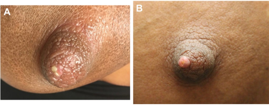
As this may transiently relieve milk obstruction in an associated ductal orifice but will also cause local tissue trauma and can lead to scarring (Figure):
This scarring can result in permanent occlusion of the nipple orifice
Patients should be instructed not to attempt to squeeze out a bleb or pick at it with their fingernails, as this can cause bleeding and further trauma
Nipple bleb at presentation (A) and after chronic tissue trauma from frequent un- roofing (B).