My name is Rodrigo Arrangoiz I am a breast surgeon/ thyroid surgeon / parathyroid surgeon / head and neck surgeon / surgical oncologist that works at Center for Advanced Surgical Oncology in Miami, Florida.
I was trained as a surgeon at Michigan State University from (2005 to 2010) where I was a chief resident in 2010. My surgical oncology and head and neck training was performed at the Fox Chase Cancer Center in Philadelphia from 2010 to 2012. At the same time I underwent a masters in science (Clinical research for health professionals) at the University of Drexel. Through the International Federation of Head and Neck Societies / Memorial Sloan Kettering Cancer Center I performed a two year head and neck surgery and oncology / endocrine fellowship that ended in 2016.
Mi nombre es Rodrigo Arrangoiz, soy cirujano oncólogo / cirujano de tumores de cabeza y cuello / cirujano endocrino que trabaja Center for Advanced Surgical Oncology en Miami, Florida.
Fui entrenado como cirujano en Michigan State University (2005 a 2010 ) donde fui jefe de residentes en 2010. Mi formación en oncología quirúrgica y e n tumores de cabeza y cuello se realizó en el Fox Chase Cancer Center en Filadelfia de 2010 a 2012. Al mismo tiempo, me sometí a una maestría en ciencias (investigación clínica para profesionales de la salud) en la Universidad de Drexel. A través de la Federación Internacional de Sociedades de Cabeza y Cuello / Memorial Sloan Kettering Cancer Center realicé una sub especialidad en cirugía de cabeza y cuello / cirugia endocrina de dos años que terminó en 2016.
Is radiation delivered locally to the resected part of the breast in the setting of lumpectomy
The benefits of APBI include:
Reduced treatment time and the potential to spare radiation to healthy tissue
Evidence to date suggests that survival and local recurrence with APBI is as effective as whole-breast irradiation:
However, careful patient selection is key
Patients suitable for APBI include:
Those ≥ 50 years old
Negative margins (by at least 2mm)
Tis or T1 tumors
Ductal carcinoma in situ (DCIS):
Screen detected
Low to intermediate nuclear grade
≤ 2.5cm
Margins of resection ≥ 3mm
APBI is considered cautionary in:
Patients 40 to 49 years old if all other criteria for suitability are met
Margins < 2mm
DCIS ≤ 3 cm and patients ≥ 50 years if patient has no unsuitable factors and at least 1 of these factors:
Size 2.1 to 3.0cm, T2 tumors, margins <2 mm, limited / focal lymph-vascular space invasion, ER– tumors, clinically unifocal tumors between 2.1-3.0 cm, invasive lobular histology, pure DCIS ≤3cm if criteria for suitability not fully met and EIC ≤3cm.
Patients are deemed unsuitable:
If they are below age 40
Between the ages of 40 to 49 years and do not meet the criteria for cautionary
Positive margins
> 3cm of DCIS
Current ASTRO guidelines do not recommend low energy IORT off prospective study, and electron IORT only for those patients with suitable risk factors
References
Correa C, Harris EE, Leonardi MC, et al. Accelerated partial breast irradiation: executive summary for the update of an ASTRO evidence-based consensus statement. Pract RadiatOncol. 2017;7(2):73-79.
Smith BD, Arthur DW, Buchholz TA, et al. Accelerated partial breast irradiation consensus statement from the American Society for Radiation Oncology (ASTRO). Int J Radiat Oncol Biol Phys. 2009;74(4):987-1001.
The efficacy of elective nodal irradiation (ENI) versus elective neck dissection:
Showed no obvious differences:
As reported by Barkley in a major retrospective experience of neck management in 596 patients with oropharynx, larynx, or hypopharynx carcinoma treated at MD Anderson Cancer Center:
Two hundred thirteen were oropharynx, 202 larynx, and 181 hypopharynx
Of these, 226 were N0 whereas 370 were node positive
Patients underwent radiation therapy (n = 292), surgical management (n = 199), or combined modality therapy (n = 105) of the neck
All patients had a minimum follow-up of 2 years and a median follow-up of 4 years
Among the electively treated patients:
Regional control was greater than 90% regardless of the treatment approach as long as comprehensive neck treatment was implemented
However, partial treatment of the neck resulted in 15%, 35%, and 20% regional failure after radiation, surgery, or combined therapy, respectively
Proton therapy for accelerated partial breast irradiation:
Remains investigational in the treatment of breast cancer
An initial phase 1 / 2 trial evaluating protons for partial breast irradiation:
Found high rates of acute skin toxicity, including:
79% moderate to severe skin color changes
22% moderate / severe desquamation
Nevertheless, physicians and patients alike reported satisfaction with cosmesis
A subsequent phase II trial involving 50 patients had minimal treatment related toxicity and excellent local control at 4-year follow-up, and post-treatment complications were thought to be less than those with more invasive techniques
References
Bush DA, Slater JD, Garberoglio C, Do S, Lum S, Slater JM. Partial breast irradiation delivered with proton beam: results of a phase II trial. Clin Breast Cancer. 2011;11(4):241-245.
Kozak KR, Smith BL, Adams J, et al. Accelerated partial-breast irradiation using proton beams: initial clinical experience. Int J Radiat Oncol Biol Phys. 2006;66(3):691-698.
Verma V, Mishra MV, Mehta MP. A systematic review of the cost and cost-effectiveness studies of proton radiotherapy. Cancer. 2016;122(10):1483-1501.
Preservation of all four parathyroid glands during total thyroidectomy is a critically important operative goal:
But this objective is not always attainable due to the extent of thyroid disease
Plus variations in the anatomical locations and blood supply of the parathyroid glands
Avoiding parathyroid damage:
First requires that the surgeon is able to recognize parathyroid tissue accurately:
The parathyroid glands are difficult to distinguish from other cervical tissues because of their small sizeand similar coloration compared to thyroid, fat, and lymph nodes
The time-honored key to parathyroid identification:
Has been a proactive anticipatory visual approach and use of surgical landmarks
Recent promise for improved parathyroid identification has arisen through:
The intraoperative stimulation of parathyroid tissue fluorescence in the presence of a contrast agent or photosensitizer (indocyanine green, amino levulinic acid hydrochloride [5-ALA], methylene blue)
Intraoperative detection with near-infrared fluorescence imaging
More recent still has been the successful detection of label-free parathyroid autofluorescence with near-infrared fluorescence spectroscopy
A gentle capsular dissection:
That reflects the perithyroidal fatty tissues off the surface of the thyroid allows for preservation of the parathyroid blood supply
This technique requires:
Dissection immediately on the surface of the thyroid gland:
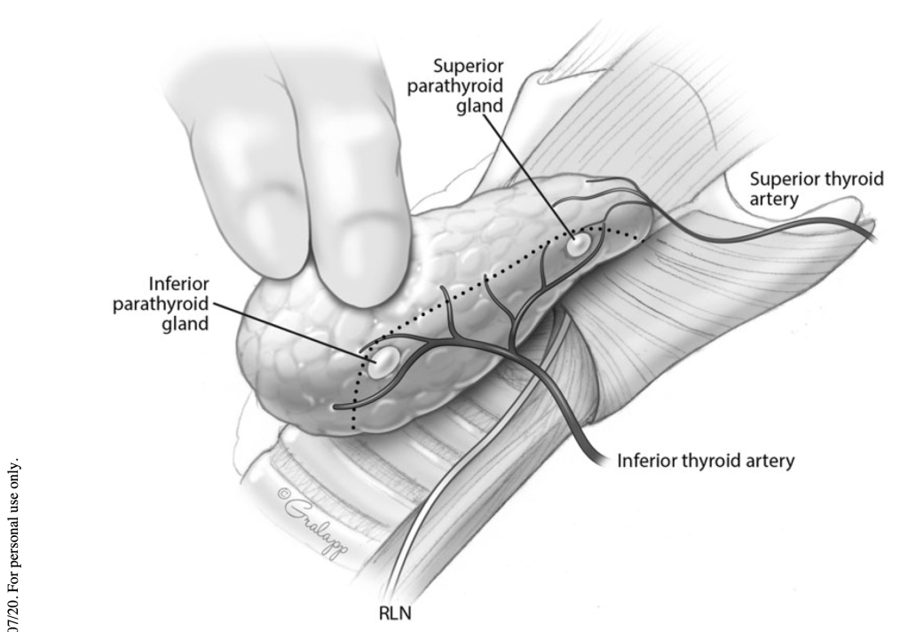
Medial or anterior to the parathyroids (Figure)
Plane of capsular dissection (dotted line) during thyroidectomy, dividing vasculature medial (distal) to the parathyroid glands in order to allow preservation of the parathyroid blood supply
The importance of staying as distal to the parathyroid gland(s) as possible when dissecting cannot be overstated
Utilization of loupe magnification (2.5 X):
Has been found to significantly reduce the rate of:
Inadvertent parathyroid gland removal:
3.8% vs. 7.8%
Postoperative biochemical hypocalcemia:
20.6% vs. 33.9%; p = 0.028
Postoperative clinical hypocalcemia:
12.7% vs. 33%; p < 0.001
The use of energy devices for vessel sealing during thyroidectomy is another relevant surgical technical factor:
These energy devices generate a zone of collateral thermal spread within the tissues:
Necessitate an optimal 3 to 5 mm distance of separation between the instrument and the parathyroid gland in order to avoid thermal injury
Interestingly, it is not essential to visualize all four parathyroid glands during thyroidectomy to reduce the incidence of postoperative hypocalcemia:
Sheahan et al:
Reported that patients with zero to two parathyroid glands identified during thyroidectomy had a significantly lower incidence of clinical hypocalcemia compared to patients who had three to four parathyroid glands visualized:
3.2% vs. 17.1%; p = 0.02
In this study, the observed differences in biochemical hypocalcemia were not significant (16.1% vs. 28.1%; p = 0.13), and the incidence of inadvertent parathyroidectomywas similar (9.7% vs. 9.4%; p=1.0)
Thomusch et al:
Demonstrated that during thyroidectomy, at least two parathyroid glands should be identified and preserved in order to avoid permanent hypoPT
The inferior parathyroid glands embryologically develop along with the thymus:
And as such may be separated enough from the inferior pole of the thyroid to make their visual identification without dissection more difficult yet their preservation more likely during thyroidectomy
Thyroid cancer surgery has an increased risk of hypoPT when a central lymph node dissection is performed:
The superior parathyroid glands are at lower risk of injury or inadvertent removal than the inferior parathyroid glands:
Since most of the central neck lymph node metastases are generally located in the more inferior paratracheal and pretracheal areas
Sometimes, a small inferior parathyroid vein:
May be seen to course lateral and anterior to the carotid artery:
When identified, it is important to preserve this vein:
Which can also be followed to facilitate identification of the inferior parathyroid gland
The blood supply to an ectopic intrathymic parathyroid gland is more difficult to preserve
Central neck lymph node dissection that is ipsilateral to the primary thyroid cancer should usually be performed first:
Then, the risk of contralateral central neck lymph node metastasis must be weighed against the risk of hypoPT when deciding whether to proceed with further nodal dissection
Parathyroid autotransplantation (PA):
The identified parathyroid glands should be assessed for devascularization, and a decision made whether to perform PA:
In order to maximize the amount of retained functional parathyroid tissue
Venous congestion:
May be alleviated by sharp scoring of the parathyroid gland capsule:
Which may result in prompt normalization or improvement in color
Ischemia (arterial insufficiency) of a parathyroid gland:
May be subtle and difficult to detect, as the gland may appear only slightly pale to normal in color
A common surgical dilemma is whether autotransplantation of persistently or progressively discolored parathyroid glands is appropriate:
Promberger et al. found that patients with discolored parathyroids only had transiently impaired function:
They recommended PA:
Only if there was clear evidence of ischemia or an inadequate blood supply
PA is accomplished by:
First storing the excised parathyroid in iced saline while a sliver of the parathyroid tissue is submitted for frozen section confirmation
The parathyroid gland is then minced into 1 mm fragments that are autotransplanted by direct implantation or injection into either intramuscular or subcutaneous pockets, within the sternocleidomastoid muscle or elsewhere
The aim of PA is to reduce the risk of permanent hypoPT
Of note, much of the literature reporting on PA is focused on glands from patients with underlying hyperparathyroidism, and their observations may not extrapolate to the transplantation of devascularized parathyroids when preoperative parathyroid function was normal
Lo and Lam reported a higher incidence of postoperative hypocalcemia in patients who underwent PA during thyroidectomy compared to those who did not (21.4% vs. 8.1%; p < 0.01), but permanent hypoPT only occurred in the patients who did not undergo PA (1.8%):
However, in a different study, the same investigators found that routine PA was associated with a higher incidence of postoperative hypocalce- mia, and did not lead to a significant reduction in the incidence of permanent hypoPT when compared to a policy of selective PA
A large Australian study examined the clinical outcomes after autotransplantation of zero, one, two, or three parathyroid glands:
As the number of autotransplanted parathyroid glands increased, the incidence of temporary hypoPT increased respectively ( p < 0.05), but the incidence of permanent hypoPT was similar at less than 1% ( p = NS), respectively
Is the most common complication of bilateral and re-operative thyroid operations
The true incidence of postoperative hypoPT is debatable:
Because of significant heterogeneity in how it has been studied:
Different time points after surgery, diverse electrolyte supplementation protocols, thyroid operations of variable aggressiveness, by surgeons of varying expertise, and for a broad array of indications are further confounded by variable use of clinical criteria (symptomatic vs, asymptomatic hypocalcemia), biochemical criteria (serum parathyroid hormone [PTH] and / or calcium and / or ionized calcium), and treatment criteria (requirement for calcium and / or vitamin D supplementation)
According to a recent meta-analysis:
The median incidence of temporary hypoPT following thyroidectomy ranges from:
19% to 38%
The median incidence of permanent hypoPT following thyroidectomy ranges from:
0% to 3%
It is critically important for the thyroid surgeon to employ strategies for minimizing and preventing hypoPT, including:
Carrying out the most appropriate extent of thyroidectomy for a specific patient
Background:
The short half-life of PTH (3 to 5 minutes), along with the fragile nature of the parathyroid glands:
Sets the stage for their functional derangement following manipulation
The etiology of hypoPT:
Is related to dissection or removal of the vulnerable parathyroid glands during central neck operations:
Resulting in a decline in circulating PTH
Definitions
Biochemical hypoPT:
Is defined as a low intact PTH level:
Below the lower limit of the laboratory standard (usually 12 pg/mL):
Ranges of normal PTH values vary:
Depending upon the laboratory
Accompanied by hypocalcemia:
Hypocalcemia is a total serum calcium level that is less than the lower limit of the center-specific reference range
Transient serum calcium values outside the normal reference range:
May reflect dynamic changes in electrolytes and state of hydration:
Rather than true hypocalcemia
Hypocalcemia may occur independent of hypoPT, but untreated hypoPT always leads to hypocalcemia, even though time lag can range from hours to days.
Clinical hypoPT:
Is defined as biochemical hypoPT that is accompanied by symptoms and/or signs of hypocalcemia
Parathyroid insufficiency, or relative hypoPT:
May occur after central neck surgery
Typically is manifested by clinical symptoms of hypoPT that require medical treatment:
Despite measured laboratory values within normal ranges
Transient or temporary hypoPT:
Is defined as occurring for less than six months after surgery:
Permanent hypoPT:
Is defined as occurring beyond six months after surgery
Mechanisms:
The mechanisms that underlie hypoPT:
Are related to:
Disruption of parathyroid gland arterial supply
Disruption of parathyroid gland venous drainage
Mechanical injury
Thermal or electrical injury
Intentional or inadvertent partial or complete removal
Normal parathyroid function requires a rich blood supply:
A normal parathyroid gland is composed of up to 30% capillary cells
Parathyroid blood supply is both delicate and complex:
Requires close attention during thyroidectomy to ensure its preservation
While the inferior thyroid artery is typically the dominant blood vessel that supplies the parathyroid glands:
Laser Doppler flowmetry has shown that:
The superior thyroid artery and vessels within the thymo-thyroid cord (ligament) can dominate in some individuals
Impaired PTH secretion results in postoperative hypocalcemia through:
Inhibition of bone resorption
Reduction of 1,25-dihyroxyvitamin D synthesis by the kidneys
Reduced intestinal absorption of calcium
Symptoms and signs:
Hypocalcemia causes:
Neuromuscular excitability and cardiac electrical instability:
Due to a reduced nerve and muscle cell depolarization threshold
Its most common early symptoms are:
Paresthesias, or numbness and tingling, of the perioral region and the fingertips
Muscle stiffness, cramps, and spasms are also common
Neuropsychiatric symptoms include:
Confusion, anger, depression, lightheadedness, and irritability
More sustained muscle contraction may lead to:
Laryngospasm
More severe neural excitability:
May lead to seizures
Signs of hypocalcemia include:
Observed or elicited tetany:
Classic bedside findings are:
A positive Chvostek sign:
Facial muscle twitching upon tapping the preauricular region over the facial nerve:
Present at baseline in up to 25% of people
A positive Trousseau sign:
Flexion of the wrist, thumb, and metacarpophalangeal joints and hyperextension of the fingers, upon brachial artery occlusion by inflation of a blood pressure cuff above systolic blood pressure)
Cardiovascular signs observed with progressive hypocalcemia include:
Prolongation of the QT interval that can result in torsades de pointes:
A form of ventricular tachycardia that may degenerate into ventricular fibrillation.
Risk factors:
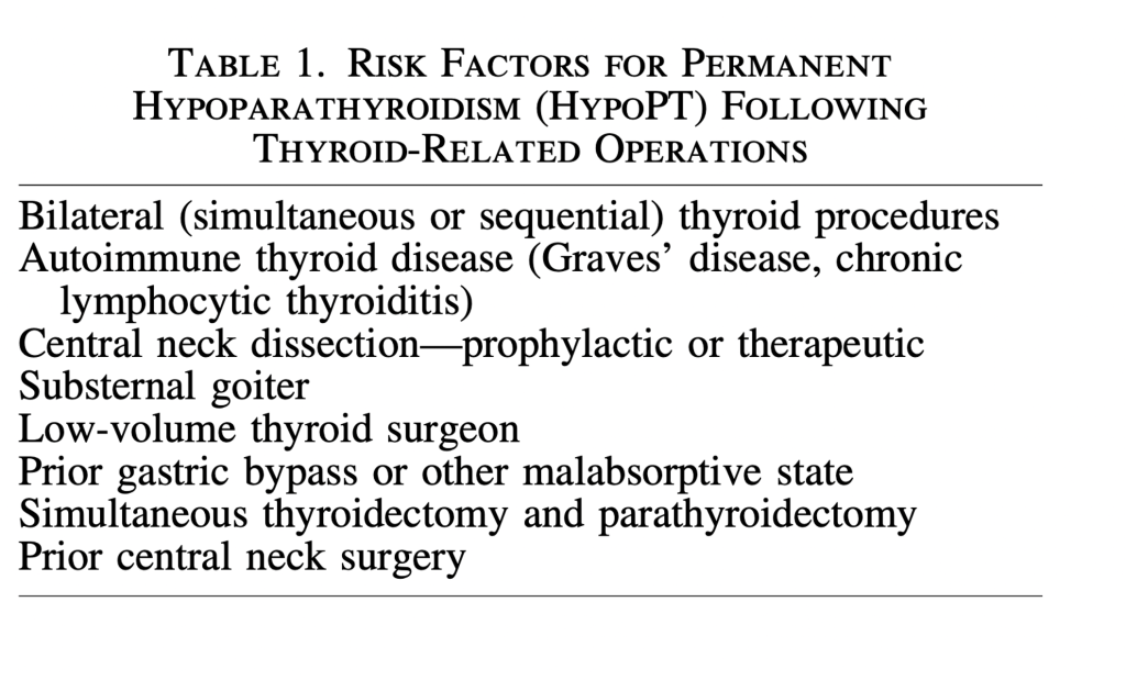
HypoPT may follow any simultaneous or staged bilateral central neck operation
Risk factors for both temporary and permanent hypoPT are presented in the following Table:
A prior partial thyroid operation creates a potentially increased risk of hypoPT during completion thyroidectomy:
Due to unknown status (presence or viability) of the parathyroid glands in the previously operated neck
The most straightforward way to avoid hypoPT:
Is to limit the extent of thyroidectomy to a unilateral approach
Though the historical rationale for a near-total or subtotal thyroidectomy
Instead of a total thyroidectomy, is in part preservation of the parathyroid glands:
It has never been adequately studied whether this actually reduces the risk of hypoPT
Parathyroid autotransplantation (PA):
At the time of thyroidectomy has been associated with an increased risk of temporary hypoPT
Paradoxically, routine PA may be associated with a reduced risk of permanent hypoPT
While data supporting propyhylactic PA are not definitive:
The risk of permanent hypoPT is very low in patients who have undergone autotransplantation of at least one parathyroid gland
Preoperative vitamin d deficiency:
When the planned thyroid operation is bilateral:
Preoperative testing of baseline serum calcium, PTH, and 25-hydroxy vitamin D blood levels can be helpful
If the baseline calcium is low normal, or below normal:
The risk of hypoPT is increased:
It may be appropriate to initiate scheduled oral calcium supplementation preoperatively
If the baseline calcium level is elevated:
Then the PTH level should be measured in order to evaluate for occult primary hyperparathyroidism, which could be definitively treated during thyroidectomy
A preoperatively elevated PTH level is commonly due to secondary hyperparathyroidism from vitamin D deficiency:
Vitamin D increases the absorption of calcium from the intestinal tract, and supplementation may be helpful to patients with hypoPT:
Assuming no underlying malabsorptive condition is present
Vitamin D also increases bone resorption and decreases renal excretion of calcium and phosphate
Vitamin D deficiency can be:
Severe – below the lowest recordable level, less than 10 ng/mL
Moderate – 10 to less than 20 ng/ mL
Mild (20 to 30 ng/mL
To optimize postoperative oral calcium absorption:
It is prudent to treat vitamin D deficiency preoperatively:
The Food and Drug Administration (FDA) approved regimen is:
50,000 IU of vitamin D3 (cholecalciferol) weekly or 6000 IU daily for eight weeks
More aggressive regimens and other vitamin D supplements are available, but their utilization should be considered off-label
Not all studies have substantiated improved postoperative calcium levels with higher preoperative vitamin D levels:
Lang et al. found the rate of clinically significant hypocalcemia after total thyroidectomy to be similar in patients with severe, moderate, and mild vitamin D deficiency, whereas Al-Khatib et al. found that severe 25-hydroxyvitamin D deficiency was an independent predictor of hypoPT in patients undergoing total thyroidectomy:
However, a large meta-analysis reported that the perioperative PTH level, the preoperative vitamin D level, and postoperative changes of calcium were biochemical predictors of post-thyroidectomy hypocalcemia
Given the present evidence, it would appear preferable to diagnose vitamin D deficiency and initiate appropriate corrective supplementation prior to surgery
In cases of elective bilateral thyroid surgery:
It may be prudent to delay surgery in order to correct severe vitamin D deficiency
References:
Ho TW, Shaheen AA, Dixon E, Harvey A 2011 Utilization of thyroidectomy for benign disease in the United States: a 15-year population-based study. Am J Surg 201:570–574.
Hauch A, Al-Qurayshi Z, Randolph G, Kandil E 2014 Total thyroidectomy is associated with increased risk of complications for low- and high-volume surgeons. Ann Surg Oncol 21:3844–3852.
Brandi ML, Bilezikian JP, Shoback D, Bouillon R, Clarke BL, Thakker RV, Khan AA, Potts JT Jr 2016 Management of hypoparathyroidism: summary statement and guide- lines. J Clin Endocrinol Metab 101:2273–2283.
Bollerslev J, Rejnmark L, Marcocci C, Shoback DM, Sitges-Serra A, van Biesen W, Dekkers OM 2015 Euro- pean society of endocrinology clinical gideline: treatment of chronic hypoparathyroidism in adults 2015. Eur J En- docrinol 173:G1–20.
Stack BC Jr, Bimston DN, Bodenner DL, Brett EM, Dralle H, Orloff LA, Pallota J, Snyder SK, Wong RJ, Randolph GW 2015 American Association of Clinical Endocrinologists and American College of Endocrinology disease state clinical review: postoperative hypoparathyroidism—definitions and management. Endocr Pract 21:674–685.
Lorente-Poch L, Sancho JJ, Mun ̃ oz-Nova JL, Sa ́ nchez- Vela ́zquez P, Sitges-Serra A 2015 Defining the syndromes of parathyroid failure after total thyroidectomy. Gland Surg 4:82–90.
Mehanna HM, Jain A, Randeva H, Watkinson J, Shaha A 2010 Postoperative hypocalcemia—the difference a defi- nition makes. Head Neck 32:279–283.
Edafe O, Antakia R, Laskar N, Uttley L, Balasubramanian SP 2014 Systematic review and meta-analysis of predic- tors of post-thyroidectomy hypocalcaemia. Br J Surg 101: 307–320.
Shoback DM, Bilezikian JP, Costa AG, Dempster D, Dralle H, Khan AA, Peacock M, Raffaelli M, Silva BC, Thakker RV, Vokes T, Bouillon T 2016 Presentation of hypoparathyroidism: etiologies and clinical features. J Clin Endocrinol Metab 101:2300–2312.
Anastasiou OE, Yavropoulou MP, Papavramidis TS, Tzouvara C, Triantafyllopoulou K, Papavramidis S, Yo- vos JG 2012 Secretory capacity of the parathyroid glands after total thyroidectomy in normocalcemic subjects. J Clin Endocrinol Metab 97:2341–2346.
Nawrot I, Woz ́ niewicz B, Tołłoczko T, Sawicki A, Go ́ rski A, Chudzin ́ski W, Wojtaszek M, Grzesiuk W, Sladowski D, Karwacki J, Zawitkowska T, Szmidt J 2007 Allo- transplantation of cultured parathyroid progenitor cells without immunosuppression: clinical results. Transplan- tation 83:734–740.
Johansson K, Ander S, Lennquist S, Smeds S 1994 Human parathyroid blood supply determined by laser-Doppler flowmetry. World J Surgery 18:417–420.
Shoback D 2008 Clinical practice. Hypoparathyroidism. New Engl J Med 359:391–403.
Kiernan CM, Schlegel C, Kavalukas S, Isom C, Peters MF, Solorzano CC 2016 Does concomitant thyroidectomy in- crease risks of parathyroidectomy? J Surg Res 203:34–39.
Thomusch O, Machens A, Sekulla C, Ukkat J, Lippert H, Gastinger I, Dralle H 2000 Multivariate analysis of risk factors for postoperative complications in benign goiter surgery: prospective multicenter study in Germany. World J Surg 24:1335–1341.
Chiang FY Lin JC, Wu CW, Lee KW, Lu SP, Kuo WR, Wang LF 2006 Morbidity after total thyroidectomy for benign thyroid disease: comparison of Graves’ disease and non-Graves’ disease.Kaohsiung J Med Sci 22:554–559.
Ebrahimi H, Edhouse P, Lundgren CI, McMullen T, Sidhu S, Sywak M, Delbridge L 2009 Does autoimmune thyroid disease affect parathyroid autotransplantation and surviv- al? ANZ J Surg 79:383–385.
Viola D, Materazzi G, Valerio L, Molinaro E, Agate L, Faviana P, Seccia V, Sensi E, Romei C, Piaggi P, Torre- grossa L, Sellari-Franceschini S, Basolo F, Vitti P, Elisei R, Miccoli P 2015 Prophylacti central compartment lymph node dissection in papillary thyroid carcinoma: clinical
implications derived from the first prospective renadomized controlled single institution study. J Clin Endocrinol Metab 100:1316–1324.
Lang BHH, Ng SH, Lau LLH, Cowling BJ, Wong KP, Wan KY 2013A systematic review and met-analysis of prophylactic central neck dissection on short-term locor- egional recurrence in papillary thyroid carcinoma after total thyroidectomy. Thyroid 23:1087–1098.
Wang TS, Cheung K, Farrokhyar F, Roman SA, Sosa JA 2013 A meta-analysis of the effect of prophylactic central compartment neck dissection on locoregional recurrence rates in pateints with papillary thyroid cancer. Ann Surg Oncol 20:3477–3483.
Zetoune T, Keutgen X, Buitrago D, Aldailami H, Shao H, Mazumdar M, Fahey III TJ, Zarnegar R 2010 Prophylactic central neck dissection and local recurrence in papillary thyroid cancer: a meta-analysis. Ann Surg Oncol 17:3287– 3293.
Giordano D, Valcavi R, Thompson GB, Pedroni C, Renna L, Gradoni P, Barbieri V 2012 Complications of central neck dissection in patients with papillary thyroid carci- noma: results of a study on 1087 patients and review of the literature. Thyroid 22:911–917.
deCarvalho A, Chulam TC, Kowalski LP 2015 Long- term results of observation vs prophylactic selective le- vel VI neck dissection for papillary thyroid carcinoma at a cancer center. JAMA Otolaryngol Head Neck Surg 141:599–606.
Kwan WY, Chow TL, Choi CY, Lam SH 2015 Compli- cation rates of central compartment dissection in papillary thyroid cancer. ANZ J Surg 85:274–278.
Moo TA, Umunna B, Kato M, Butriago D, Kundel A, Lee JA, Zarnegar R, Fahey TJ III 2009 Ipsilateral versus bilat- eral central neck l ymph node dissection in papillary thyroid carcinoma. Ann Surg 250:403–408.
Barczyn ́ski M, Konturek A, Stopa M, Nowak W 2013 Prophylactic central neck dissection for papillary thyroid cancer. Br J Surg 100:410–418.
Testini M, Gurrado A, Avenia N, Bellantone R, Biondi A, Brazzarola P, Calzolari F, Cavallaro G, De Toma G, Guida P, Lissidini G, Loizzi M, Lombardi CP, Piccinni G, Portincasa P, Rosato L, Sartori N, Zugni C, Basile F 2011 Does mediastinal extension of the goiter increase mor- bidity of total thyroidectomy? A multicenter study of 19,662 patients. Ann Surg Oncol 18:2251–2259.
Randolph GW, Shin JJ, Grillo HC, Mathisen D, Katlic MR, Kamani D, Zurakowski D 2011 The surgical man- agement of goiter: Part II. Surgical treatment and results. Laryngoscope 121:68–76.
Karakas E, Osei-Agyemang T, Schlosser K, Hoffmann S, Zielke A, Rothmund M, Hassan I 2008 The impact of parathyroid auto transplantation during bilateral surgery for Graves’ disease on postoperative hypocalcemia. En- docr Regul 42:39–44.
Sosa JA, Bowman HM, Tielsch JM, Powe NR, Gordon TA, Udelsman R 1998 The importance of surgeon expe- rience for clinical and economic outcomes from thyroid- ectomy. Ann Surg 228:320–330.
Kandil E, Noureldine SI, Abbas A, Tufano RP 2013 The impact of surgical volume on patient outcomes following thyroid surgery. Surgery 154:1346–1352.
Was a randomized controlled trial comparing completion axillary lymph node dissection (ALND) versus axillary radiotherapy (ART):
In women with clinical T1 to T2, N0 who were found to have a positive sentinel lymph node upon undergoing sentinel lymph node biopsy
Axillary RT was to levels I to III and SCV fossa:
To 50 Gy / 25 fractions
The primary endpoint was:
Non-inferiority of 5-year axillary recurrence
The 5-year axillary lymph node recurrence rate:
As 0.43% in the ALND group and 1.19% in the ART group (p=non-significant), with authors concluding that ALND and ART offer excellent and comparable axillary control in this set of patients
Of note, non-randomized patients with a negative sentinel lymph node biopsy had a similar axillary recurrence rate of 0.8%
Disease-free and overall survival were similar between the two arms
Lymphedema:
Was increased in those receiving ALND (23% vs. 11%, p<0.001 with clinical signs; 13% vs. 5%, p=0.0009 with arm circumference > 10%)
This result echoes findings of the ACOSOG Z0011 trial with low rates of axillary recurrence while omitting ALND
Those offering their expert opinion have agreed that results from these two randomized trials support the omission of ALND in patients with a clinically node negative axilla and a low burden of node positive disease found upon sentinel lymph node dissection
References
Donker M, Van Teinhoven G, Straver M, et al. Radiotherapy or surgery of the axilla after a positive sentinel node in breast cancer (EORTC 10981-22023 AMAROS): a randomised, multicentre, open-label, phase 3 non-inferiority trial. Lancet Oncol. 2014;15(12):1303-1310.
Giuliano AE, McCall L, Beitsch P, et al. Locoregional recurrence after sentinel lymph node dissection with or without axillary dissection in patients with sentinel lymph node metastases: the American College of Surgeons Oncology Group Z0011 randomized trial. Ann Surg. 2010;252(3):426-432.
Mahmoud O, Haffty B. Regional nodal management in the light of the AMAROS trial. Ann Transl Med. 2015; 3(7):88.
Is extremely rare and is seen in less than 1% of patients who receive radiotherapy for breast cancer
Polgár et al:
Found an incidence of 0.46 per 1,000 cases at their institution
Radiation-induced angiosarcoma:
Was associated with a latency period of 6 to 8 years in that study
Additional studies have demonstrated an interval of 3 to 12 years:
With a median time to development of 6 years
Presentation often includes:
A bruise-like appearance in the treated region:
So a high index of suspicion is needed to make the diagnosis
Treatment includes:
Surgery with or without radiation:
But radiation is not contraindicated
Smith et al. treated patients with hyperfractionated accelerated re-irradiation therapy with or without surgery:
Utilizing three radiation therapy treatments each day:
With a minimum interfraction interval of 4 hours, 5 days a week:
At 1 Gy per fraction, to total doses of 45 Gy, 60 Gy, and 75 Gy for areas with a moderate risk for subclinical disease, a high risk for subclinical disease, and gross disease, respectively
Toxicity was minimal
Median survival was 7 years:
10-year cause specific was 71%
These numbers are better than the 5-year overall survival of 40% and local control rates of approximately 30% reported by Depla et al
References
Abbott R, Palmieri C. Angiosarcoma of the breast following surgery and radiotherapy for breast cancer. Nat Clin Pract Oncol. 2008;5(12):727-736.
Polgár C, Orosz Z, Szerdahelyi A, et al. Postirradiation angiosarcoma of the chest wall and breast: issues of radiogenic origin, diagnosis, and treatment in two cases. Oncology. 2001;60(1):31-34.
Smith TL, Morris CG, Mendenhall NP. Angiosarcoma after breast-conserving therapy: long-term disease control and late effects with hyperfractionated accelerated re-irradiation (HART). Acta Oncol. 2014;53(2):235-241.
Koerner F. Sarcoma. In: Hoda S, Brogi E, Koerner F, Rosen PP, eds. Rosen’s Breast Pathology. 4th ed. Philadelphia, PA: LWW, Wolters Klumer; 2014:1118–1126.
Depla AL, Scharloo-Kareis CH, de Jong MA, et al. Treatment and prognostic factors of radiation- associated angiosarcoma (RAAS) after primary breast cancer: a systematic review. Eur J Cancer. 2014;50(1):1779-1788.
Tri-modality therapy for patients with breast cancer can result in complications
In women undergoing adjuvant radiation therapy, treatment of the regional nodes:
Is associated with higher rates of lymphedema
The published MA-20 trial:
Found an 8.4% rate of lymphedema with regional nodal irradiation compared with 4.5% without
As a part of the toxicity discussion a patient has with her provider, it is worth noting that there is no strong evidence of worse lymphedema:
When comprehensive post mastectomy RT is delivered using hypofractionation versus conventional fractionation:
Analysis of the START trials found no difference in shoulder stiffness (11% standard fractionation vs.14% hypofractionation), difficulty raising arm (17% standard fractionation vs. 14% hypofractionation), lymphedema (11% standard fractionation vs. 6% hypofractionation)
Shoulder dysfunction:
Can also be seen in patients requiring regional nodal irradiation:
With data documenting significant differences in shoulder function in patients with a history of breast cancer
Breast edema:
Can be noted acutely during radiotherapy as well as chronically following treatment:
With toxicity rates of 5%
There is evidence supporting an increased risk of ischemic heart disease in the years following breast radiotherapy:
Albeit with some criticism as these conclusions were largely based on antiquated radiation techniques
No data suggest an increased risk of pulmonary embolus with radiation for breast cancer
References
Shah C, Vicini FA. Breast cancer-related arm lymphedema: incidence rates, diagnostic technique, optimal management, and risk reduction strategies. Int J Radiat Oncol Biol Phys. 2011;81(4):907-914.
Whelan TJ, Olivotto IA, Pareulekar WR, et al. Regional nodal irradiation in early-stage breast cancer. N Engl J Med. 2015;373(4):307-316.
Haviland JS, Manino M, Griffin C, et al. Late normal tissue effects in the arm and shoulder following lymphatic radiotherapy: results from the UK START (Standardisation of Breast Radiotherapy) trials. Radiother Oncol. 2018;126(1):155-162.
Harrington S, Padua D, Battaglini C, et al. Comparison of shoulder flexibility, strength, and function between breast cancer survivors and healthy participants. J Cancer Surviv. 2011;5(2):167-174.
Hille-Betz U, Baske B, Bremer M, et al. Late radiation side effects, cosmetic outcomes, and pain in breast cancer patients after breast-conserving surgery and three-dimensional conformal radiotherapy: risk modifying factors. Strahlenther Onkol. 2016;192(1):8-16.
Darby SC, Ewertz M, McGale P. Risk of ischemic heart disease in women after radiotherapy for breast cancer. New Engl J Med. 2013;368(11):987-998.
Most women with breast cancer who undergo breast-conserving surgery receive whole-breast irradiation
The MA20 trial examined whether the addition of regional nodal irradiation to whole-breast irradiation improved outcomes
The MA-20 trial:
Was a randomized trial that included women with:
Node-positive or high-risk node-negative breast cancers
Randomizing patients to radiation therapy with or without comprehensive regional nodal irradiation:
Which includes internal mammary nodes, supraclavicular nodes, axillary nodes
High-risk node negative patients:
Were those with a tumor > 5 cm, or > 2 cm and < 10 lymph nodes dissected with at least one of the following:
Grade 3, ER negative, or lymphovascular space invasion
A total of 1,832 women were included and with 10-year follow-up
Results:
The median follow-up was 9.5 years
At the 10-year follow-up, there was no significant between-group difference in survival:
With a rate of 82.8% in the nodal-irradiation group and 81.8% in the control group (hazard ratio, 0.91; 95% confidence interval [CI], 0.72 to 1.13; P=0.38)
The rates of disease-free survival:
Were 82.0% in the nodal-irradiation group and 77.0% in the control group (hazard ratio, 0.76; 95% CI, 0.61 to 0.94; P=0.01)
Patients in the nodal-irradiation group had higher rates of grade 2 or greater acute pneumonitis (1.2% vs. 0.2%, P=0.01) and lymphedema (8.4% vs. 4.5%, P=0.001)
Conclusion of the trial:
Among women with node-positive or high-risk node-negative breast cancer, the addition of regional nodal irradiation to whole-breast irradiation did not improve overall survival but reduced the rate of breast-cancer recurrence
References
Whelan TJ, Olivotto IA, Pareulekar WR, et al. Regional nodal irradiation in early-stage breast cancer. N Engl J Med. 2015;373(4):307-316.
Poortmans PM, Collette S, Kirkove C, et al. Internal mammary and medial supraclavicular irradiation in breast cancer. N Engl J Med. 2015;373(4):317-327.
Patients with left-sided breast cancers in particular:
Are candidates for cardiac sparing breast radiation techniques
Multiple techniques exist to reduce cardiac dose and include:
Active breathing coordinator techniques
Prone breast irradiation
Intensity-modulated radiation therapy
Accelerated partial breast irradiation
None, however, is considered superior to another and the choice of technique employed depends on multiple patient factors
Active breathing coordinator techniques:
Has been used consistently as a method to reduce heart dose:
By increasing the distance from the chest wall to the heart
Prone breast irradiation:
Allows for the breast to fall away from the chest, potentially improving cardiac dose particularly for larger breasted patients
Intensity-modulated radiation therapy:
Can create segments within the beam to limit heart dose
Accelerated partial breast irradiation:
Limits the volume treated to the area surrounding the lumpectomy cavity and thus spares the heart
Regional nodal irradiation:
Would treat the axilla and / or internal mammary nodes and therefore is not cardiac sparing
Hypofractionated whole-breast irradiation:
On its own is a difference in dose regimen, not a cardiac-sparing technique
Abdominal compression can be used to limit respiratory motion but is not traditionally used as a cardiac sparing technique
References
Shah C, Badiyan S, Berry S, et al. Cardiac dose sparing and avoidance techniques in breast cancer radiotherapy. Radiother Oncol. 2014;112(1):9-16.
Eldredge-Hindy H, Lockamy V, Crawford A, et al. Active breathing coordinator reduces radiation dose to the heart and preserves local control in patients with left breast cancer: report of a prospective trial. Pract Radiat Oncol. 2015;5(1):4-10.
Mulliez T, Veldeman L, Speleers B, et al. Heart dose reduction by prone deep inspiration breath hold in left-sided breast irradiation. Radiother Oncol.2015;114(1):79-84.
Yim J, Suttie C, Bromley R, Morgia M, Lamoury G. Intensity modulated radiotherapy and 3D conformal radiotherapy for whole breast irradiation: a comparative dosimetric study and introduction of a novel qualitative index for plan evaluation, the normal tissue index. J Med RadiatSci. 2015;62(3):184-191.



 Rodrigo Arrangoiz MS, MD, FACS, FSSO