My name is Rodrigo Arrangoiz I am a breast surgeon/ thyroid surgeon / parathyroid surgeon / head and neck surgeon / surgical oncologist that works at Center for Advanced Surgical Oncology in Miami, Florida.
I was trained as a surgeon at Michigan State University from (2005 to 2010) where I was a chief resident in 2010. My surgical oncology and head and neck training was performed at the Fox Chase Cancer Center in Philadelphia from 2010 to 2012. At the same time I underwent a masters in science (Clinical research for health professionals) at the University of Drexel. Through the International Federation of Head and Neck Societies / Memorial Sloan Kettering Cancer Center I performed a two year head and neck surgery and oncology / endocrine fellowship that ended in 2016.
Mi nombre es Rodrigo Arrangoiz, soy cirujano oncólogo / cirujano de tumores de cabeza y cuello / cirujano endocrino que trabaja Center for Advanced Surgical Oncology en Miami, Florida.
Fui entrenado como cirujano en Michigan State University (2005 a 2010 ) donde fui jefe de residentes en 2010. Mi formación en oncología quirúrgica y e n tumores de cabeza y cuello se realizó en el Fox Chase Cancer Center en Filadelfia de 2010 a 2012. Al mismo tiempo, me sometí a una maestría en ciencias (investigación clínica para profesionales de la salud) en la Universidad de Drexel. A través de la Federación Internacional de Sociedades de Cabeza y Cuello / Memorial Sloan Kettering Cancer Center realicé una sub especialidad en cirugía de cabeza y cuello / cirugia endocrina de dos años que terminó en 2016.
Immediately upon identification of a suspicious thyroid nodule
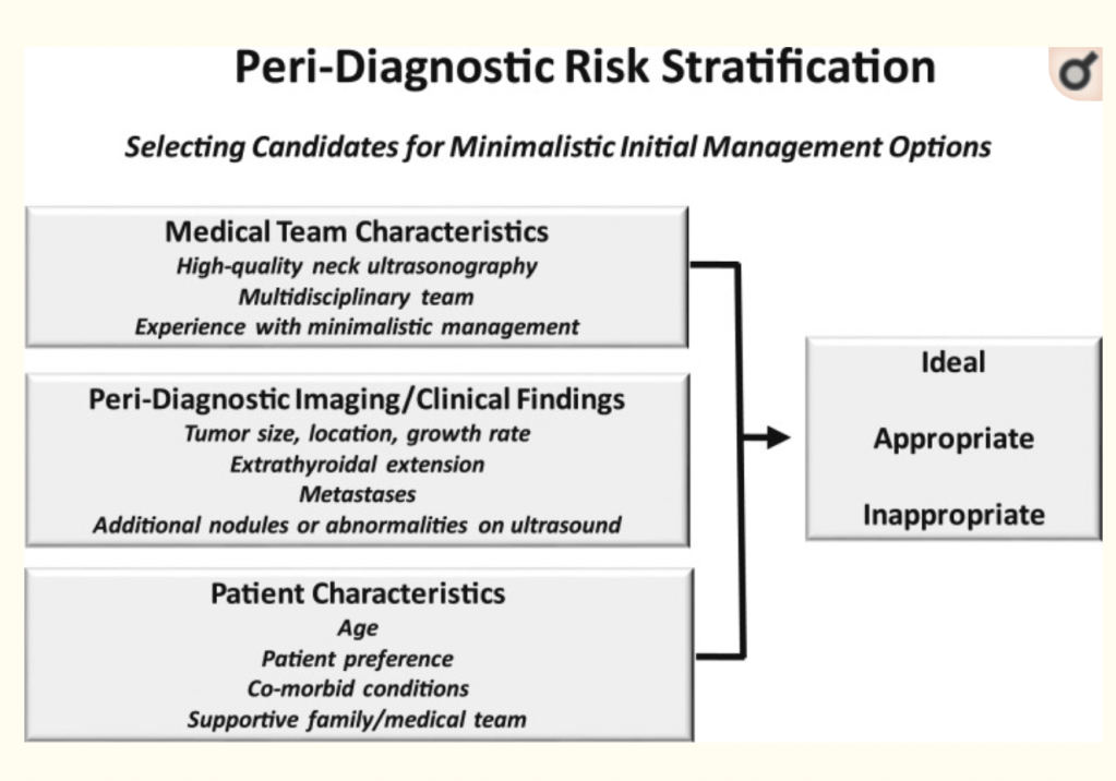
In the absence of a validated peri-diagnostic risk-stratification system, I use a clinical framework that incorporates:
Tumor imaging characteristics, medical team characteristics, and patient preferences to risk stratify patients as:
Ideal
Appropriate
Inappropriate for minimalistic initial management options:
Such as active surveillance or thyroid lobectomy
This clinical framework address the key factors that differentiate actionable from non-actionable disease
Peri-diagnostic risk stratification considers medical team characteristics, imaging/clinical findings, and patient characteristics to classify patients as ideal, appropriate, or inappropriate for a minimalistic initial management approach.
The administration of neoadjuvant chemotherapy (NAC):
Offers several advantages in locally advanced breast cancer:
It allows for downstaging the disease:
Which can potentially allow for less extensive surgery in the breast and axilla
It also provides information regarding the responsiveness of the cancer to systemic therapy while the tumor remains in vivo:
Which can guide the course of therapy
Administering chemotherapy in the neoadjuvant vs. adjuvant setting:
Does not change overall survival:
As demonstrated in the National Surgical Adjuvant Breast and Bowel Project (NSABP) B-18 and NSABP B-27 trials
The patient’s response to chemotherapy:
However, does offer prognostic information:
Particularly in patients with hormone receptor negative (HR-) disease
Patients who achieve pathologic complete response (pCR):
Which is typically defined as no residual invasive disease in the breast or axilla:
Appear to have improved event-free survival (EFS) and overall survival (OS) compared with patients with residual disease
This finding was demonstrated by a recent meta-analysis that included 36 studies including 5,768 patients with HER2 positive breast cancer:
This correlation was strongest in patients with HR- disease
Further, among patients with HER2 positive disease that do not have a pCR:
The degree of residual cancer burden appears to correlate with outcomes
Patients with HER2 positive tumors:
May complete up to 1 year of HER2-targeted therapy with trastuzamab ± pertuzamab
When planning surgery:
The pre-treatment volume does not need to be excised if the tumor has responded to chemotherapy:
However if multifocal disease is present, the satellite lesion(s) should be localized and excised with the index lesion
When considering the appropriateness for breast conservation following NAC, the distance between the lesions, location, and breast size must be considered
Placement of clips in the index lesion and any satellite lesions prior to initiation of NAC is critical for appropriate surgical planning post-NAC
References:
Rastogi P, Anderson SJ, Bear HD. Preoperative chemotherapy: updates of National Surgical Adjuvant Breast and Bowel Project Protocols B-18 and B-27. J Clin Oncol. 2008; 10;26(5):778-785.
Broglio KR, Quintana M, Foster M, et al. Association of pathologic complete response to neoadjuvant therapy in HER2-positive breast cancer with long-term outcomes: a meta-analysis. JAMA Oncol. 2016;2(6):751-760.
Symmans WF, Wei C, Gould R, et al. Long-term prognostic risk after neoadjuvant chemotherapy associated with residual cancer burden and breast cancer subtype. J Clin Oncol. 2017;35(10):1049-1060.
Boughey JC, Peintinger F, Meric-Bernstam F, et al. Impact of preoperative versus postoperative chemotherapy on the extent and number of surgical procedures in patients treated in randomized clinical trials for breast cancer. Ann Surg.2006;244(3):464-470.
Is a very rare special histological type of breast cancer:
Accounting for approximately 0.1% of all breast tumors
It is usually triple negative
It is much less likely to have nodal involvement
Is more common in postmenopausal women:
Most cases are in females
The median age of onset is:
Between 50 and 60 years
With a mean age of 66
The typical clinical feature is:
A single breast tumor / mass:
Multiple nodules are rare
Most ACCs are located:
Under the areola or in the upper outer quadrants
ACC of the breast has no characteristic imaging findings:
Ultrasound features are those of:
A hypoechoic solid or heterogeneous mass
On mammography:
The case may present as a lobulated mass with sharp or un sharp margins
Nevertheless, these clinical and radiographic features may be similar to any breast cancer:
Thus making their precise diagnosis difficult for radiologists
Histologically:
ACC of the breast typically consists of a dual-cell population of:
Luminal and myoepithelial-basal cells:
Which are generally negative for estrogen receptor (ER), progesterone receptor (PR), and human epidermal growth factor receptor 2 (HER2)
In addition, some studies have also reported some HR-positive ACC cases:
The significance of a positive hormone receptor status is not known:
Compared with ACC with negative HR expression, the clinical characteristics and prognosis of this type of ACC are also unknown
Distant metastases are rare:
However, the lung is the most common site
It has a better prognosis than infiltrating ductal triple negative breast cancer:
With a 5-year overall survival rate of 88%
As prognosis is good:
Accurate preoperative diagnosis is important in the determination of suitable treatment
References
Treitl D, Radkani P, Rizer M, El Hussein S, Paramo JC, Mesko TW. Adenoid cystic carcinoma of the breast, 20 years of experience in a single center with review of literature. Breast Cancer. 2018;25(1)28-33.
Welsh JL, Keeney MG, Hoskin TL, et al. Is axillary surgery beneficial for patients with adenoid cystic carcinoma of the breast? J Surg Oncol. 2017;116(6):690-695.
Kulkarni N, Pezzi CM, Greif JM, et al. Rare breast cancer: 933 adenoid cystic carcinomas from the National Cancer Data Base. Ann Surg Oncol. 2013;20(7):2236-2241.
Kshirsagar AY, Wader JV, Langade YB, Jadhav KP, Zaware SU, Shekhar N. Adenoid cystic carcinoma of the male breast. Int Surg (2006) 91(4):234–6.
Pang W, Wang Z, Jin X, Zhang Q. Adenoid cystic carcinoma of the breast in a male: A case report. Med (Baltimore) (2019) 98(32):e16760. doi: 10.1097/MD.0000000000016760
Tang W, Peng WJ, Gu YJ, Zhu H, Jiang TT, Li C. Imaging Manifestation of Adenoid Cystic Carcinoma of the Breast. J Comput Assist Tomogr (2015) 39(4):523–30. doi: 10.1097/RCT.
Torrao MM, da Costa JM, Ferreira E, da Silva MV, Paiva I, Lopes C. Adenoid cystic carcinoma of the breast. Breast J (2007) 13(2):206.
Marchio C, Weigelt B, Reis-Filho JS. Adenoid cystic carcinomas of the breast and salivary glands (or ‘The strange case of Dr Jekyll and Mr Hyde’ of exocrine gland carcinomas). J Clin Pathol (2010) 63(3):220–8. doi: 10.1136/jcp.2009.073908
National Comprehensive Cancer Network (NCCN) guidelines:
Recommend surgical management:
For local control for women with early stage invasive breast cancer
Several studies have shown an equivalence in overall and / or breast cancer-specific survival rates:
For breast conservation with radiation compared to mastectomy among early stage breast cancer patients
For patients with ER positive disease:
Endocrine therapy with tamoxifen or aromatase inhibitors is prescribed after surgery:
A systematic review evaluated the efficacy of primary endocrine therapy alone versus surgery in women over 70 years old with operable tumors:
The review reported similar survival between the two groups, but women treated with surgery had lower rates of local failure when compared to endocrine therapy alone
The authors concluded that primary endocrine therapy should be reserved for women who are unfit for surgery or decline surgery
Sentinel node biopsy:
Has become the standard method for staging the axilla in women with early stage breast cancer:
Who are clinically node negative
Axillary dissection is only performed in women with:
Fisher B, Anderson S, Bryant J, et al. Twenty-year followup of a randomized trial comparing total mastectomy, lumpectomy, and lumpectomy plus irradiation for the treatment of invasive breast cancer. New Engl J Med. 2002;347(16):1233-1241.
Litiere S, Werutsky G, Fentiman IS, et al. Breast-conserving therapy versus mastectomy for stage I-II breast cancer: 20 year followup of the EORTC 10801 phase 3 randomized trial. Lancet Oncol. 2012;13(4):412-419.
Veronesi U, Cascinelli N, Mariani L, et al. Twenty-year follow-up of a randomized study comparing breast-conserving surgery with radical mastectomy for early breast cancer. New Engl J Med. 2002;347(16):1227-1232.
Is characteristically spontaneous, unilateral, or bloody
Physiologic discharge:
Is non-spontaneous, bilateral, and milk
The most common causes for pathologic nipple discharge:
Are benign:
Intraductal papillomas
Duct ectasia
The presence of abnormal clinical findings on imaging or physical exam:
Is associated with increased risk of malignancy:
38% vs. 2%
Contemporary workup for nipple discharge includes:
Mammography
Evaluation of the retroareolar region with ultrasound
Patients with normal findings on mammography, ultrasound, and physical exam:
Can be further evaluated with breast MRI:
As it is highly sensitive and specific for cancer
Surgical management of nipple discharge includes:
Excision of a single duct or central duct apparatus:
Depending on the number of ducts involved
References:
Li GZ, Wong SM, Lester S, Nakhlis F. Evaluating the risk of underlying malignancy in patients with pathologic nipple discharge. Breast J. 2018;24(4):624-627.
de Paula IB, Campos AM. Breast imaging in patients with nipple discharge. Radiol. Bras. 2017;50(6):383-388.
Yilmaz R, Bender O, Celik Yabul F, Dursun M, Tunaci M, Acunas G. Diagnosis of nipple discharge: value of magnetic resonance imaging and ultrasonography in comparison with ductoscopy. Balkan Med J. 2017;34(2):119-126.
Three important areas form the foundation for the evolving use of altered fractionation:
Tissue response
Duration of treatment
Fraction size and number
Acutely responding tissues:
Are rather active in ongoing cellular proliferation
Most tumors (except perhaps prostate cancer, breast cancers, and melanoma) and some normal tissues such as skin, mucous membranes, and gastrointestinal epithelium:
Share this characteristic:
These tissues are most affected by the overall treatment duration rather than by the size or number of fractions used
Late-responding tissues:
Have a low proliferative rate and include the spinal cord, brain, bone, and cartilage
These tissues are most affected by the:
Size and number of fractions rather than by treatment duration:
Therefore are spared by decreasing the dose per fraction of radiation delivered
Because most tumors consist of rapidly dividing cells:
Local tumor control is strongly dependent on the overall treatment duration rather than on the size or number of fractions
When squamous cell carcinoma of the head and neck is exposed to radiation:
The less radiosensitive cells within the lesion:
Can undergo rapid proliferation:
Approximately 3 to 5 weeks after treatment commences
This accelerated repopulation can overwhelm the ongoing treatment effects of radiation:
Which ultimately can lead to local failure
The clinical significance of this phenomenon is that even with significant regression of the primary tumor mass:
Local failure still ultimately could result from proliferation of these resistant clones
Therefore it is essential to complete treatment in as short a time as possible so that accelerated repopulation is minimized:
Increasing the chance for local control
For this reason, split-course radiation:
Which incorporates a treatment break during the course of radiotherapy is not recommended
Based on the aforementioned principles:
The goal of altered fractionation schemes:
Is to improve the therapeutic ratio by maximizing the tumoricidal effect and minimizing acute and late toxicities while using readily available low-LET radiation
Two major categories of altered fractionation schemes exist:
Hyperfractionation
Accelerated fractionation
They share basic radiobiological principles yet have their own particular features (Table)
Accelerated fractionation:
Is the strategy of choice for rapidly proliferative tumors
Accelerated fractionation is based on the concept that the shortened overall treatment time:
Would reduce the opportunity for accelerated repopulation effectively
Hyperfractionation:
Is preferred for slowly proliferating tumors
Hyperfractionation improves the therapeutic ratio primarily through:
Redistribution of tumor cells into more radiosensitive phases as a result of multiple fractions
Differential sparing of late-responding normal tissues because of a decrease in the size of the dose per fraction
Current consensus guidelines from the American Society of Breast Surgeons:
Do not recommend CPM for women with sporadic breast cancers
A Cochrane review of 8 studies evaluating patients who underwent CPM:
Concluded that while CPM reduces risk of contralateral breast cancer:
It is not associated with improved survival
Reasons for not recommending CPM include:
A low estimated risk of cancer in the contralateral breast (2% to 6% over 10 years)
Increased complication rates
Studies showing that CPM does not improve survival or recurrence from the index cancer
References:
Lostumbo L, Carbine N, Wallace J, Ko H. Prophylactic mastectomy for the prevention of breast cancer. Cochrane Database Syst Rev 2004(4):CD002748.
Boughey JC, Attai DJ, Chen SL, et al. Contralateral prophylactic mastectomy consensus statement from the american society of breast surgeons: additional considerations and a framework for shared decision making. Ann Surg Oncol. 2016;23(10):3106-3111
Is a clinical syndrome in women with invasive breast cancer that is characterized by:
Erythema and edema (peau d’orange) of a third or more of the skin of the breast
The differential diagnosis includes:
Cellulitis of the breast or mastitis
Because most IBC cases are first seen by healthcare providers not necessarily familiar with IBC:
The absence of complete response to a trial of antibiotic therapy should heighten suspicion of IBC and prompt further investigation:
Further trial of antibiotics is not warranted in the absence of clinical signs of infection and previous adequate antibiotic therapy
Workup includes physical exam and imaging:
Imaging may not reveal a mass:
But thickening of the skin is frequently seen
The most common signs of IBC on mammography include:
Thickening of the skin (84%)
Trabecular thickening (81%)
Asymmetric focal density (61%)
Microcalcifications (56%)
Mammography is the least sensitive diagnostic tool available for IBC:
Whereas ultrasound and MRI are more sensitive:
In a series published by Yang, et al., sonography demonstrated a mass or architectural distortion in 95% of patients with associated global skin and subcutaneous thickening and dilated lymphatics
MRI can also show skin thickening and is more sensitive than mammography in detecting an underlying mass:
The same series by Yang and colleagues found that a primary breast lesion was present in every MRI obtained in patients with IBC as either nonmass or mass-like enhancement
IBC is a clinical diagnosis:
Dermal biopsy confirmation is not mandatory:
Dermal lymphatic invasion is seen only in approximately 60% of IBC cases:
It is neither required, nor sufficient by itself for a diagnosis of inflammatory breast cancer
Treatment is multidisciplinary trimodality therapy consisting of neoadjuvant chemotherapy, modified radical mastectomy, and local regional radiation:
Yet, the median survival at 5 years for patients presenting with primary IBC is still only approximately 55%
References
Yang WT, Le-Petross HT, Macapinlac H, et al: Inflammatory breast cancer: PET/CT, MRI, mammography, and sonography findings. Breast Cancer Res Treat. 2008;109(3):417-426.
Somio G, Jones V. Inflammatory breast cancer. In: Klimberg S, Bland K, eds. The Breast: Comprehensive Management of Benign and Malignant Disease. Philadelphia, PA: Wolters Kluwer Health/Lippincott Williams & Wilkins; 2011:832-838.
Larger fraction size (600 to 800 cGy) compared with conventional fractionation (180 to 200 cGy)
Fractions delivered several days apart
Lower total dosage (2100 to 3200 cGy) than conventional fractionation (7000 cGy)
Shortened overall treatment duration compared with conventional fractionation
Hypofractionation:
Is the administration of high-dose-per-fraction (HDPF) radiation:
In which only one or two fractions are given per week
This technique has evolved for the treatment of malignant melanoma:
Which generally is perceived as being radioresistant
Conventional fractions of 200 cGy delivered 5 days a week:
Allow normal tissues and tumor cells to recover during the intervals between fractions
Experimental in vitro data have shown that malignant melanoma cells are better at repairing radiation-induced sublethal damage compared with other cells:
This finding may explain the long-standing notion that melanoma is intrinsically “radioresistant”
HDPF regimens:
Deliver higher doses of radiation per fraction (600 cGy twice a week or 800 cGy once weekly):
With the aim of overcoming the reparative capacity of the tumor cells by increasing the damage per fraction
In retrospective analyses, response rates have been shown to correlate with dose per fraction but not with the total dose delivered:
However, a prospective randomized trial (RTOG 83–05) found no therapeutic advantage in a comparison of HDPF (800 cGy once a week up to a total dose of 3200 cGy) and conventional fractionation (250 cGy daily, 5 days a week, for a total of 5000 cGy):
Although no therapeutic advantage was seen, the shorter delivery time of HDPF radiation allows earlier initiation of systemic therapies if applicable
Moderately hypofractionated radiation (225 cGy per fraction):
Has demonstrated superior results for early-stage larynx cancers treated with radiotherapy alone:
This is currently considered the standard of care in this setting
Additionally, a regimen commonly referred to as quad shot:
Which was originally developed for advanced pelvic tumors:
Is sometimes applied for palliation of tumors in the head and neck
This involves cycles of a 1480 cGy course of radiotherapy delivered in four fractions over the course of 2 days:
Which can be repeated multiple times over a period of weeks or months depending on the treatment response
Aside from the demonstrated efficacy of this regimen, it also allows significant advantages in terms of patient convenience in the palliative setting
Has been a significant concern for breast cancer patients undergoing axillary surgery
The development of BCRL is associated with:
Significantly lower physical and psychosocial well-being and increased health care utilization
The risk of BCRL:
Is a function of the extent of axillary intervention:
Ranging from about 12% following a sentinel node biopsy to about 30% after an axillary lymph node dissection (ALND)
The highest risk of BCRL (51%) has been reported in patients with inflammatory breast cancer:
Who receive trimodality therapy (neoadjuvant taxane-containing chemotherapy, modified radical mastectomy, and adjuvant radiation):
Therefore, adjuvant radiotherapy is associated with an increased risk of BCRL
The value of routine screening for BCRL in patients at risk is controversial
There is growing evidence that subclinical lymphedema:
Defined as relative volume change of the affected arm of 5% to 10% compared to the baseline measurement:
Is strongly associated with the development of more symptomatic BCRL:
Which correlates with a relative volume change of greater than 10%
Consequently, identifying patients with subclinical lymphedema is a potential opportunity for early intervention and long-term improvement in quality of life
Furlan et al prospectively evaluated 85 breast cancer patients (n=40 had an ALND and n=45 had a sentinel node biopsy) by obtaining serial circumferential arm measurements preoperatively, then 1 month, 3, 6, 12, and 24 months after surgery:
Study results showed that the earliest signs of subclinical lymphedema were detected no sooner than the 6-month assessment, and those with subclinical lymphedema were promptly referred for decongestive therapy
An international randomized trial comparing bioimpedance spectroscopy (BIS) and tape measurement to detect subclinical lymphedema:
Showed that BIS had a higher sensitivity and was associated with an earlier referral for decongestive therapy
In the same study, earlier administration of decongestive therapy was associated with a lower risk of progression to symptomatic BCRL
The practical aspects of implementing BCRL screening with BIS versus tape measurements and other techniques warrant further study
References
Coriddi M, Kim LN, Haglich K, et al. The impact of lymphedema on patient-reported outcomes after breast reconstruction: a preliminary propensity score-matched analysis. Ann Surg Oncol. 2023;30(5):3061-3071. doi: 10.1245/s10434-022-12994-z
Cheville A, Lee M, Moynihan T, et al. The impact of arm lymphedema on healthcare utilization during long-term breast cancer survivorship: a population-based cohort study. J Cancer Surviv. 2020;14(3):347-355. doi: 10.1007/s11764-019-00851-0
Bucci LK, Brunelle CL, Bernstein MC, et al. Subclinical lymphedema after treatment for breast cancer: risk of progression and considerations for early intervention. Ann SurgOncol. 2021;28(13):8624-8633. doi: 10.1245/s10434-021-10173-0
Farley CR, Irwin S, Adesoye T, et al. Lymphedema in inflammatory breast cancer patients following trimodal treatment. Ann Surg Oncol. 2022;29(10):6370-6378. doi: 10.1245/s10434-022-12142-7
Furlan C, Matheus CN, Jales RM, Derchain SFM, Bennini JR Jr, Sarian LO. Longitudinal, long-term comparison of single-versus multipoint upper limb circumference periodical measurements as a tool to predict persistent lymphedema in women treated surgically for breast cancer: an optimized strategy to early diagnose lymphedema and avoid permanent sequelae in breast cancer survivors. Ann Surg Oncol. 2021;28(13):8665-8676. doi: 10.1245/s10434-021-10290-w
Ridner SH, Dietrich MS, Boyages J, et al. A comparison of bioimpedance spectroscopy or tape measure triggered compression intervention in chronic breast cancer lymphedema prevention. Lymphat Res Biol. 2022;20(6):618-628. doi: 10.1089/lrb.2021.0084



 Rodrigo Arrangoiz MS, MD, FACS, FSSO