Bone Disease and Primary Hyperparathyroidism

  • Bone disease, including:
    • Osteopenia, osteoporosis, and osteitis fibrosa cystica:
      • Is found in approximately 15% of patients with primary hyperparathyroidism at the time of diagnosis
    • Increased bone turnover:
      • As found in patients with osteitis fibrosa cystica:
        • Can be determined by documenting:
          • Elevated alkaline phosphatase concentration
    • Advanced PHPT with osteitis fibrosa cystica:
      • Now occurs in less than 5% of patients
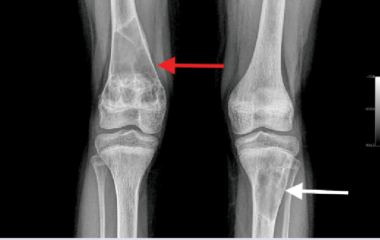
    • The classic manifestation of PHPT bone disease is:
      • Osteitis fibrosa cystica:
        • Which is characterized clinically by bone pain and radiographically by subperiosteal bone resorption
      • It has pathognomonic radiological findings:
        • Which are best seen on radiographs of the hands:
          • And are characterized by subperiosteal resorption:
            • Most evident in the radial aspect of the middle phalanx of the second and third fingers
          • Bone cysts
          • Tuft formation of the upper part of the distal phalanges
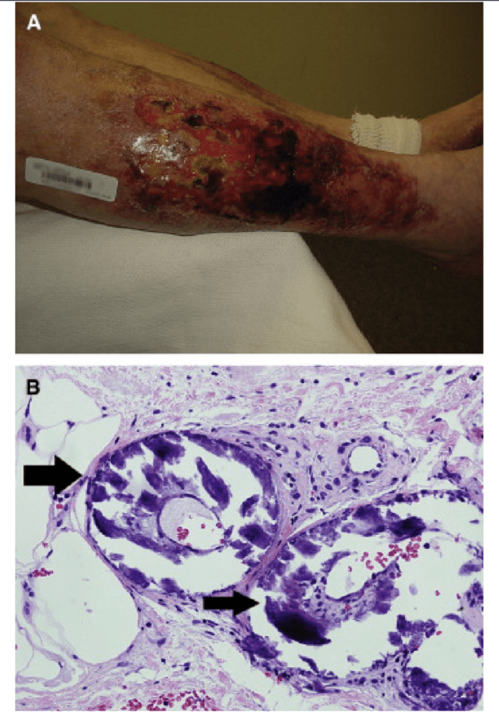
        • Brown or osteoclastic tumors of the long bones:
          • Result from excess osteoclast activity:
            • And consist of collections of osteoclasts intermixed with fibrous tissue and poorly mineralized woven bone
          • The brown coloration is due to:
            • Hemosiderin deposition
        • Tapering of the distal clavicle
      • Patients with normal serum alkaline phosphatase levels:
        • Rarely have clinically apparent osteitis fibrosa cystica
  • PHPT typically results in a loss of bone volume:
    • At the cortical bone sites such as:
      • The radius
    • Relative preservation of cancellous bone:
      • As found in vertebral bodies
      • Patients with primary hyperparathyroidism, however:
        • May also have osteoporosis in the lumbar spine that dramatically improves after parathyroidectomy
  • Fractures are also more common in patients with primary hyperparathyroidism, and the incidence of fractures:
    • Also decreases after parathyroidectomy:
      • Bone disease is correlated with serum PTH and vitamin D levels

#Arrangoiz #ParathyroidSurgeon #ParahthyroidExpert #Hyperparathyroidism #HeadandNeckSurgeon #EndocrineSurgery #MountSinaiMedicalCenter #MSMC #Miami #Mexico

Criteria for Performing a Limited Parathyroidectomy

  • Sestamibi characteristics:
    • Radioactive uptake by the adenoma:
      • Should be different from the thyroid gland
    • Rest of the study should not have any abnormality
  • Patients greater than 25 years of age
  • No family history of:
    • PHPT or
    • Multiple endocrine neoplasia (MEN)
  • Absence of goiter
  • No history of lithium intake
  • No history of radiation to the neck
  • No history of diseases of the:
    • Pancreas, adrenal gland and pituitary gland

#Arrangoiz #ParathyroidSurgeon #ParathyroidExpert #Hyperparathyroidism #PrimaryHyperparathyroidism #CancerSurgeon #EndocrineSurgery #Teacher #Surgeon #HeadandNeckSurgeon #SurgicalOncologist #ParathyroidAdenoma #Hypercalcemia #ElevatedCalciumLevels #Miami #MountSinaiMedicalCenter #MSMC #Mexico #Hialeah

Secondary Hyperparathyroidism

  • Secondary hyperparathyroidism:
    • Is overproduction of parathyroid hormone (PTH):
      • Due to a nonparathyroid cause:
        • Usually a result of renal failure
    • Renal failure decreases the production of 1,25(OH)2 D3:
      • Impairing intestinal absorption of calcium:
        • Leading to hypocalcemia:
          • Which increases PTH secretion by the parathyroid glands:
            • Leading to diffuse or nodular hyperplasia
    • Hyperphosphatemia:
      • From decreased renal excretion also contributes to PTH secretion:
        • The resulting calcium level is low or normal
    • Less common causes of secondary hyperparathyroidism include:
      • Osteomalacia
      • Long-term lithium therapy
      • Vitamin D deficiency
      • Malabsorption syndromes
      • Secondary hyperparathyroidism has beenv increasingly described following bariatric surgery:
        • Most commonly Roux-en-Y gastric bypass and duodenal switch:
          • Due to prolonged vitamin D deficiency:
            • It occurs despite vitamin D replacement
  • The pathophysiology of secondary hyperparathyroidism:
    • Is multifactorial and can result from:
      • Phosphorous retention
      • Altered vitamin D metabolism and resistance
      • Altered metabolism of PTH
      • Impaired calcemic response to PTH
      • Possible genetic mutations
  • The condition most commonly occurs in patients:
    • With a history of chronic renal failure
  • Gastric bypass:
    • Has also been an increasingly recognized cause of altered vitamin D metabolism
  • Patients will commonly have:
    • An elevated PTH level and normal or low serum calcium:
      • In such a setting, vitamin D levels should be measured:
        • If low, treated for a minimum of 6 weeks with supplemental vitamin D
  • Secondary hyperparathyroidism:
    • Is most commonly managed medically with the use of:
      • Calcimimetic agents:
        • For example cinacalcet
      • Phosphate binders
      • Adequate calcium intake
      • Vitamin D replacement
  • Cinacalcet:
    • Works by binding the calcium-sensing receptors on the chief cells of the parathyroid gland:
      • Increasing its sensitivity to extracellular calcium
  • Surgical treatment is indicated in patients with:
    • Renal osteodystrophy
    • Calciphylaxis:
      • Calciphylaxis is a rare complication of secondary hyperparathyroidism resulting in acute deposition of calcium in tissues and skin necrosis:
        • That may lead to a systemic inflammatory response:
          • It is associated with a high rate of mortality (87 to 93%)
      • Calcium phosphate product of ≥ 70
      • Soft tissue calcium deposition and tumoral calcinosis
      • Calcium level greater than 11 mg/dL with an inappropriately high level of PTH
  • Renal osteodystrophy:
    • Is a major issue in hemodialysis patients:
      • The aluminum present in the dialysate bath:
        • Accumulates in the bone:
          • Contributes to the development of osteomalacia
  • Osteitis fibrosa cystica:
    • A type of renal osteodystrophy:
      • Is characterized by marrow fibrosis and increased bone turnover
    • Bone cysts, osteopenia, and decreased bone strength develop
    • To halt the progression of this disease process:
      • These patients with secondary hyperparathyroidism are treated surgically
  • Calciphylaxis:
    • Is a rare vascular disorder:
      • In which calcium is deposited in the media of small- to medium-sized arteries
        • As a result, ischemic damage:
          • To the dermal and epidermal structures develops
    • The ulcerated lesions:
      • Are extremely painful and can become infected with subsequent sepsis, and eventually death
    • Patients with early signs of calciphylaxis:
      • Should undergo an urgent parathyroidectomy:
        • Although there is some evidence that aggressive management of serum calcium and parathyroid levels:
          • With cinacalcet may be beneficial
    • Care should be taken in wound care management:
      • Because aggressive debridement can lead to chronic nonhealing wounds:
        • Since wound healing is very poor in these patients
  • Uremic pruritus:
    • Is characterized by severe itching:
      • That is thought to result from increased deposition of calcium salt in the dermis:
        • Without the visible lesions of calciphylaxis
    • Parathyroidectomy:
      • Seems to alleviate these symptoms and halts progression to the more serious skin and vascular complications seen with calciphylaxis
 (a) Patient with chronic renal failure and calciphylaxis of the lower extremities. Lesions are superficial
erosions with islands of intervening granulation tissue and areas of eschar. The surrounding skin was
violaceous and very tender to palpation (b). Characteristic histologic findings in calciphylaxis.
Photomicrograph (original magnification × 400) showing three blood vessels with surrounding
mineralization, indicated by arrows. 

#Arrangoiz #ParathyroidSurgeon #ParathyroidExpert #EndocrineSurgery #HeadandNeckSurgeon #MSMC #Miami #MountSinaiMedicalCenter #Mexico #Teacher #Surgeon

Clinical Manifestations of Primary Hyperparathyroidism (PHPT)

  • Despite what most of the literature reports:
    • PHPT is symptomatic in more than 95% of the cases if proper attention is payed to the subtle symptoms and signs that this disease can produce due to the fluctuating calcium levels
  • The “classic” pentad of:
    • Kidney stones
    • Painful bones
    • Abdominal groans
    • Psychic moans. and
    • Fatigue overtones
      • Are rarely seen today since the advent and general use of automated blood analyzers in the early 1970s
  • At this point in time most patients present with:
    • Fatigue (# 1 symptom)
    • General malaise
    • Decrease levels of energy
    • Anxiety
    • Irritability leading to decrease social interaction
    • Depression (10% of cases)
    • Memory loss
    • Decrease concentration
    • Decrease ability to learn new things
    • Decrease ability to complete daily tasks at home
    • Decrease ability to complete daily tasks at work
    • Decrease social interaction
    • Insomnia
    • Arthralgia’s (32% of the cases)
    • Myalgia’s (14% to 41% of the cases)
    • Bone pain
    • Muscle weakness (specially proximal muscle groups)
    • Intermittent headaches
    • Polydipsia
    • Polyuria
    • Nocturia
    • Nausea (24% of the cases)
    • Anorexia (15% of the cases)
    • Non-specific abdominal pain
    • Heartburn (30% of the cases)
    • Constipation (33% of the cases)
    • Palpitations, arrhythmias (usually atrial fibrillation)
    • Elevated blood pressure
    • Thinning of the hair (specially in women in the frontal region)
    • Pruritus
  • Patients with PHPT also tend to score lower than healthy controls when evaluated by general multidimensional health assessment tools:
    • Such as the Medical Outcomes Study Short-Form Health Survey (SF-36) and other specific questionnaires
  • PHPT that is truly “asymptomatic” is a rare occurrence:
    • Seen in less than 5% of patients:
      • This is important when talking about management based on current guidelines
  • Patients with PHPT have some degree of renal dysfunction or symptoms in approximately 80% of the cases. The renal manifestations implicated with PHPT are decreased glomerular filtration rate, hypercalciuria, nephrolithiasis, nephrocalcinosis, impaired urinary concentrating ability sometimes leading to polyuria, polydipsia, and nocturia, reduced fractional phosphate reabsorption leading to hypophosphatemia, and increased urinary exertion of magnesium [66]. Nephrolithiasis was previously reported in approximately40% to 80% of patients [77], but now occur only in about 20% to 25% of the cases [78]. The pathophysiology is thought to be related to the filtered load of calcium in the glomerulus that increases proportionately with the degree of hypocalcaemia [79]. Most renal stones in patients with PHPT are composed of calcium oxalate, although slightly alkaline urine may favor the precipitation of calcium phosphate stones [79,80]. Stone formers are more likely to be hypercalciuric, but less than one-third of the hypercalciuric patients with PHPT actually develop renal stones [80]. Hypercalciuria is not a predictor of nephrolithiasis in patients with PHPT and is no longer considered as an indication for surgery [81]. At the present
  • Skeletal manifestations includingosteopenia, osteoporosis, and osteitis fibrosacystica, are found in approximately 15% of patients with PHPT [88,89]. PHPT is linked with a reduction in bone mineral density (BMD), particularly in the cortical bone, such as in the distal third of the radius [88,89]. In the lumbar region, composed all most exclusively by trabecular bone, and in the femoral region, composed by cortical and trabecular bone, the decrease in BMD is less severe . Osteitis fibrosacystica, a skeletal manifestation that is rarely seen today (seen in less than five percent of patients with PHPT), is caused by an increase in bone turnover and can be determined by finding an elevated serum alkaline phosphatase level [91]. The radiologic findings seen in patients with PHPT with bone disease are characterized by subperiosteal resorption (most obvious on the radial aspect of the middle phalanx of the second and third fingers), bone cysts, and tufting of the distal phalanges [92], which are best evaluated on plain x-rays of the hands. Brown or osteoclastic tumors (accumulations of osteoclasts and fibrous tissue) and bone cysts also may be present [93]. Brown tumors have a slightly greater incidence in PHPT than in secondary HPT (3% versus 2%) [94]. In patients with chronic kidney disease, persistent and excessive urinary calcium elimination can lower serum calcium level and lead to an increase in PTH secretion. This results in mobilization of calcium from the bones through rapid osteoclastic turnover of bone to maintain normal serum calcium levels . In regions where bone loss is exceptionally fast, hemorrhage, and reparative granulation tissue, with active, vascular, proliferating fibrous tissue may replace the normal marrow contents, resulting in a brown tumor. Hemosiderin imparts the brown color(hence the name of the lesions). The skull also may be affected and appears mottled with a loss of definition of the inner and outer cortices . Patients with normal serum alkaline phosphatase levels almost never have clinically apparent osteitis fibrosacystica. Bone disease correlates with serum PTH and vitamin D levels. The most frequent gastrointestinal manifestations of PHPT are constipation, heartburn, nausea and anorexia that occur in 33%, 30%, 24% and 15% of cases, respectively [69]. A significant reduction in patient symptoms is seen after parathyroidectomy. The precise Pathophysiology is not fully known. Variations in gene expression secondary to sustained stimulation of PTH
  • Nephrocalcinosis, which refers to renal parenchymal calcification, is found in less than five percent of patients and is more likely to lead to renal dysfunction [82]. The incidence of hypertension is variable, anywhere between 30% to 50% of patients with PHPT. Hypertension appears to be more common in older patients and correlates with the magnitude of renal dysfunction and, in contrast to other symptoms, is least likely to improve after parathyroidectomy [84]. Another plausible explanation of the origin of hypertension in patients with PHPT is the synthesis of parathyroid hypertensive factor that triggers an increase in blood pressure. The elevated levels of PTH is also linked with the disruption in the renin-angiotensin- aldosterone system.

Medical Management and Surgical Indications of Secondary Hyperparathyroidism

  • The primary treatment for secondary hyperparathyroidism:
    • Is medical using:
      • Calcium supplementation
      • Phosphate binders
      • Vitamin D
      • Calcitriol
      • Calcimimetics:
        • Such as cinacalcet
  • Therapy is targeted to:
    • Reduce serum calcium and phosphate:
      • With improvement in medical management through the advent of:
        • Vitamin D analogs and cinacalcet:
          • Rates of parathyroidectomy for secondary hyperparathyroidism have declined
  • Indications for parathyroidectomy include:
    • Failure of medical management, including:
      • Persistent hypercalcemia
      • Persistent hyperphosphatemia
      • PTH less than 800 pg/mL
      • Additionally, uncontrollable symptoms including:
        • Intractable pruritus
        • Skeletal pain or fractures
        • Progressive extraskeletal calcifications
  • Removal of the parathyroid glands in secondary hyperparathyroidism:
    • Has been shown to quickly lower calcium, phosphorus, and PTH:
      • It is associated with greater survival in patients on hemodialysis
  • It is estimated that 0.5% to 2% of patients with end-stage renal disease:
    • Require parathyroidectomy for secondary hyperparathyroidism
  • Additional indications for surgery include:
    • Patient preference and inability to establish routine medical follow-up
  • Calciphylaxis:
    • Is a rare complication of secondary hyperparathyroidism resulting in acute deposition of calcium in tissues and skin necrosis:
      • That may lead to a systemic inflammatory response and is associated with a high rate of mortality (87% to 93%) Figure
Patient with chronic renal failure and calciphylaxis of the lower extremities. Lesions are superficial erosions with islands of intervening granulation tissue and areas of eschar. The surrounding skin was violaceous and very tender to palpation (b). Characteristic histologic findings in calciphylaxis. Photomicrograph (original magnification × 400) showing three blood vessels with surrounding mineralization, indicated by arrows.

Failure to Accurately Diagnose Primary Hyperparathyroidism

Dr. Rodrigo Arrangoiz is a board-certified surgical oncologist who subspecializes in breast cancer and head and neck cancer. Dr. Arrangoiz earned his medical degree at the Anahuac University Medical School in Mexico City, Mexico and graduated Suma Cum Laude. He completed his internship and residency in general surgery at Michigan State University, where he was named chief resident during his fifth year of residency. Dr. Arrangoiz also completed a complex surgical oncology, head and neck fellowship at the Fox Chase Cancer Center in Philadelphia and at the same time he undertook a master’s in science (Clinical Research for Health Care Professionals) at Drexel University in Philadelphia. Dr. Arrangoiz also participated in a two-year global online fellowship in head and neck surgery and oncology through the International Federation of Head and Neck Societies / Memorial Sloan Kettering Cancer Center.

Dr. Arrangoiz has participated in multiple courses and academic congresses as a lecturer and guest professor and has also participated in several publications on topics related to his specialty that include oral cavity cancer, hyperparathyroidism, thyroid cancer, breast cancer, endocrine tumors, squamous cell carcinoma of the head and neck, and more. He is board certified by the American Board of Surgery, the Mexican Board of General Surgery and the Mexican Board of Oncology.

He is a member of various medical associations such as the American College of Surgeons, American Thyroid Association, American Head and Neck Society, American Medical Association, American Society of Clinical Oncology, Association of Academic Surgeons, Society of Surgical Oncology, The Society of Surgery of the Alimentary Tract, Society of American Gastrointestinal Endoscopic Surgeons, and the American Society of Breast Surgeons, among others.

He is currently an Assistant Professor at the Columbia University Division of Surgical Oncology at Mount Sinai Medical Center

Specialty:

Head and Neck Surgery
Thyroid and Parathyroid Surgery
Breast Surgery
Complex Surgical Oncology

Areas of Clinical Interest:

Malignant thyroid disease (papillary, follicular, medullary, anaplastic thyroid cancer, thyroid lymphoma, and metastatic disease to the thyroid gland) benign thyroid diseases (goiter, multinodular goiter, substernal goiter, hyperthyroidism), hyperparathyroidism / hypercalcemia, benign and malignant breast diseases, head and neck surgery and head and neck cancer.

#Arrangoiz #ParathyroidSurgeon #ParathyroidExpert #Hyperparathyroidism #PrimaryHyperparathyroidism #CancerSurgeon #EndocrineSurgery #Teacher #Surgeon #HeadandNeckSurgeon #SurgicalOncologist #ParathyroidAdenoma #Hypercalcemia #ElevatedCalciumLevels #Miami #MountSinaiMedicalCenter #MSMC #Mexico #Hialeah 

Calcium and Hyperparathyroidism

  • What if my calcium level is normal but PTH level is elevated?
    • And other causes of secondary hyperparathyroidism (renal calcium loss and GI malabsorption) have been ruled out:
      • In some patients the total serum calcium (albumin corrected) these patients may be determined to have normocalcemic hyperparathyroidism:
        • Many consider it to be an early form of traditional primary Hyperparathyroidism (PHPT)
  • More info:

#Arrangoiz #ParathyroidSurgeon #ParathyroidExpert #Hyperparathyroidism #PrimaryHyperparathyroidism #CancerSurgeon #EndocrineSurgery #Teacher #Surgeon #HeadandNeckSurgeon #SurgicalOncologist #ParathyroidAdenoma #Hypercalcemia #ElevatedCalciumLevels #Miami #MountSinaiMedicalCenter #MSMC #Mexico #Hialeah

Hyperparathyroidism

  • Most parathyroid glands are the size of a grain of rice.
  • Enlarged parathyroid glands can be 5 times that size!

#CheckYourCalcium #Arrangoiz #ParathyroidSurgeon

Radioactive Iodine Therapy and Survival Among Patients With Intermediate-Risk Differentiated Thyroid Cancer: A SEER Analysis.

  • Study Overview:
    • Study:
      • Zeng R, et al. Radioactive Iodine Therapy and Survival Among Patients With Intermediate-Risk Differentiated Thyroid Cancer: A SEER Analysis
    • Journal:
    • Database:
      • SEER (2010 to 2015)
    • Population:
      • Patients with intermediate-risk DTC after total thyroidectomy (no distant metastases)
    • Design:
      • Retrospective cohort study using stabilized inverse probability of treatment weighting (sIPTW) to adjust for selection bias
    • Key Findings:
      • Cancer-Specific Survival (CSS):
        • RAIT was associated with a 41% reduction in thyroid cancer–specific mortality:
          • Hazard Ratio (HR): 0.59 (95% CI, 0.44–0.80; p = 0.0019)
      • Overall Survival (OS):
        • Significantly better in patients who received RAIT (p < 0.0001)
      • Age Stratification (using AJCC 8th edition cutoff of 55 years):
        • In patients ≥ 55 years;
          • RAIT was significantly associated with improved CSS (p = 0.0045)
        • Age ≥55 was an independent negative prognostic factor (HR ~8.9)
  • Clinical Implications for Thyroid Surgeons:
    • Level of Evidence:
      • Level II:
        • Well-adjusted large retrospective cohort
    • Key Recommendation:
      • In intermediate-risk DTC:
        • RAIT should be strongly considered, especially in patients ≥55 years or those with multiple adverse features:
          • Such as microscopic extrathyroidal extension, lymph node involvement
    • Clinical Application:
      • Supports risk-adapted RAIT strategies as outlined in ATA 2015 guidelines
  • Expert Takeaway:
    • This SEER-based 2024 study reinforces that RAIT offers a survival advantage in intermediate-risk DTC, especially for older patients
    • The findings provide crucial evidence to support more precise, individualized RAI recommendations within a multidisciplinary framework
  • References:
    • Haugen BR, et al. 2015 American Thyroid Association Management Guidelines for Adult Patients with Thyroid Nodules and Differentiated Thyroid Cancer. Thyroid. 2016;26(1):1–133. doi:10.1089/thy.2015.0020
    • Zeng R, et al. Radioactive Iodine Therapy and Survival Among Patients With Intermediate-Risk Differentiated Thyroid Cancer: A SEER Analysis. Thyroid. 2024;34(6):700–710. doi:10.1089/thy.2023.0656 PMID: 38809346

18F-FCH-PET/CT in Hyperparathyroidism

👉The diagnostic performance of 18F-FCH-PET/CT may be better than that of dual-phase dual-isotope parathyroid scintigraphy or neck US in normocalcemic or hypercalcemic primary hyperparathyroidism.

👉https://www.ncbi.nlm.nih.gov/pubmed/30094743/

👉Rodrigo ARRANGOIZ MS, MD, FACS miembro de Mount Sinai Medical Center y pionero en MEXICO de la paratiroidectomia mínimamente invasiva radioguiada

#Arrangoiz #ParathyroidExpert #ParathyroidSurgeon #Hiperparatiroidism #Hipercalcemia #HeadandNeckSurgeon #MountSinaiMedicalCenter #MSMC #Miami #Mexico #Hiperparatiroidismo #ExpertoenParatiroides