Primary Hyperparathyroidism and Pregnancy

  • Hyperparathyroidism can be diagnosed during pregnancy and should be closely monitored to prevent complications associated with hypercalcemia.
  • Mild hypercalcemia in gestational PHPT is generally not associated with an increased risk of obstetrical complications.
  • https://academic.oup.com/jcem/article/100/5/2115/2829737

#Arrangoiz #ParathyroidSurgeon #ParathyroidExpert #HeadandNeckSurgeon #CancerSurgeon #EndocrineSurgery #MountSinaiMedicalCenter #MSMC #Miami #Mexico

Embryology of the Parathyroid Glands

  • During the fifth to sixth week of intrauterine development:
    • The embryonic pharynx is marked:
      • Externally by:
        • Four branchial clefts of ectoderm origin
      • Internally by:
        • Five branchial pouches of endoderm origin
  • The branchial apparatus:
    • Is made up by the branchial clefts and branchial pouches:
      • Together with the branchial arches of mesoderm origin:
        • Found in between them
    • This apparatus undergoes normal involution:
      • Leaving behind some derivatives which include the thyroid gland, parathyroid glands, thymus, ultimobranchial body, Eustachian tube, middle ear, and external auditory canal
  • The parathyroid glands:
    • Develop as epithelial thickenings of the dorsal endoderm of the third and fourth branchial pouches
  • The superior parathyroid glands:
    • Are derived from the fourth branchial pouch:
      • Which also gives rise to the thyroid gland
  • The inferior parathyroid glands:
    • Are derived from the third branchial pouch:
      • Which also gives rise to the thymus
  • The parathyroid glands:
    • Remain intimately connected with their respective branchial pouch derivatives
  • The normal anatomic location of the superior parathyroid glands:
    • Is more constant than the inferior parathyroid glands:
      • With 80% of the superior glands being found near the posterior aspect of the thyroid gland at the junction of the upper and middle portion of the thyroid lobes:
        • At the level of the cricoid cartilage:
          • Each gland with its own capsule of connective tissue
    • Roughly one percent of the superior parathyroid glands;
      • May be found in the paraesophageal or retroesophageal space
    • Enlarged superior glands may descend in the tracheoesophageal groove and come to lie below the inferior parathyroid glands
    • Truly ectopic superior parathyroid glands:
      • Are extremely rare:
        • But may be localized to the middle or posterior mediastinum or in the aortopulmonary window 
  • During intrauterine development, the thymus and the inferior parathyroid glands migrate caudally in the neck:
    • The most common location for the inferior parathyroid glands:
      • Is within a distance of 1 cm from a point centered where the inferior thyroid artery and the recurrent laryngeal nerve (RLN) cross
    • Approximately 15% to 50% of the inferior glands:
      • Are found in the thymus
    • The position of the inferior parathyroid glands:
      • However, tends to be more variable:
        • Due to their longer migratory route
    • Undescended inferior glands:
      • May be found near the skull base, angle of the mandible, or above the superior parathyroid glands along with an undescended thymus
  • The frequency of intrathyroidal glands:
    • Is approximately 2% 
  • There are normally two pairs of parathyroid glands (inferior and superior)
    • The parathyroid gland:
      • Is oval or bean-shaped (Figure)
      • It typically measures 6 mm × 4 mm × 2 mm
      • Weighs 40 mg to 60 mg
  • Most people have four parathyroid glands:
    • Akerström et al, in a series of 503 autopsies:
      • Identified four parathyroid glands in 84% of the cases
      • Supernumerary glands were found in:
        • 13% of the cases:
          • Most commonly in the thymus
        • In the literature, the incidence of supernumerary glands:
          • Is anywhere between 3% and 13%
    • Only in three percent of the cases less than four parathyroid glands are identified
  • The superior glands usually are dorsal to the RLN at the level of the cricoid cartilage:
    • Whereas the inferior parathyroid glands are located ventral to the nerve
Schematic view of the pharynx of an 8- to 10-mm embryo. Locations of the thyroid, parathyroid, and thymic tissues in a 13- to 14-mm embryo 
The lower parathyroids are derived from the third branchial pouch and migrate with the thymus, whereas the upper parathyroids are derived from the fourth branchial pouch and lie in close proximity to the ultimobranchial bodies.

Intraoperative Parathyroid Hormone

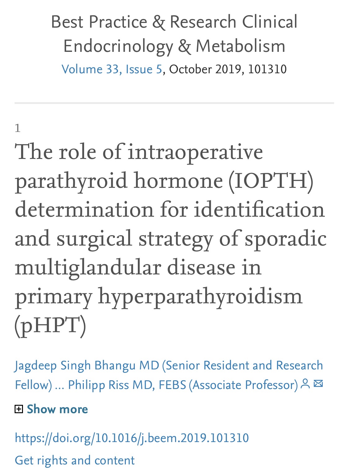
👉The first report for using intraoperative parathyroid hormone (IOPTH) level as an adjunct to guide removal of hyperfunctioning parathyroids was published by Dr. G. Irvin the 3rd et al. in 1993.

👉There are many criteria, however, all require judgement to balance risk of removing multiple glands with risk of recurrent / persistent disease, as outlined in this review.

https://www.sciencedirect.com/science/article/abs/pii/S1521690X19300612?via%3Dihub

Dr. Rodrigo Arrangoiz is a board-certified surgical oncologist who subspecializes in breast cancer and head and neck cancer. Dr. Arrangoiz earned his medical degree at the Anahuac University Medical School in Mexico City, Mexico and graduated Suma Cum Laude. He completed his internship and residency in general surgery at Michigan State University, where he was named chief resident during his fifth year of residency. Dr. Arrangoiz also completed a complex surgical oncology, head and neck fellowship at the Fox Chase Cancer Center in Philadelphia and at the same time he undertook a master’s in science (Clinical Research for Health Care Professionals) at Drexel University in Philadelphia. Dr. Arrangoiz also participated in a two-year global online fellowship in head and neck surgery and oncology through the International Federation of Head and Neck Societies / Memorial Sloan Kettering Cancer Center.

Dr. Arrangoiz has participated in multiple courses and academic congresses as a lecturer and guest professor and has also participated in several publications on topics related to his specialty that include oral cavity cancer, hyperparathyroidism, thyroid cancer, breast cancer, endocrine tumors, squamous cell carcinoma of the head and neck, and more. He is board certified by the American Board of Surgery, the Mexican Board of General Surgery and the Mexican Board of Oncology.

He is a member of various medical associations such as the American College of Surgeons, American Thyroid Association, American Head and Neck Society, American Medical Association, American Society of Clinical Oncology, Association of Academic Surgeons, Society of Surgical Oncology, The Society of Surgery of the Alimentary Tract, Society of American Gastrointestinal Endoscopic Surgeons, and the American Society of Breast Surgeons, among others.

Specialty:

Head and Neck Surgery
Thyroid and Parathyroid Surgery
Breast Surgery
Complex Surgical Oncology

Areas of Clinical Interest:

Malignant thyroid disease (papillary, follicular, medullary, anaplastic thyroid cancer, thyroid lymphoma, and metastatic disease to the thyroid gland) benign thyroid diseases (goiter, multinodular goiter, substernal goiter, hyperthyroidism), hyperparathyroidism / hypercalcemia, benign and malignant breast diseases, head and neck surgery and head and neck cancer.

#Arrangoiz #ParathyroidSurgeon #ParathyroidExpert #Hyperparathyroidism #PrimaryHyperparathyroidism #CancerSurgeon #EndocrineSurgery #Teacher #Surgeon #HeadandNeckSurgeon #SurgicalOncologist #ParathyroidAdenoma #Hypercalcemia #ElevatedCalciumLevels #Miami #MountSinaiMedicalCenter #MSMC #Mexico #Hialeah

Long-term Consequences of Primary Hyperparathyroidism

  • Shorter life span
  • Increase risk of developing cardiovascular disease
  • Increase risk of developing cerebrovascular disease
  • Increase risk of developing a malignancy
  • Increase risk of developing bone disease
  • Increase risk of developing renal disease
  • Decrease quality of life

#Arrangoiz #ParathyroidSurgeon #ParathyroidExpert #EndocrineSurgery #EndocrineSurgery #HeadandNeckSurgery #MountSinaiMedicalCenter #MSMC #Miami #Mexico #HeadandNeckSurgeon #CancerSurgeon

Gastrointestinal Manifestations of Primary Hyperparathyroidism (PHPT)

  • The most frequent gastrointestinal manifestations of PHPT are:
    • Constipation:
      • That occurs in 33% of the cases
    • Heartburn:
      • That occurs in 30% of the cases
    • Nausea:
      • That occurs in 24% of the cases
    • Anorexia:
      • That occur in 15% of the cases
  • A significant reduction in patient symptoms:
    • Is seen after parathyroidectomy
  • The precise pathophysiology is not fully known:
    • Variations in gene expression secondary to sustained stimulation of PTH result:
      • In gut dysmotility:
        • That often leads to constipation and dyspepsia
  • PHPT has been associated with increased incidence of malignancies:
    • Especially of the colon and rectum
  • PHPT has been associated with peptic ulcer disease:
    • The incidence varies between:
      • 5% to 30 % of the cases
    • In animal models:
      • Elevated gastric levels have been shown to result from PTH infusion into blood vessels supplying the stomach:
        • Independent of its effects on serum calcium
  • An increased incidence of pancreatitis has been reported in patients with PHPT:
    • PHPT as a cause of acute pancreatitis was first described by Cope et al, in 1957
    • In retrospective series:
      • The incidence of acute pancreatitis in patients with PHPT:
        • Has varied from 1% to 12%
    • In a study by Jacob et al:
      • They showed a 28-fold increase in the risk of developing pancreatitis in patients with PHPT compared to the general population
      • After removing all other causes:
        • The average serum calcium level seems to be the only predictive factor for pancreatitis development
      • In the diagnostic work-up of acute pancreatitis:
        • PHPT should be included in the differential diagnosis:
          • Although PHPT is found in less than 1% of individuals who present with acute pancreatitis
      • The mechanism of origin that leads to pancreatitis:
        • Seems to be related more to the hypercalcemia than to the PHPT
      • Experimental studies have validated that calcium ions cause calculus deposition within the pancreatic ducts:
        • With subsequent obstruction and inflammation
      • Calcium can also trigger the pancreatitis cascade:
        • By promoting conversion of trypsinogen to trypsin
  • Patients with PHPT also have an increased incidence of cholelithiasis:
    • Presumably due to PTH inhibition of:
      • Gallbladder wall emptying, hepatic bile secretion and sphincter Oddi dysmotility, as well as modification of bile composition (increase in biliary calcium):
        • Which leads to the formation of calcium bilirubinate stones

#Arrangoiz #ParathyroidSurgeon #ParathyroidExpert #HeadandNeckSurgeon #EndocrineSurgery #MountSinaiMedicalCenter #Miami #Mexico #Teacher #Surgeon #Parathyroidectomy #Hypercalcemia #ElevatedCalcium #MSMC

Differential Diagnosis of Hypercalcemia

  • Primary hyperparathyroidism
  • Malignancy:
    • Hematologic (multiple myeloma)
    • Solid tumors (PTHrP)
  • Endocrine diseases:
    • Hyperthyroidism:
      • Mild hypercalcemia occurs in up to 15% to 20% of thyrotoxic patients:
        • Due to a thyroid hormone mediated:
          • Increase in bone resorption:
            • It typically resolves following correction of hyperthyroidism
    • Addisonian crisis:
      • Hypercalcemia occurs in occasional patients with Addisonian crisis:
        • Multiple factors appear to contribute to the hypercalcemia including:
          • Increased bone resorption
          • Volume contraction and increased proximal tubular calcium reabsorption
          • Hemoconcentration
          • Perhaps increased binding of calcium to serum proteins
      • Cortisol administration reverses the hypercalcemia within several days
      • Hypercalcemia has also been reported in patients with secondary adrenal insufficiency due to lymphocytic hypophysitis:
        • The increased release of calcium from bone occurs:
          • Despite appropriate suppression of PTH and calcitriol release:
            • And appears to be mediated, at least in part, by:
              • Thyroid hormone via a process normally inhibited by glucocorticoids
    • Acromegaly
    • Pheochromocytoma:
      • Hypercalcemia is a rare complication of pheochromocytoma
      • It can be due to:
        • Concurrent hyperparathyroidism (in MEN type IIa) or
        • To the pheochromocytoma itself:
          • Appears to be due to:
            • Tumoral production of PTH-related protein
          • Serum PTH-related protein concentrations in these patients can be reduced by alpha-adrenergic blockers:
            • Suggesting a mediating role for alpha-stimulation
  • Vipoma
  • Milk alkali syndrome:
    • In the absence of renal failure:
      • Hypercalcemia can be induced by a:
        • High intake of milk or more commonly, calcium carbonate:
          • Leading to the Milk-Alkali-Syndrome:
            • Hypercalcemia
            • Metabolic alkalosis
            • Renal insufficiency
      • The metabolic alkalosis augments the hypercalcemia:
        • By directly stimulating calcium reabsorption in the distal tubule:
          • Thereby diminishing calcium excretion
      • A calcium-induced decline in renal function:
        • Due to renal vasoconstriction and, with chronic hypercalcemia:
          • Leads to structural injury:
            • This can also contribute to the inability to excrete the excess calcium
      • Renal function usually returns to baseline after cessation of milk or calcium carbonate intake:
        • But irreversible injury can occur in patients who have prolonged hypercalcemia
      • Milk-alkali syndrome accounted for:
        • 8.8% of hypercalcemia cases between 1998 and 2003
  • Granulomatous diseases:
    • Sarcoidosis
    • Tuberculosis
    • Berylliosis
    • Histoplasmosis
    • Wegeners Granulomatosis
      • Mechanism:
        • Increased calcitriol:
          • Activation of extra-renal 1-alpha-hydroxylase
  • Medications:
    • Thiazide diuretics:
      • Thiazide diuretics reduce urinary calcium excretion:
        • And therefore can cause mild hypercalcemia (up to 11.5 mg/dL [2.9mmol/L]):
      • In addition, some patients with PHPT may be prescribed thiazides:
        • Which may elevate the serum calcium further and thereby unmask the hyperparathyroidism:
          • Following discontinuation of the drug:
            • These individuals remain hypercalcemic:
              • Although perhaps less so, and are found to have surgically proven hyperparathyroidism:
                • Thus, if a patient taking a thiazide diuretic is found to be hypercalcemic, the drug should be withdrawn, if possible, and calcium and PTH assessed three months later:
                  • Persistent hypercalcemia (with elevated or high-normal PTH) after drug is withdrawal suggests that the thiazide has unmasked primary hyperparathyroidism
  • Lithium:
    • Patients receiving chronic lithium therapy often develop mild hypercalcemia:
      • Most likely due to increased secretion of PTH:
        • Due to an increase in the set point at which calcium suppresses PTH release
        • The hypercalcemia usually, but not always:
          • Subsides when the lithium is stopped
            • Lithium can also unmask previously unrecognized mild primary hyperparathyroidism (PHPT)
    • Conversely, lithium can also raise serum PTH concentrations without raising serum calcium concentrations
  • Teriparatide
  • Abaloparatide
  • Theophylline toxicity
  • Vitamin A poisoning:
    • Hypervitaminosis A:
      • In which there is prolonged ingestion of more than 50,000 International Units per day or the administration of retinoic acid to patients with certain tumors (as either cis-retinoic acid or all-trans retinoic acid):
        • Retinoic acid causes a dose-dependent increase in bone resorption:
          • Resulting in an overall incidence of hypercalcemia of approximately:
            • 30%
        • All-trans retinoic acid:
          • Inhibits cell growth in part by downregulation of interleukin-6 receptors:
            • The subsequent rise in serum interleukin-6 concentrations:
              • May be responsible for increased bone resorption and hypercalcemia
  • Vitamin D poisoning
  • Increased calcium intake:
    • A high calcium intake alone is rarely a cause of hypercalcemia:
      • Because the initial elevation in serum calcium concentration:
        • Inhibits both the release of parathyroid hormone (PTH) and in turn the synthesis of calcitriol:
          • In patients who also have reduced urinary excretion, however:
            • Increased intake can cause hypercalcemia:
              • This combination of high calcium intake and low urine calcium excretion occurs in two clinical situations:
                • Chronic kidney disease
                • The milk-alkali syndrome
  • Chronic kidney disease:
    • Renal failure alone, although associated with decreased calcium excretion:
      • Does not lead to hypercalcemia because of the:
        • Calcium-lowering effects of concurrent hyperphosphatemia and decreased calcitriol synthesis:
          • However, hypercalcemia is not unusual in patients who are given:
            • Calcium carbonate or calcium acetate to bind dietary phosphate:
              • Particularly if they have adynamic bone disease or are also treated with calcitriol (or another form of vitamin D):
                • In an attempt to reverse both hypocalcemia and secondary hyperparathyroidism
  • Benign hypocalcuric hypercalcemia
  • Paget’s disease of the bone:
    • With immobilization
  • Immobilization
  • Administration of estrogens or anti-estrogens (Tamoxifen):
    • In patients with breast cancer or bone metastases
  • Rhabdomyolysis:
    • With acute renal failure:
      • Hypercalcemia has been described during the diuretic phase of acute renal failure, most often in patients with rhabdomyolysis:
        • Hypercalcemia in this setting is primarily due to:
          • The mobilization of calcium that had been deposited in the injured muscle
        • Correction of hyperphosphatemia (induced by the rise in glomerular filtration rate), mild secondary hyperparathyroidism induced by the renal failure, and an unexplained increase in serum calcitriol concentrations:
          • All appear to contribute to the hypercalcemia

#Arrangoiz #ParathyroidSurgeon #ParathyroidExpert #HeadandNeckSurgeon #EndocrineSurgery #MountSinaiMedicalCenter

Primary hyperparathyroidism (PHPT) in Pregnancy

Primary hyperparathyroidism in pregnancy is of concern primarily for its potential effect on the fetus and neonate. 

Complications of primary hyperparathyroidism in pregnancy include:

  • Spontaneous abortion, low birth weight, supravalvular aortic stenosis, and neonatal tetany:

    • The latter condition is a result of fetal parathyroid gland suppression by high levels of maternal calcium, which readily cross the placenta during pregnancy.

    • Infants with this condition, used to hypercalcemia in utero, have functional hypoparathyroidism after birth and can develop hypocalcemia and tetany in the first few days of life.

Calcium levels vary in the pregnant patient due to the physiological changes that occur.

  • Carella and Gossain stated that calcium concentrations greater than 10.1 mg/dL during the second or third trimester should prompt an evaluation of PHPT.

In a retrospective patient series in the Norman Parathyroid Clinic in Florida, investigators examined pregnant patients with fetal loss and PHPT:

  • They found that patients with calcium levels of 10.7 mg/dL were associated with pregnancy loss, but most pregnancies continued to term.

  • Calcium levels  greater than 11.4 mg/dL were associated with higher levels of fetal loss, and 72% of fetal loss occurred at or above this level.

Surgery is the definitive treatment for PHP.T

  • However, since surgery for PHPT has inherent potential risks for the pregnant patient, it is often viewed as the last resort.

  • However, given the increasing evidence that supports a higher morbidity and mortality associated with calcium levels of  greater than 11.4 mg/dL, surgical intervention is recommended in patients with levels  greater 11.0 mg/dL, particularly in patients with prior pregnancy loss.

Gestational age plays a role in determining surgical candidacy:

  • Traditionally, surgery is reserved for patients in the second trimester, given the higher risk in the first and third trimesters.

  • First-trimester surgery is avoided due to incomplete organogenesis, and third-trimester surgery has been discouraged because it is associated with a higher risk of preterm labor.

    • In addition, there is a reported 58% fetal mortality associated with third-trimester parathyroidectomy.

    • This mortality rate includes all postoperative complications for the infant, such as premature delivery, intrauterine growth retardation, infant hypocalcemia, neonatal death, and stillbirth.

    • It is impossible to differentiate surgical complications from complications of prolonged hypercalcemia related to the underlying disease process.

Rodrigo Arrangoiz MS, MD, FACS

Cirugía Oncológica

Fox Chase Cancer Center
Tumores de Cabeza y Cuello / Cirugía Endocrina
Fox Chase Cancer Center
Tumores de Cabeza y Cuello / Cirugía Endocrina
IFHNOS / Memorial Sloan Kettering Cancer Center
Maestría en Ciencias de Investigación
Drexel University
Certificado por el Colegio Americano de Cirugía
Fellow del Colegio Americano de Cirugía
Fellow de la Sociedad de Cirugia Oncológica
#hyperparathyroidism
#primaryhyperparathyroidism
#hypercalcemia
#pregnancy
#endocrinesurgeon
#headandnecksurgeon

Coronary Artery Disease is More Severe in Hyperparathyroidism

👉Patients with PHPT have higher coronary calcium scores than population-based controls.

👉Read more at : https://www.surgjournal.com/article/S0039-6060(19)30612-9/fulltext

#MountSinaiMedicalCenter #MSMC #Mexico #Miami #CancerSurgeon #Arrangoiz

#ParathyroidSurgeon

#ParathyroidExpert

#HeadandNeckSurgeon

Recurrent or Persistent Hyperparathyroidism

  • Recent cure rates of hyperparathyroidism following parathyroidectomy:
    • Are reported between 92% and 97%
  • When cure is not obtained, it is due to either:
    • Recurrent or persistent disease (Figure)
  • Persistent hyperparathyroidism:
    • Is defined as hypercalcemia and elevated parathyroid hormone (PTH) levels:
      • During the 6-month period after parathyroidectomy:
        • It occurs in 0.4% to 7.8% of patients
  • Recurrent hyperparathyroidism:
    • Is defined as hypercalcemia and elevated PTH levels that occur after normal values have been obtained for 6 months after parathyroidectomy and may be slightly less common than persistent disease:
      • Occurring in 0.4% to 4.8% of patients
  • It is most common in:
    • Familial hyperparathyroidism
  • Advances in imaging modalities and surgical techniques have decreased the rate of persistent disease significantly over the past two decades
  • Persistent disease is considered a technical error:
    • Most commonly due to a missed single parathyroid adenoma
  • A rare but notable cause of persistent or recurrent hyperparathyroidism is parathyromatosis:
    • A condition in which multiple benign hyperfunctioning parathyroid tissues are
      scattered throughout the neck and superior mediastinum and that occurs due to surgical implants from the fragmentation of abnormal parathyroid tissue in the previous operation
  • Other causes are detailed in Table 1 (Table 1)
  • Additional studies have shown that specific clinical factors are associated with an increased or decreased risk of having persistent hyperparathyroidism:
    • Negative, equivocal, or discordant imaging including ultrasonography and sestamibi is associated with a higher likelihood of persistent disease
    • Whereas intraoperative PTH (IOPTH) monitoring, with 50% decrease in serum PTH after removal of parathyroid tissue, increased weight of the affected gland, and surgery performed at high-volume centers is associated with successful initial parathyroidectomy
Causes of Altered Calcium Metabolism after Parathyroidectomy
  • Work-up and Diagnosis:
    • When a patient presents with recurrent or persistent hyperparathyroidism, all other causes of hyperparathyroidism including:
      • Vitamin D deficiency, renal insufficiency, medications such as lithium resulting in hypercalcemia, and renal calcium leak need to be ruled out and treated if identified
    • Additional work-up should also be done to evaluate for familial causes of hyperparathyroidism
    • The operative reports, imaging studies, and pathology reports associated with prior operation(s) should be obtained and meticulously reviewed
    • Confirmation of the location(s) of abnormal parathyroid glands:
      • Using at least two imaging modalities preoperatively is recommended in all cases of recurrent or persistent disease
    • Imaging modalities include ultrasonography, sestamibi with or without single-photon emission CT, and four-dimensional CT and should include evaluation of the mediastinum
    • When multiple imaging techniques are used:
      • 95% of abnormal glands can be identified
    • Evaluation of the vocal cords:
      • To assess for any previous recurrent laryngeal nerve injury resulting in paralysis is recommended for all neck reoperations
    • Fine-needle aspiration (FNA):
      • May be used preoperatively to confirm presence of abnormal parathyroid tissue when noninvasive imaging studies are unclear but should be used with caution and
        only in select patients:
        • As FNA carries the risk of hematoma, parathyromatosis, and cancer seeding
      • Selective PTH venous sampling (SVS) has also been described to regionalize recurrent or persistent disease when other imaging modalities fail and has been reported as successful in up to 94% of cases:
        • SVS is performed by obtaining serial samples of PTH from the superior vena cava, brachiocephalic, internal jugular, vertebral, thymic, and thyroid veins, after which levels are compared to identify the approximate location of the hyperfunctioning gland
  • Indications for Surgery:
    • Reoperation is associated with significantly increased risk of:
      • Hypoparathyroidism (5% to 20%)
      • Recurrent laryngeal nerve injury (10% to 15%)
    • Therefore, it is recommended to have a higher
      threshold before recommending reoperation for recurrent or persistent disease
    • Indications for reoperation:
      • Significant life-limiting symptoms
      • Nephrolithiasis
      • Osteoporosis
      • Hypercalcemia
      • Decrease in renal function
    • If surgery is indicated, early reexcision in the case of persistent disease:
      • Is not recommended, rather revision should be delayed by 4 to 6 months to avoid a surgical bed affected by inflammation and scarring
  • Operative Approach:
    • The goal of resection:
      • Is to remove the remaining hyperfunctioning parathyroid tissue while preserving enough to avoid hypoparathyroidism
    • Due to increased scar tissue in a reoperative neck, care is taken to avoid injury to surrounding structures, including the recurrent laryngeal nerve
    • For most patients, resecting the residual hyperfunctioning parathyroid tissue:
      • Is possible through a standard, anterior cervical incision
    • A lateral approach for targeted glands in the
      tracheoesophageal groove or sternocleidomastoid:
      • Is an excellent approach to avoid scar tissue
    • Glands within the mediastinum:
      • Most often lie within the thyrothymic ligament
      • They can be gently pulled up through a cervical incision successfully, even in the reoperative neck
    • A thoracic approach:
      • Is recommended for those with abnormal parathyroid tissue of 6 cm or greater identified below the superior aspect of the clavicle
    • IOPTH is recommended:
      • As it significantly improves the ability to confirm adequate resection of recurrent disease intraoperatively:
        • With sensitivity of up to 100%
    • Additional strategies have been reported to localize parathyroid tissue and minimize injury to surrounding structures in a reoperative neck, including:
      • The use of the gamma probe after injection of 99m technetium-methoxy isobutyl isonitrile preoperatively, ultrasonography-guided methylene blue injection, near-infrared autofluorescence imaging, indocyanine green fluorescence angiography, and recurrent laryngeal nerve monitoring:
      • Previously, IV infusion of methylene blue was
        used by some to identify parathyroid tissue; however, this is no longer recommended due to associated toxicities
    • Parathyroid autotransplantation and cryopreservation are often recommended following resection of the affected parathyroid tissue in reoperative parathyroidectomies:
      • Especially if the abnormal gland is the last viable parathyroid tissue, multiple glands have been previously removed, or there is concern for possible devascularization of the
        remaining glands
      • A case where cryopreservation may not be necessary is that of a missed adenoma
        where other parathyroid glands have not been disturbed in either the initial operation or re-exploration
  • Follow-up postoperatively includes monitoring of serum PTH and calcium with the treatment of hypocalcemia if present

Primary Hyperparathyroidism

  • Primary Hyperparathyroidism (PHPT):
    • Occurs when one (or more) of the four parathyroid glands grows into a tumor and behaves inappropriately by constantly making excess parathyroid hormone (PTH)
    • The parathyroid glands grow into a tumor and produce too much parathyroid hormone (PTH):
      • Which removes calcium from the bones
  • In Primary Hyperparathyroidism (PHPT) the tumor (Adenoma):
    • Has lost its control mechanism and makes large amounts of parathyroid hormone (PTH):
      • Thus, even when the serum calcium level is high:
        • When the normal response of the parathyroid glands should be to stop making any PTH at all:
          • One of the parathyroid glands keeps making the parathyroid hormone
  • One important thing to remember as you read about PHPT:
    • Is that the abnormal PTH production is caused by a benign parathyroid tumor, called ADENOMA:
      • It is not a cancer:
        • However this disease (PHPT) can increase the occurrence of other cancers:
          • Breast Cancer
          • Prostate Cancer
          • Renal Cancer
          • Skin Cancer
          • Colon and rectal cancer
  • The most common cause of excess hormone production (primary hyperparathyroidism):
    • Is the development of a benign tumor in one of the parathyroid glands:
      • This enlargement of one parathyroid glands is called a:
        • Parathyroid adenoma:
          • Which accounts for roughly 85% to 90% of all patients with primary hyperparathyroidism:
            • The other 10% to 15% will have more than one bad gland (mulitiglandular disease):
              • Doble Adenomas:
                • 6% to 9% of the cases
              • Triple Adenomas:
                • Less than 0.3% of the cases
              • Parathyroid Gland Hyperplasia:
                • 3% of the cases
  • This out of control (autonomously functioning) parathyroid gland:
    • Is essentially never cancerous (virtually 99% of them are benign tumors):
      • However, PHPT slowly causes damage to the body:
        • Because it induces an abnormally high level of calcium in the blood:
          • Which can slowly destroy a number of tissues
  • Parathyroid adenomas:
    • Typically are much bigger than the normal “grain-of-rice” parathyroid and will frequently be about the size of an olive or grape:
      • Thus, the typical patient with primary hyperparathyroidism will be cured of the disease when this “grape” size tumor is removed
    • Up to 30% will have more than one tumor
    • This tumor / adenoma will almost always continue to grow until it is removed:
      • As it grows, it makes more PTH and will make you feel worse (symptomatic)
    • This tumor is constantly making parathyroid hormone (PTH):
      • Which is what takes the calcium out of your bones and makes the calcium in your blood too high
Algorithm for Diagnosis of Primary Hyperparathyroidism *Borderline serum Calcium and PTH levels may need to be measured several times to establish the diagnosis, as levels will fluctuate over time. ** Common secondary causes of elevated PTH levels include chronic kidney disease, vitamin D deficiency, medications, renal hypercalciuria, malabsorption syndromes

#Arrangoiz #ParathyroidSurgeon #ParathyroidExpert #Hyperparathyroidism #PrimaryHyperparathyroidism #CancerSurgeon #EndocrineSurgery #Teacher #Surgeon #HeadandNeckSurgeon #SurgicalOncologist #ParathyroidAdenoma #Hypercalcemia #ElevatedCalciumLevels #Miami #MountSinaiMedicalCenter #MSMC #Mexico #Hialeah