Wound Healing

  • Phases and Timelines:
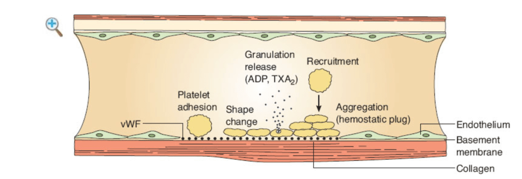
    • Hemostasis (minutes – hours):
      • Platelets → fibrin clot:
        • Releases PDGF, TGF-β, vWF, fibrinogen, Factor V, thrombospondin
    • Inflammatory phase:
      • Last roughly from day 0 to day 3/4:
        • Can last up to ~7 days in larger / contaminated wounds
      • PMNs dominate first 48 hours:
        • Macrophages peak ~ 48 hours to 72 hours
      • Key signals:
        • TNF-α, IL-1, IL-6, TGF-β, PDGF
    • Proliferative / Regenerative Phase:
      • From rougly day 3 to day 21
      • Fibroblasts:
        • Lay down type III collagen
      • Endothelial cells:
        • Neovascularization
      • Keratinocytes:
        • Re-epithelialization:
          • Approximalty 1 mm / day, (range 0.5 mm to 1.5 mm)
      • Growth factors:
        • PDGF, FGF (including KGF / FGF-7), EGF, VEGF, TGF-β
    • Remodeling / Maturation phase:
      • From 3 weeks to 12 to 18 months
      • Collagen Type III → type I collagen:
        • Collagen fibers re-align, cross-link; decreased cellularity and vascularity
  • Who Arrives When?
    • Immediate: 
      • Platelets
    • Hours to Day 2: 
      • PMNs
    • Day 2 to 4: 
      • Macrophages:
        • Switch from M1 → M2 phenotype to drive repair
    • Day 3+:
      • Peak ~ 1 to 2 weeks): 
        • Fibroblasts and endothelial cells
    • ~ Day 5+: 
      • Lymphocytes:
        • Modulate later phases
  • Matrix and Collagen:
    • Provisional matrix: 
      • Fibrin + fibronectin + hyaluronic acid:
        • Scaffold for cells
    • Fibronectin (from fibroblasts, platelets):
      • Chemotactic for macrophages
      • Anchors fibroblasts
    • Collagen synthesis requires: 
      • α-ketoglutarate, O₂, Fe²⁺, vitamin C for prolyl/lysyl hydroxylases:
        • Hydroxylation → triple-helix stability
      • Cross-linking: 
        • Lysyl oxidase (Cu²⁺-dependent) forms intermolecular cross-links:
          • Equals tensile strength
      • Triple helix motif: 
        • Glycine is every 3rd amino acid (Gly-X-Y; X/Y often Pro/Hyp).
    • Collagen types (surgical high-yield):
      • Type I: 
        • Skin, tendon, bone:
          • Predominant in healed wounds
      • Type II: 
        • Cartilage
      • Type III: 
        • Granulation tissue, vessels, early wound
      • Type IV: 
        • Basement membranes
      • Type V: 
        • Widespread
        • Enriched in cornea / placenta
    • Drugs: 
      • d-Penicillamine (and β-aminopropionitrile:
        • Impair cross-linking
    • Tensile Strength (what really matters for primary closure):
      • ~ 10% by end of week 1
      • ~ 20%to 30% by 3 weeks:
        • Collagen content near max by ~ 3 to 4 weeks
      • ~ 50% to 60% by 6 to 8 weeks
      • 80% by ~ 3 months:
        • Plateau (never returns to 100%) through 6 to 12 months
  • Epithelialization and Contraction:
    • Epithelialization sources: 
      • Hair follicles (#1), wound edges, sweat glands
    • Rate ≈ 1 mm / day
    • Requires healthy granulation bed
    • Myofibroblasts (α-SMA+, gap junctions):
      • Drive wound contraction:
        • Greatest where skin is lax:
          • Perineum, scrotum
        • Least on scalp / tight skin
  • Open wounds (secondary intention): 
    • Epithelial integrity is key; until closed
    • Leak protein-rich exudate and are prone to colonization
  • Practical Timelines:
    • Suture removal (general guide; adjust for tension / vascularity):
      • Face: 5 to 7 days
      • Scalp: 7 to 10 days
      • Trunk / Upper limb: 10 to 14 days
      • Lower limb / Joints/Back: 12 to 14 days
    • Bowel strength:
      • Submucosa is the strength layer:
        • Anastomosis weakest at day 3 to 5:
          • Collagenolysis > synthesis
    • Peripheral nerve regeneration: ~ 1 mm/day (range: 0.5 mm to 3 mm/day)
  • Essentials to Optimize Healing:
    • Moist wound environment:
      • Avoid desiccation
    • Oxygen delivery: 
      • Correct anemia, optimize perfusion, pain control, stop nicotine (vasoconstriction), consider revascularization if PAD
    • Transcutaneous oxygen measurement (TCOM) targets:
      • Greater than 40 mm Hg predicts healing
      • 25 to 40 mm Hg borderline
      • < 25 mm Hg poor
    • Edema control: 
      • Elevation, compression (if ABI adequate)
    • Debridement: 
      • Remove necrotic tissue / biofilm:
        • Consider enzymatic or surgical debridement
    • Infection control: 
      • 10⁵ CFU/g tissue (not per cm²) impairs healing:
        • Debride +/- topical / systemic therapy as indicated
    • Nutrition: 
      • 1.2 to 1.5 g/kg/day protein
      • Adequate calories, vitamin C, zinc (short-term if deficient), copper, arginine / glutamine if malnourished
  • Impediments and Meds (nuance):
    • Diabetes:
      • Impaired neutrophil chemotaxis / killing
      • Microvascular disease and neuropathy → pressure injury
    • Tight peri-op glycemic control helps.
      Steroids: 
      • Blunt inflammation and collagen synthesis → ↓ tensile strengt
      • Vitamin A 25,000 IU/day for 1 to 2 weeks can reverse steroid effects (avoid in pregnancy / liver disease)
    • Cytotoxics / antimetabolites (5-FU, MTX) and calcineurin inhibitors (cyclosporine, tacrolimus):
      • Impair early proliferative phase
      • Biggest impact within ~2 weeks of injury
    • Radiation: 
      • Endarteritis obliterans, fibroatrophy
      • Consider HBOT for selected compromised grafts / flaps
    • Smoking / nicotine: 
      • Vasoconstriction, ↑ COHb, ↓ oxygen delivery – cessation is critical
  • Specific Clinical Entities:
    • Keloids: 
      • Extend beyond original borders
      • Familial predisposition (higher in patients of African / Asian ancestry)
      • Treatment:
        • Intralesional triamcinolone ± 5-FU, silicone, pressure therapy
        • Consider post-excision RT in select cases
    • Hypertrophic scars: 
      • Confined within original wound
      • Common in high-tension areas (shoulders, presternal, joints)
      • Treatment as above:
        • Often regress with time
    • Pyoderma gangrenosum: 
      • Neutrophilic dermatosis
      • Avoid aggressive debridement (pathergy)
      • Treatment: steroids / immunosuppressants
    • Epidermolysis bullosa: 
      • Structural protein defects:
        • Examples – keratin, laminin, collagen VII
      • Supportive care; wound-care expertise
    • Diabetic foot ulcers: 
      • Most commonly plantar metatarsal heads and heel
    • Charcot midfoot deformity shifts pressure to plantar midfoot:
      • Treatment: 
        • Off-loading (total contact cast), debridement, infection control, revascularization if needed
    • Leg ulcers: 
      • ~ 70% to 80% venous, 10% to 15% arterial, rest mixed
      • Use ABI before compression:
        • Unna boot or multilayer compression for venous disease
    • Scar revision: 
      • Delay roughly 12 to 18 months for maturation
    • Infants vs fetal healing: 
      • Fetal wounds (early gestation) may heal scar-lessly
      • Infants still scar (often less conspicuously)
  • Primary vs Secondary Intention:
    • Primary intention: 
      • Tensile strength hinges on collagen deposition and cross-linking
    • Secondary intention: 
      • Epithelial integrity over a healthy granulation bed is paramount
    • Delayed primary (tertiary) closure: 
      • Leave open initially for contaminated wounds:
        • Close once clean – reduces infection risk:
          • Ensure no residual infection to avoid abscess
  • Platelet Granules and Aggregation:
    • Alpha granules: 
      • PDGFTGF-βvWFfibrinogenFactor VthrombospondinP-selectin, β-thromboglobulin
    • Dense granules: 
      • ADP / ATPserotoninCa²⁺, (± epinephrine)
    • Key aggregation / activation mediators: 
    • It’s β-thromboglobulin in platelets:
      • Binds thrombin
    • PF4 (CXCL4):
      • Is a chemokine that neutralizes heparin and modulates coagulation

How to Assess Family History for Breast Cancer?

  • In assessing a family history, consider how likely it is that there is a deleterious mutation, and how likely it is that your patient could carry that mutation
  • Factors to consider include:
    • Age at diagnosis
    • What degree relative is affected
    • If a cancer type is associated with a hereditary syndrome
  • Cancers suggestive of a hereditary mutation include:
    • Those occurring in young patients:
      • For example premenopausal cancers
    • Those occurring in 1st or 2nd degree relatives
    • Multiple generations with cancer
    • Multiple cancers in a single person
    • The presence of cancers associated with certain syndromes
  • References
    • Daly MB, Axilbund JE, Buys S, Crawford B, Farrell CD, Friedman S, et al. Genetic/familial high-risk assessment: breast and ovarian. J Natl Compr Canc Netw. 2010;8(5):562-594.
    • Domchek SM, Gaudet MM, Stopfer JE, Fleischaut MH, Powers J, Kauff N, et al. Breast cancer risks in individuals testing negative for a known family mutation in BRCA1 or BRCA2. Breast Cancer Res Treat. 2010;119(2):409-414.
    • Streff H, Profato J, Ye Y, Nebgen D4, Peterson SK5, Singletary C, et al. Cancer Incidence in first- and second-degree relatives of BRCA1 and BRCA2 mutation carriers. Oncologist. 2016;21(7):869-874.
#Arrangoiz #Doctor #Surgeon #CancerSurgeon #SurgicalOncologist #BreastSurgeon #MountSinaiMedicalCenter #MSMC #Miami #Mexico #BreastCancer

Head and Neck Squamous Cell Carcinoma – Positive Deep Margin + ENE+ → Best Adjuvant Plan?

  • Positive deep margin + ENE(+):
    • Best adjuvant plan: 
      • Cisplatin–Post-op RT (CRT):
        • Why: 
          • These are the classic high-risk pathologic features that derived clear benefit from adding concurrent high-dose cisplatin to adjuvant RT in the two landmark randomized trial:
            • EORTC 22931 and RTOG 9501, and in their comparative analyses
  • What the trials showed:
    • EORTC 22931 (Bernier et al., NEJM 2004):
      • Population: 
        • Resected stage III to IV HNSCC with high-risk features:
          • Definition broader than RTOG 9501
      • Arms: 
        • RT alone (66 Gy) vs RT + cisplatin 100 mg / m² q3wk ×3
      • 5-yr outcomes (Kaplan–Meier):
        • Overall survival: 
          • 53% (CRT) vs 40% (RT) (significant)
        • Progression-free survival:
          • 47% (CRT) vs 36% (RT) (significant)
        • Locoregional control: 
          • Improved with CRT
      • Interpretation: 
    • RTOG 9501 (Cooper et al., NEJM 2004; 10-yr update 2012):
      • Population: 
        • Resected HNSCC with prespecified high-risk factors
      • Arms: 
        • RT 60 Gy/6 wk vs RT + cisplatin 100 mg / m² on days 1, 22, 43
      • Entire cohort (10-yr KM): 
        • OS and DFS not significantly different overall
        • LRF numerically lower with CRT but not significant
      • Crucial prespecified subset:
        • Positive margins and / or ENE(+):
          • Locoregional failure: 
            • 33.1% (RT) vs 21.0% (CRT)p = 0.02
          • Disease-free survival: 
            • 12.3% (RT) vs 18.4% (CRT)p = 0.05
          • Overall survival: 
            • 19.6% (RT) vs 27.1% (CRT), trend in favor of CRT
      • Interpretation: 
    • Combined / Comparative analyses (what ties it together):
      • Bernier et al., Head & Neck 2005 (comparative look at EORTC 22931 and RTOG 9501):
        • Concluded that the greatest and most consistent benefit from adjuvant CRT accrues to patients with:
          • Extranodal extension and / or positive marginsPubMed+1
      • Updated combined analysis (Zumsteg et al., 2025, Annals of Oncology—abstract): 
        • Pooling EORTC 22931 + RTOG 9501:
          • Shows OS improvement with postoperative CRT overall:
            • ENE and margin status were prognostic but not strictly predictive:
              • Patients without these features may still benefit
          • While cancer-specific mortality fell with CRT:
            • But some benefit was offset by other-cause mortalit:
  • Practical plan for positive deep margin + ENE(+):
    • Adjuvant CRT:
      • RT with cisplatin 100 mg / m² q3wk ×2 to 3:
        • Goal cumulative ≥ 200 mg / m²:
      • If cisplatin-ineligible: 
        • Acknowledge that randomized survival benefit in this setting is with cisplatin:
          • Alternatives (e.g., RT alone or RT+cetuximab) are considered when cisplatin cannot be given:
            • But are not proven equivalent post-op (See NCCN.) NCCN
  • Bottom line: 
    • For positive deep margin plus ENE(+):
      • The weight of EORTC 22931RTOG 9501 (10-yr), and subsequent comparative work:
        • Supports adjuvant cisplatin-RT as the best plan to maximize locoregional control and survival
Rodrigo Arrangoiz, MD (Oncology Surgeon)

BRCA 1 and BRCA 2 Genes

  •  BRCA 1 and BRCA 2 are genes:
    • That produce tumor suppressor proteins:
      • Which help repair damaged DNA
    • They are the most common gene alterations:
      • Seen in the hereditary breast cancer population
    • They are associated with an increased risk of breast cancer estimated to be:
      • 55% to 70% for BRCA 1 carriers by age 70
      • 45% to 70% in BRCA 2 carriers by age 70
    • While both BRCA 1 and BRCA 2 mutations are associated with an increased risk of breast cancer:
      • BRCA 1 breast cancers more commonly occur in:
        • Younger
        • Premenopausal women
        • Are more likely to be triple negative
        • High grade lesions
    • BRCA 1 is associated with a higher risk of ovarian cancer compared to BRCA 2:
      • With a lifetime risk of 40% to 45% in BRCA 1 carriers compared to 15% to 20% in BRCA 2 carriers
    • BRCA 2 breast cancers more closely resemble the sporadic breast cancer pattern:
      • With a predominance of hormone receptor positive cancers in women greater than 50 years
  • CHEK 2 and PALB 2 are moderate penetrance genes:
    • That are less common than BRCA mutations
    • Similar to BRCA 2 deleterious mutations:
      • CHEK 2 and PALB 2 mutations are associated with:
        • Hormone receptor positive postmenopausal breast cancer
  • References:

What the CREATE-X Trial Showed?

  • The CREATE-X trial:
    • Studied adjuvant capecitabine in patients with HER2-negative breast cancer:
      • Who had residual invasive disease after neoadjuvant chemotherapy:
        • Did not achieve a pathological complete response
    • It found that adding capecitabine:
      • Improved disease-free survival (DFS) and overall survival (OS) compared to observation in that specific patient population
    • The effect was more pronounced in certain subgroups:
      • Particularly patients with triple-negative breast cancer
    • In long-term follow-up, for example:
      • 5-year DFS was higher in capecitabine arm vs control
  • So the key message of the Create -X trial:
    • In patients who have residual disease after neoadjuvant chemotherapy (i.e. higher risk of relapse):
      • Adding capecitabine may provide a survival benefit
  • Is CREATE-X Still Applicable Today?
    • Yes:
      • But one must interpret in light of modern advances
    • Some important considerations:
      • Population and treatment context have changed:
        • In CREATE-X, many patients did not receive newer therapies that are more commonly used today (e.g. immunotherapy, targeted agents)
        • The neoadjuvant regimens used then may differ from current ones (some now include platinum drugs, immunotherapy, etc.)
        • Thus, whether the magnitude of benefit from capecitabine is identical in today’s more aggressive or modern regimens is uncertain
  • Evolving standard of care:
    • In triple-negative breast cancer (TNBC):
      • Immunotherapy (checkpoint inhibitors) is now incorporated into neoadjuvant and adjuvant settings in many protocols:
        • Some patients receive pembrolizumab or other immune agents in the neoadjuvant plus adjuvant phase
    • In patients with germline BRCA mutations:
      • Adjuvant PARP inhibitors (e.g. olaparib) have been shown to improve outcomes in high-risk disease, including non-pCR settings
    • Newer antibody-drug conjugates (ADCs) and other novel therapies are being tested in residual disease settings:
      • Sacituzumab govitecan in SASCIA trial:
        • That could potentially surpass capecitabine in benefit
  • Subgroup-specific relevance:
    • The benefit in the original CREATE-X was strongest in certain subtypes (especially TNBC)
    • For hormone receptor–positive, HER2-negative disease:
      • The benefit is less clear or more modest
    • Some meta-analyses and reviews suggest that in modern TNBC:
      • The role of capecitabine is still valid:
        • Especially for patients with residual disease after standard therapy
  • Ongoing trials and unanswered questions:
    • As newer therapies emerge, trials are ongoing to compare or combine capecitabine with immunotherapy or other agents in the post-neoadjuvant (residual disease) setting:
      • One open question is whether capecitabine adds incremental benefit on top of modern therapies (immunotherapy, PARP inhibitors) or whether it’s supplanted in certain subgroups
  • My Bottom Line / Practical View:
    • Yes, clinicians often still use the CREATE-X findings as a rationale for giving adjuvant capecitabine in patients with residual disease after neoadjuvant chemotherapy:
      • Particularly in HER2-negative / triple-negative cases, when no more effective alternative is clearly indicated:
        • But, the decision must be individualized:
          • Consider what neoadjuvant therapy was used (did it include immunotherapy or platinum?)
          • Consider patient risk factors, subtype (TNBC vs HR+), mutation status (BRCA), comorbidities, etc
          • Consider newer options that may be more beneficial (e.g. PARP inhibitors in BRCA carriers, or ADCs if approved in that setting)

The Study of Tamoxifen and Raloxifene (STAR) Trial

  • The NSABP P-2, or STAR trial:
    • Enrolled 19,747 postmenopausal women with a 5-year Gail risk assessment score of ≧ 1.66% for the development of invasive breast cancer at 5 years
    • The women were randomized to receive:
      • 20 mg of tamoxifen plus placebo or 60 mg of raloxifene plus placebo
    • The updated results of the STAR trial (median follow-up 81 months):
      • Reported more cases of invasive breast cancer in the raloxifene group than the tamoxifen group (risk ratio [RR]: 1.24; 95% confidence interval [CI]: 1.05–1.47):
        • Demonstrating that raloxifene is about 76% as effective as tamoxifen in reducing breast cancer risk
      • There were significantly fewer cases of invasive uterine cancer with raloxifene compared to tamoxifen (RR: 0.55; 95% CI, 0.36–0.83)
      • Thromboembolic events occurred less often in the raloxifene group (RR: 0.75; 95% CI: 0.6–0.93)
      • There were fewer cataracts and cataract surgeries in the women taking raloxifene (RR: 0.79; 95% CI: 0.68–0.92)
      • Importantly, there was no significant difference in mortality between the two groups
  • References
    • Vogel VG, Costantino JP, Wickerham DL, Cronin WM, Cecchini RS, Atkins JN, et al; for the National Surgical Adjuvant Breast and Bowel Project (NSABP). Effects of tamoxifen vs raloxifene on the risk of developing invasive breast cancer and other disease outcomes: the NSABP Study of Tamoxifen and Raloxifene (STAR) P-2 trial. JAMA. 2006;295(23):2727-2741.
    • Vogel VG. The NSABP Study of Tamoxifen and Raloxifene (STAR) trial. Expert Rev Anticancer Ther. 2009;9(1):51-60.
    • Mamounas EP, Wicherham DL, Fisher B, Geyer CE, Julian TB, Wolmark N. The NSABP experience. In: Kuerer HM, ed. Kuerer’s Breast Surgical Oncology. New York, NY: McGraw-Hill Companies; 2010:475-508.

#Arrangoiz #BreastSurgeon #CancerSurgeon #SurgicalOncologist #BreastCancer #NSABPP2 #Raloxifen #Tamoxifen

What is the RxPONDER Trial in Breast Cancer?

  • Full name:
    • “Rx for Positive Node, Endocrine Responsive Breast Cancer”:
      • SWOG S1007
  • Population:
    • Women with hormone receptor (HR)-positive, HER2-negative breast cancer:
      • With 1 to 3 positive axillary lymph nodes:
        • Who have had surgery
    • All had a 21-gene Recurrence Score (RS) of:
      • ≤ 25
  • Key question:
    • Among patients with 1 to 3 positive nodes and RS ≤ 25:
      • Which patients benefit from adding adjuvant chemotherapy to endocrine therapy vs endocrine therapy alone? 
  • Trial Design:
    • Multi-center trial:
      • 632 sites across 9 countries (USA, Canada, Mexico, Colombia, Ireland, France, Spain, South Korea, Saudi Arabia):
        • Enrolled over 5,000 women
    • Enrollment:
      • ~ 5,083 women assigned
      • ~ 5,015 analyzed
      • Approximately two-thirds were postmenopausal
      • One-third premenopausal
    • Treatment arms:
      • Randomized to endocrine therapy alone vs chemotherapy + endocrine therapy
    • Chemotherapy regimens included:
      • Taxane and / or anthracycline
    • Recurrence Score stratifications within ≤ 25:
      • RS categories 0 to 13 vs 14 to 25 used for some analyses
    • Also stratified by menopausal status, number of nodes (1 vs 2 to 3), tumor size / grade
    • Follow-up:
      • Median ≈ 5.1 years for primary results

RxPONDER (SWOG S1007): Five-Year Outcomes
HR-positive / HER2-negative, 1 to 3 positive nodes, Recurrence Score ≤ 25
  • Other data:
    • Overall survival at 5 years among premenopausal women:
      • 98.6% with chemotherapy + endocrine therapy vs 97.3% endocrine therapy only (absolute ~ 1.3%) – HR ~ 0.47; P ≈ 0.032
    • Among postmenopausal women, OS was virtually identical:
      • 96.2% vs 96.1% (chemo vs no chemo) – HR ~ 0.96; not statistically significant
    • Also:
      • The benefit in premenopausal women:
        • Was consistent across RS levels (0 to 13 and 14 to 25):
          • Though absolute benefit tended to be larger in those with RS 14 to 25
      • The benefit was also seen irrespective of number of positive nodes (1 vs 2 to 3):
        • But, again, the magnitude of benefit varied
  • Secondary / Extended Analyses and Modeling:
    • A modeling study (Wojcik et al., 2024) simulated 10-year distant recurrence-free survival, life-years, and quality-adjusted life-years (QALYs) for women like those in RxPONDER:
      • In premenopausal women:
        • 10-year distant RFS ~ 85.3% with chemo-endocrine therapy vs 80.1% endocrine therapy alone (absolute benefit ~ 5.6%) in the simulation
      • In postmenopausal women:
        • No meaningful benefit; distant RFS practically the same between arms
      • Modeled life-years gained:
        • ~ 2.1 years for premenopausal women; no gain or even small losses for postmenopausal receiving chemotherapy (due to toxicity and side effects) when weighed
    • Another RxPONDER analysis looked at racial / ethnic outcomes:
      • Non-Hispanic Black women had worse 5-year IDFS and DRFS compared to non-Hispanic White women despite similar RS, node numbers, and treatment:
        • Asian women had somewhat better outcomes
      • But chemotherapy efficacy didn’t differ significantly by race
  • Implications / Guidelines Impact:
    • For postmenopausal women with HR+ / HER2- disease, 1 to 3 positive nodes, and RS ≤ 25:
      • Chemotherapy can generally be omitted without compromising IDFS:
        • Endocrine therapy alone is acceptable in most
    • For premenopausal women in the same category:
      • Chemotherapy + endocrine therapy provides meaningful benefit:
        • Omission risks worse IDFS
    • The decision should consider absolute benefit, potential side effects, and patient preferences
    • Also – it is not completely certain how much of the
    • Recurrence Score:
      • Remains a useful tool in node-positive disease (for nodes 1 to 3) to stratify risk and guide therapy
    • Previously, Oncotype DX was used more in node-negative disease (TAILORx):
      • But RxPONDER expands its utility
    • The data supports more personalized treatment – sparing many postmenopausal women unnecessary chemotherapy, reducing overtreatment and its toxicities
  • Things to Remember / Caveats:
    • Follow-up duration:
      • Median ~ 5 years:
        • Longer-term data (10+ years) may reveal differential distant recurrence or survival effects:
          • Especially in HR+ disease which often has late recurrences
    • Chemotherapy regimens, adherence, patient comorbidities:
      • The trial setting may not fully reflect “real world” in all respects – e.g. older women with comorbidity may suffer more chemotherapy toxicity
    • Menopausal status matters:
      • Distinct differences in benefit. Also, in premenopausal women, part of chemotherapy’s benefit may be mediated via ovarian suppression (or ablation), which was not fully controlled for
    • Patient preferences are key:
      • Absolute benefit for many is modest; potential chemotherapy toxicities (short-term and long-term) need weighing
    • RS >25 were excluded – standard indications for chemotherapy remain for high RS or more nodes, etc
  • Key Points:
    • No significant chemotherapy benefit in postmenopausal women:
      • Absolute IDFS gain only ~ 0.6 % at 5 years, hazard ratio ~1.0
    • Clear benefit in premenopausal women:
      • Absolute IDFS gain ~ 4% to 5 %, distant recurrence reduction ~ 3 %, and a small OS improvement (~ 1 %)
      • Benefit was consistent across Recurrence Score 0 to 13 vs 14 to 25 and for 1 vs 2 to 3 positive nodes
  • Clinical implication:
    • Postmenopausal patients with RS ≤ 25 and 1 to 3 positive nodes:
      • Can usually omit adjuvant chemotherapy
    • Premenopausal patients in the same setting should be offered chemotherapy:
      • As the IDFS and DRFS advantages are clinically meaningful
  • Primary source:
    • Kalinsky K et al. N Engl J Med 2021;385:2336-2347 【NEJM doi:10.1056/NEJMoa2108873】

Create-X Trial

  • The CREATE-X study:
    • Randomly assigned 910 patients with HER2 negative breast cancer and residual disease after undergoing neoadjuvant chemotherapy:
      • To standard postsurgical treatment and capecitabine or placebo
    • The primary end point:
      • Was disease-free survival (DFS)
    • Secondary end points included:
      • Overall survival (OS)
  • DFS was longer in the capecitabine group than in the control group:
    • 74.1% vs. 67.6% of the patients were alive and free from recurrence or second cancer at 5 years
  • Among patients with triple-negative disease:
    • DFS was 69.8% in the capecitabine group versus 56.1% in the control group
    • The OS rate was 78.8% versus 70.3%
  • Residual disease after completion of neoadjuvant chemotherapy:
    • Is associated with worse outcomes
  • References
    • Masuda N, Lee SJ, Ohtani S, Im YH, Lee ES, Yokota I, et al. Adjuvant capecitabine for breast cancer after preoperative chemotherapy. N Eng J Med. 2017;376(22):2147-2159.
    • Symmans WF, Wei C, Gould R, Yu X, Zhang Y, Liu M, et al. Long-term prognostic risk after neoadjuvant chemotherapy associated with residual cancer burden and breast cancer subtype. J Clin Oncol. 2017;35(10):1049-1060.

Weekly vs q3-weekly Cisplatin in Post-Op High-Risk HNSCC: Weekly 40 mg / m² Non-Inferior (JCOG1008)

  • Design:
    • Multicenter, open-label, phase II / III noninferiority RCT:
      • In resected, high-risk LA-SCCHN:
        • Features such as positive / close margin and / or ENE
    • Randomized to:
      • q3-weekly cisplatin 100 mg m² × 2 to 3 with RT vs
        weekly cisplatin 40 mg / m² with the same RT
    • Primary (phase III): 
      • Overall survival (OS); NI margin HR 1.32 PubMed
  • Patients and follow-up:
    • 261 enrolled:
      • 132 q3-weekly
      • 129 weekly
    • Planned third interim analysis
    • Median follow-up 2.2 years at the time of the report;:
      • Updated curves included in the article figures PubMed
    • Efficacy:
      • Noninferiority met
    • OS: 
      • Weekly noninferior to q3-weekly:
        • HR 0.6999.1% CI 0.374–1.273; one-sided P for NI 0.0027 < 0.0043
      • Kaplan–Meier curves:
        • Show overlapping survival with no detriment for weekly dosing PubMed
    • Relapse-free survival (RFS): 
      • KM curves presented:
        • No signal of inferiority for weekly dosing in subgroup displays PubMed
    • Safety (clinically meaningful reductions with weekly dosing):
      • Grade ≥ 3 neutropenia: 
        • 35% weekly vs 49% q3-weekly
      • Infections (grade ≥ 3): 
        • 7% weekly vs 12% q3-weekly
      • Renal impairment and hearing impairment: 
        • Less frequent with weekly cisplatin:
          • Favors organ preservation of kidney / ear
      • Treatment-related deaths: 
        • 0 in q3-weekly
        • 2 (1.6%) in weekly arm (rare) PubMed
  • Interpreting the curves and endpoint strategy:
    • The trial was powered for OS noninferiority, not superiority:
      • The KM OS plots (and RFS plots) are consistent with therapeutic equivalence on survival while achieving lower nephro / ototoxicit:
        • A clinically relevant trade-off in the post-op population where competing risks (nutrition, wound healing, dysphagia) matter PubMed
  • External context and follow-ups:
    • Editorial perspective:
      • JCOG1008 provides some of the strongest evidence supporting weekly cisplatin as a standard alternative in the adjuvant setting: PMC
        • Subsequent / supplementary analyses continue to explore renal events and adherence:
          • Without overturning the primary conclusion of noninferior OS with improved tolerability PMC+1
  • Practical takeaways for tumor board:
    • Either schedule is acceptable:
      • For post-op high-risk patients (positive / close margin and / or ENE):
        • Weekly 40 mg / m² is a guideline-consistent alternative to 100 mg / m² q3-weekly:
          • With less nephrotoxicity and ototoxicity PubMed
    • Maintain attention to cumulative dose (aim ≥ 200 mg /m² overall when feasible) and supportive care:
      • Weekly scheduling can improve deliverability in frailer patients.:
        • General principle supported across cisplatin CRT literature MDPI
  • Key citation: 
    • Kiyota N, et alJCOG1008J Clin Oncol. 2022;40:1980–1990. 

Risk Factors for Breast Cancer

  • Contemporary breast cancer care:
    • Increasingly relies on a personalized multidisciplinary approach to treatment
  • In order to provide individual counseling of risk:
    • Several risk assessment models are available
  • The most important risk factor for the development of breast cancer is:
    • Gender:
      • The female-to-male ratio for breast cancer is:
        • 100:1
  • Multiple additional factors are associated with an increased risk of developing breast cancer, including:
    • Age, genetic predisposition, a history of proliferative breast disease, prior radiation exposure, a personal or family history of breast cancer, obesity, and hormone exposure
  • Age:
    • According to the National Cancer Institute’s Surveillance, Epidemiology, and End Results (SEER) Program:
      • The incidence of breast cancer increases rapidly:
        • During the fourth decade of life
        • After menopause:
          • The incidence continues to increase but at a much slower rate – peaking in the fifth and sixth decades of life and slowly leveling off during the sixth and seventh decades
      • Approximately one out of eight invasive breast cancers:
        • Will be found in women younger than 45 years
      • Approximately two-thirds of invasive breast cancers:
        • Are found in women older than 55 years
  • Personal and Family History of Breast Cancer:
    • A strong family history of breast cancer:
      • Has been recognized to increase a woman’s risk of breast cancer
    • The overall risk depends on:
      • The number of relatives with breast cancer
      • Their ages at diagnosis
      • Whether the disease was unilateral or bilateral
    • The highest risk is associated with:
      • A young first-degree relative with bilateral breast cancer
    • Overall, the risk of developing breast cancer is increased approximately:
      • 1.5- to 3-fold if a woman has a first-degree relative (mother or sister) with breast cancer
    • A personal history of breast cancer:
      • Is a significant risk factor for the development of cancer in the contralateral breast:
        • With an estimated risk of approximately 0.4% to 1% per year of follow-up (depending on the source)
  • Genetic Predisposition:
    • Hereditary breast cancer secondary to genetic mutations:
      • Accounts for 5% to 10% of all breast cancer
    • Several mutations have been identified to have an increased association with breast cancer risk:
      • Although to varying degrees:
        • These include BRCA1, BRCA2, PALB2, CHEK2, p53 (Li–Fraumeni syndrome), PTEN (Cowden disease), ATM, CDH1, STK11 (Peutz–Jeghers syndrome), and Lynch syndrome
    • Expanded panel genetic testing:
      • Is becoming increasingly common although the penetrance of these mutations and relative risk of breast cancer may vary:
      • Testing of an affected family member:
        • Is recommended to identify and direct testing for a specific genetic loci mutation in unaffected family members
    • Genetic testing:
      • Should be preceded by genetic counseling
    • The most widely studied and known mutations are in the BRCA1 and 2 genes:
      • BRCA1 mutations:
        • Have an estimated lifetime risk of breast cancer of 57% to 65%
        • Have an estimated lifetime risk of ovarian of 10% to 40%
        • They have an increase risk if fallopian tube, peritoneal, pancreatic cancers, and melanoma
      • BRCA2 mutation carriers:
        • Have an estimated breast cancer lifetime risk of 45% to 55%
        • Have an estimated lifetime risk of ovarian of 10% to 20%
        • They and an increase risk of pancreatic, prostate, and higher association with male breast cancers (lifetime risk approximately 5% to 10%) in addition to Fanconi anemia, a syndrome that is associated with childhood solid tumors and development of acute myeloid leukemia
      • BRCA1 and 2 mutation carriers are encouraged to undergo high-risk screening:
        • With annual mammogram alternating with breast MRI or prophylactic mastectomy for risk-reduction
  • Proliferative Breast Disease:
    • Nonproliferative breast diseases:
      • Such as adenosis, fibroadenomas, apocrine changes, duct ectasia, and mild hyperplasia:
        • Are not associated with an increased risk of breast cancer
    • Proliferative breast diseases:
      • Are associated with and increase breast cancer risk to various degrees (RR is 1.5 to 2)
        • Moderate or florid hyperplasia without atypia, papilloma, and sclerosing adenosis carry a slightly increased risk of breast cancer:
          • 1.5 to 2 times that of the general population
        • Atypical ductal hyperplasia (ADH) or atypical lobular hyperplasia (ALH):
          • Is associated with a four- to fivefold increased risk of developing breast cancer in either breast
        • Lobular carcinoma in situ (LCIS):
          • Is associated with up to an 8- to 10-fold risk of breast cancer
    • Risk factor modification with chemoprevention in the setting of high-risk lesions:
      • Is highly effective as evidenced by the findings of the NSABP P2 trial:
        • This study found chemoprevention with tamoxifen or raloxifene was associated with a significant decrease in the incidence of invasive and noninvasive breast cancer in the setting of ADH and LCIS:
          • Therefore, consideration of chemoprevention and risk assessment strategies for patients with high-risk lesions should be strongly encouraged
  • Radiation Exposure:
    • Therapeutic radiation exposure to treat disease:
      • Can be a significant cause of radiation-induced carcinogenesis
    • The highest associated risk is seen with higher doses of radiation and radiation treatment given at a young age:
      • Particularly before age 30:
        • Relative risk is 5.2
      • This has been observed in women receiving mantle irradiation for treatment of Hodgkin disease:
        • Given the elevated lifetime risk of breast cancer in this population:
          • High-risk screening with annual mammography and breast MRI is recommended
  • Endogenous Hormone Exposure:
    • The hormonal milieu at different times in a woman’s life may affect her risk of breast cancer:
      • The total duration of exposure to endogenous estrogen:
        • Is an important factor in breast cancer risk
    • Increased risk has been associated with:
      • Early age at menarche
      • Early establishment of regular ovulatory cycles
      • Nulliparity
      • Advanced age at first childbirth
      • Late menopause
    • Interestingly, women who have their first child between ages 30 and 34:
      • Have the same risk as nulliparous women:
        • Whereas women older than 35 years have a greater risk than nulliparous women
    • Obesity can also contribute to endogenous estrogen exposure:
      • Given higher rates of conversion of androgenic precursors through peripheral aromatization in adipose tissue
    • Lifestyle modification:
      • With healthy diet and regular physical activity is beneficial
  • Exogenous Hormone Exposure:
    • Exogenous hormone replacement therapy is a known risk factor for breast cancer:
      • The Women’s Health Initiative, a large-scale prospective study, was abruptly halted in 2002:
        • After interim analysis indicated hormonal replacement therapy (HRT) was associated with:
          • A 26% increase in the risk of breast cancer over a 5-year period
          • As well as an increased risk of stroke and coronary artery disease
      • HRT was found to be associated with increased bone density and fewer menopausal symptoms:
        • Which makes the ongoing use of HRT attractive to many woma
    • A meta-analysis from the Mayo Clinic by Benkhadra et al:
    • Looked at 43 randomized controlled trials and found no association between the use of HRT and cardiac death or stroke
    • Estrogen plus progesterone use:
      • Was associated with a likely increase in breast cancer mortality (relative risk [RR] 1.96 [95% confidence interval (CI) 0.98–3.94])
    • The use of estrogen alone:
      • Did not increase this risk
    • In women who started HRT at less than 60 years of age:
      • There was a reduction in all-cause mortality including cardiovascular and cancer deaths:
        • Overall, the current evidence suggests that HRT does not affect the risk of death from all causes, cardiac death, and death from stroke or cancer
      • Therefore, treating physicians should thoroughly discuss the risks and benefits of this therapy with their patients