What Do Hürthle Cells Mean in Thyroid Nodule Aspirates?

  • Clin Thyroidol 2021;33:83–86.
  • Background
    • Hürthle cells (also called “oncocytes”):
      • Are follicular thyroid cells with increased mitochondrial content:
        • Characterized by:
          • Polygonal shape
          • Abundant granular eosinophilic cytoplasm
          • Prominent nucleoli
    • Hürthle cell change:
      • Is thought to represent metaplastic change:
        • In response to cellular stress and microenvironmental factors
    • Hürthle cells are found in both benign and malignant thyroid conditions, yet they can present diagnostic challenges and confusion for both cytopathologists and clinicians
    • Cytopathologists have attempted to use the proportion of Hürthle cells, if any are found, to help classify the malignancy risk in fine-needle aspiration (FNA) samples:
      • A Hürthle-cell neoplasm is a subtype of follicular neoplasm in which the FNA consists exclusively or nearly exclusively of Hürthle cells; if surgery is performed, this difference may correspond to either a:
        • Benign Hürthle-cell adenoma or a malignant Hürthle-cell carcinoma
    • But what about all other thyroid FNAs containing fewer Hürthle cells?
      • The current study aimed to determine the risk of malignancy (ROM) across all Bethesda categories in FNAs containing Hürthle cells, subclassified according to degree of Hürthle-cell change (mild, moderate, or predominant)
      • Ultimately, this information could help inform clinicians about how to interpret cytology reports that show the presence of Hürthle cells
  • Methods
    • This retrospective analysis studied all thyroid nodules that had undergone FNA with cytology reports that included:
      • The words Hürthle cell or oncocytes
      • Used the Bethesda System for Reporting Thyroid Cytopathology (BSRTC or “Bethesda,” or analogous/ Bethesda-inferred) categorization
      • Had undergone surgical resection at the Massachusetts General Hospital between 2000 and 2013
  • Results
    • The proportion of the 300 study FNA samples in each Bethesda category (I to VI) were:
      • 4.7, 37.7, 11, 41.6, 4, and 1%, respectively
    • Of the subset of 203 samples stratified according to Hürthle-cell content:
      • 29% had mild
      • 6% moderate
      • 65% predominant Hürthle cells
    • The ROM for each Bethesda category (I to VI) based on final histopathologic diagnosis was:
      • 7%, 7%, 15%, 21%, 50%, and 100%
    • The ROM for each Hürthle-cell category based on final histopathologic diagnosis was:
      • Mild – 3%
      • Moderate – 15%
      • Predominant – 21.4%.
    • The ROM in the study cohort was calculated for the combination of Bethesda score and Hürthle-cell category and compared to the multiinstitutional control group (that was not stratified by Hürthle-cell content)
    • The ROM for the study group was found to be equal or lower for each Bethesda category, except for one:
      • Hürthle cell-predominant, Bethesda II (benign) FNAs:
        • Which had a higher rate of malignancy (27.3%) than the Bethesda II control group:
          • 9.3%, not stratified for Hürthle-cell content
    • From the entire study cohort of nodules, 16% were malignant on surgical pathology, but only half of these were Hürthle-cell carcinomas; the second half were other cancers (mostly papillary, but also follicular and medullary carcinomas)
  • Conclusions
    • In this study, the presence of Hürthle cells did not generally increase the ROM for any given Bethesda category:
      • The one exception of increased ROM was the finding of Hürthle-cell predominance (>75%) from Bethesda II (benign) FNAs
  • If cytopathologists find the interpretation of Hürthle cells within thyroid FNA samples to be challenging, no wonder clinicians are even more confounded:
    • The alias oncocyte (which conjures oncology) doesn’t help
  • The connotation of poor prognosis of malignant Hürthle cell carcinomas is at least partly attribut- able to their lower avidity for radioactive iodine compared to other differentiated thyroid cancers
  • Although Hürthle-cell carcinomas represent only 3% to 5% of all thyroid malignancies:
    • Hürthle cells are found in both nonneoplastic conditions (Hashimoto’s thyroiditis being the most common) and neoplasms (which include benign and malignant follicular and Hürthle-cell tumors and even papillary and medullary carcinomas)
  • The current study aims to determine how Hürthle-cell presence in FNA samples relates to both Bethesda classification and the risk of malignancy
  • The study began with FNA samples whose reports mentioned Hürthle cells:
    • So the ROM rates are understandably different from those predicted by the 2017 Bethesda system (both excluding and including noninvasive follicular thyroid neoplasm with papillary-like nuclear features [NIFTP] as “malignant”) as well as from the multicenter control group, neither of which selected FNAs for Hürthle cells
    • The study further integrated Hürthle-cell content (mild, moderate, or predominant) with Bethesda classification, to reveal even more stratified ROM rates
    • The net results were that Hürthle cells did not increase ROM beyond that of the underlying Bethesda category, except in one group: Bethesda II with predominance of Hürthle cells
  • This study and its extrapolation have limitations:
    • There is no mention of whether the cytopathologist was blinded to final results, nor that the study institution (and perhaps even some of the same samples) were part of the multiinstitutional control cohort
    • The diagnosis and reporting of Hürthle cells is variable among cytopathologists, and Hürthle cells may especially be underreported in otherwis unequivocally benign FNAs
    • On the other hand, only highly experienced cytopathologists would likely label Hürthle-cell–predominant (>75%) FNAs as benign (Bethesda II) as was the case in 11 patients in this cohort, three of whom proved to have malignant tumors according to surgical pathology (for a rate of 27.3%)
    • The study time frame largely preceded the development of the Bethesda system (2009) its revision (2017), although the authors describe their reporting system as “analogous” to the BSRTC prior to 2009
    • Yet they excluded 77 cases that lacked Bethesda assignments, though these cases are relevant
    • Surgical pathology specimens were not reviewed, so some may have been called “malignant” that would now be called “NIFTP” or “nonmalignant
    • The cohort was biased toward patients selected for surgery, and there may have been worrisome clinical or sonographic features that contributed to the decision to operate
    • Finally, although there were “Hürthle-cell–predominant” samples across all Bethesda categories, the majority were in the Bethesda IV group, and the majority of these (81.3%) were benign according to surgical pathology
    • If the Hürthle-cell–predominant benign (Bethesda II) FNAs had been called “Bethesda IV follicular” (Hürthle cell) neoplasms / suspicious for follicular neoplasms, as would likely occur in the community at large, the overall ROM calculations would have been altered but thus more similar to those of the Bethesda system alone
  • In summary, the presence and proportion of Hürthle cells in FNA samples does not help the clinician interpret ROM beyond that predicted by the Bethesda system itself
  • The mere mention of Hürthle cells does not increase malignancy risk from that predicted by Bethesda category
  • The quest for understanding the significance of Hürthle cells continues
#Arrangoiz #ThyroidSurgeon #ThyroidExpert #SurgicalOncologist #CancerSurgeon #ThyroidCancer #HeadandNeckSurgeon #CASO #CenterforAdvancedSurgicalOncology

Lingering Questions about Active Surveillance for Papillary Thyroid Microcarcinomas

  • Clin Thyroidol 2021;33:128–130.
  • Background
    • Autopsy studies have shown that up to one third of adults who die of other causes:
      • Have latent or unsuspected papillary thyroid microcarcinomas (PTMC):
        • That is, papillary thyroid cancers (PTCs) of sizes ≤ 1 cm and frequently quite minute
    • For PTMCs detected during life (by ultrasound and / or fine-needle aspiration [FNA]):
      • The 2015 American Thyroid Association guidelines for the management of thyroid nodules and cancer recommend:
        • Thyroid lobectomy as a definitive treatment:
          • Provided there is no evidence of extrathyroidal extension (ETE), lymph node (LN) or distant metastases, or history of high risk
    • However, the guidelines also support the non‐ surgical alternative of active surveillance (AS) in such settings
    • Unease with AS arises from the fact that a small minority of PTMCs are associated with advanced features such as:
      • LN or distant metastases, lymphovascular invasion (LVI), or ETE:
        • Features that may be difficult to detect prior to surgical extirpation and histopathologic evaluation
    • The aim of this study was to investigate the incidence of and interrelation between such advanced pathologic features in a large database of PTMC cases, which make up about 30% of all diagnosed PTCs
  • Methods
    • This retrospective cohort study used the National Cancer Database (NCDB; 2010 to 2014) and analyzed adult patients with a primary diagnosis of PTMC who had undergone thyroid surgery
    • Independent factors assessed were age, gender, race, Deyo–Charlson comorbidity score, type of thyroidectomy, LN dissection (or not), radioactive iodine therapy (or not), and hospital PTMC volume
    • The association between each of these independent factors and the risk of advanced pathologic features was tested, and a multivariable logistic-regression model incorporated the factors that showed a significant association
    • Further, the association of each factor with overall survival was assessed using log-rank and Kaplan–Meier tests.
  • Results
    • The study cohort consisted of 30,180 patients, of whom 5628 (18.65%) had at least one advanced pathologic feature (LN metastasis, ETE, LVI, or distant metastasis)
    • The median follow-up was 38.93 months
    • The 5-year overall survival (OS) was 98.45%, which was similar to the OS of patients without advanced pathologic features (98.40%)
    • Most patients (82%) had no comorbidities
    • The majority of patients (82.64%) underwent total thyroidectomy, half (52.22%) underwent concomitant neck dissection, and a quarter (25.42%) received adjuvant radioactive iodine therapy.
    • Patients with advanced pathologic features:
      • Were more likely to be young (less than 55 years old)
      • Male
      • White
      • Treated in high-PTMC-volume hospitals (P<0.05 or stronger)
    • With regard to the association of pathologic features with OS:
      • Both central and lateral LN metastases, as well as gross ETE and distant metastases:
        • Were associated with decreased OS,
        • Whereas microscopic ETE and LVI were not
    • The presence of central or lateral LN metastasis or gross ETE:
      • Was also associated with distant metastasis
  • Conclusions
    • Since AS does not yield the opportunity for histopathologic evaluation of PTMC and related LNs:
      • Advanced pathologic features may go undetected and untreated in some patients (18.65% of patients in this cohort)
    • The authors propose that for PTMC, thyroid lobectomy offers both therapeutic and diag‐ nostic advantages for such patients and likely results in improved survival
  • Large cancers grow from smaller ones, and even knowing that one has a small (or “micro”) cancer is different from having a microscopic cancer found in one’s thyroid after death
  • The current study recommends diagnostic and therapeutic thyroid lobectomy for seemingly isolated PTMCs:
    • Owing to the limited ability to detect features, through clinical and radiographic examination, that have been associated with worse prognosis
  • Even more limited is the ability to predict future behavior of small cancers:
    • Are they truly indolent tumors?
    • Or are they potentially aggressive cancers that were just caught early?
    • In this NCDB cohort of over 30,000 patients with PTMC who underwent surgery:
      • So-called advanced features were identified in 18.65% of patients:
        • 8% had central LN metastases
        • 4% had lateral LN metastases
        • 6.7% had ETE
        • 4% had LVI
        • 0.4% had distant metastases
    • Yet the high (98.5%) 5-year survival rate was the same for patients with no versus those with any advanced pathologic features (although median follow-up in this study was just over 3 years)
    • The NCDB does not provide data on recurrence (or persistence) rate
  • Preoperative findings that led to surgery for these patients is unknown, but these findings likely contributed to selection bias
  • The time frame of the study (2010 to 2014) overlapped with the 7th edition of the AJCC staging system and 2009 ATA guidelines for the management of thyroid nodules and cancer, which promoted total thyroidectomy (rather than lobectomy) and suggested prophylactic central neck dissection for patients with PTC
  • Newer (2015) ATA guidelines and the 2017 8th edition AJCC staging system have helped to deescalate therapy through evidence-based deemphasis of certain features as “aggressive”
  • Time will tell how much further we have to go to achieve deescalation and deferred intervention (AS) or whether the pendulum will swing back, as suggested here
  • We remain hampered by the lack of biologic predictors, such as genetic markers, that portend bad behavior, independent of tumor size at the time of detection
  • This limitation is further aggravated by operator dependence and variable quality of neck ultrasonography
  • Perhaps the most impactful observation was that younger patients were more likely to harbor advanced pathologic features:
    • A finding that parallels the observation that under AS, PTMC is most likely to progress in younger patients
  • One caveat is that while refraining from FNA biopsy of suspicious but subcentimeter thyroid nodules:
    • Remember that a small percentage of such nodules may harbor medullary, rather than papillary carcinoma
    • A serum calcitonin test in such patients might go far in detecting and preventing progression of a truly more aggressive cancer
    • The current ATA guidelines for the management of medullary thyroid cancers advise that routine measurement of serum calcitonin concentrations for patients with thyroid nodules should be decided on an individual basis

#Arrangoiz #CancerSurgeon #ThyroidSurgeon #ThyroidExpert #EndocrineSurgery #HeadandNeckSurgeon #CASO #CenterforAdvancedSurgicalOncologist

Routine Parathyroid Autotransplantation?

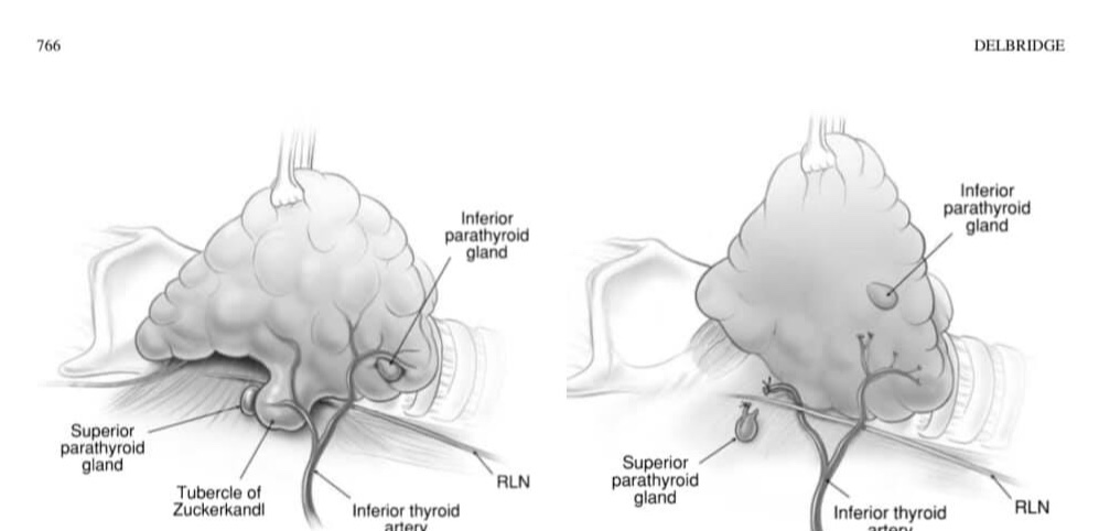
👉Although most authors still favour the approach of selective (‘ready’) parathyroid autotransplantation, routine autotransplantation, a philosophical approach based on the understanding that while parathyroid autotransplantation is very effective, the viability of in situ vascularized parathyroids remains unpredictable with late ischaemia always a possibility.

👉The routine autotransplantation of at least one parathyroid gland during every total thyroidectomy, while unnecessary in most cases, provides insurance in cases where late ischaemia of the remaining glands actually occurs.

👉The parathyroid gland chosen for routine autotransplantation is either one determined to be non-viable on the basis of anatomical location or a failed knife-test, or else the least viable appearing of the remaining in situ glands.

👉Routine autotransplantion will be associated with an increase in the rate of temporary hypocalcaemia in the short term but has, in theory, the potential to reduce permanent hypoparathyroidism to zero.

#Arrangoiz

#ThyroidSurgeon

#ParathyroidSurgeon

#ThyroidExpert

#ParathyroidExpert

#HeadandNeckSurgeon

#Teacher

Radiofrequency Ablation for Papillary Thyroid Microcarcinoma Is Safe and Effective in Long-Term Follow-up

  • Clin Thyroidol 2021;33:121–123.
  • Background
    • Low-risk papillary thyroid microcarcinomas (PTMCs) have been shown to have an:
      • Indolent course
    • This observation has led to alternative management approaches such as:
      • Active surveillance (AS) rather than standard treatment with surgery
    • Studies examining AS have shown promising results, with a low incidence of disease progression such as:
      • Tumor enlargement (5.3%)
      • Lymph node involvement (1.6%)
      • No evidence of distant metastasis
    • However, delayed surgeries during AS do occur:
      • Likely driven by patient and physician:
        • Anxiety
    • Therefore ultrasound-guided thermal ablation, particularly radiofrequency ablation (RFA):
      • Has been proposed as an alternative therapy in this setting
    • A recent meta-analysis of 11 cohorts:
      • Has shown RFA to be safe and effective in the management of PTMC over a relatively short follow-up period of up to two years
    • As clinicians consider the management of PTMC:
      • Longer-term data on the durability of RFA outcomes are needed
    • This study was a single-institution report of the authors’ experience with RFA in a cohort of patients with PTMC
  • Methods
    • This was a retrospective study of 84 PTMCs in 74 patients at a single center in South Korea
    • The study population was derived from a previously reported cohort of 133 patients who underwent RFA after September 2008 and had a follow-up of longer than 5 years
    • Participants had PTMC, with exclusion of aggressive subtypes on cytology, while imaging was used to exclude gross extrathyroidal extension, cervical adenopathy and distant metastases
    • All participants had either a contraindication to surgery or refused it
    • The RFA procedure was performed under local anesthesia by a single radiologist, and the approach was trans-isthmic, using the moving-shot technique
    • After RFA, patients were followed using ultrasound, interpreted by the same radiologist who performed the procedure, as well as clinical evaluations at 1, 6, and 12 months and annually thereafter for a minimum of 5 years
    • The outcomes of interest included PTMC volume changes, newly developed PTMCs, lymph node involvement, distant metastasis, delayed surgery during the follow-up period, and RFA safety, defined by immediate and long-term complications
  • Results
    • The study cohort consisted of 74 adults with a mean age of 46 years
    • There were 84 PTMCs treated with RFA, with 62% of them measuring < 5 mm (range, 3 to 10 mm maximum diameter)
    • Only 13 lesions required a second RFA session because of incomplete ablation during the first RFA session (average, 1.2 RFA sessions / patient)
    • Over a mean (±SD) follow-up period of 72±18 months:
      • There were no cases of tumor progression, lymph node or distant metastases, or delayed surgery
    • Complete tumor disappearance was noted by 60 months in all PTMCs:
      • With 98.8% disappearance by 36 months
    • Four new PTMCs developed in three participants’ remaining thyroid glands:
      • These were also treated effectively with RFA
    • Minor complications occurred in 4.1% of participants and included:
      • Hematomas and first-degree burns
    • Major complications occurred in 1.4% and included:
      • Voice changes, with recovery by 2 months
  • Conclusions
    • In adults with low-risk PTMCs, RFA has been shown to be effective and safe in over 5 years of follow-up
    • This approach has led to complete disappearance of all treated lesions and prevention of local tumor progression, metastasis, or delayed surgery, all with minimal risk of complication
  • This study is the first to report on long-term follow-up of a sizable cohort of PTMC patients treated with RFA and found excellent efficacy and safety
  • Previous reports were limited by a short duration of follow-up—a significant limitation considering the indolent course of PTMC
  • However, there are some notable considerations:
    • First, these data reflect experience at a single center with a single and highly experienced ultrasound and RFA operator, thus limiting external validity to centers with similar expertise:
      • Experience with RFA is paramount given the reported doubling in ablated lesion volume early-on post ablation
        • Due to ablation of parenchyma surrounding PTMCs:
          • Which needs to be separated from residual tumor presence with need for repeat RFA
          • Thus, without adequate expertise regarding interpretation of post-RFA imaging, unnecessary procedures might be performed
    • Second, the favorable outcomes with regard to tumor recurrence and LN metastasis are in contrast with some previous reports:
      • This is likely related to selection of less aggressive tumors:
        • As evidenced by cytologic criteria and small tumor size (greater than 62% of participants had a tumor size less than 0.5 cm)
      • Since thyroid cancer screening is no longer pursued, it is likely that the median size of RFA-treated PTMCs will increase, hence the need for revisiting the efficacy of the procedure periodically
  • Overall, these results are certainly promising, but their application to all PTMCs remains limited
  • In our practice, we do not routinely biopsy thyroid nodules less than 10 mm, in line with current American Thyroid Association (ATA) guidelines
  • For greater adoption of RFA, this approach would need to be reconsidered
  • At the same time, as RFA is being considered as an alternative to AS and surgery, patient-centered outcomes such as anxiety, pain, quality of life, and cost will be essential in future studies
  • Currently, without direct comparison between RFA, AS, and surgery:
    • RFA should be individualized to patient-specific situations:
      • Small low-risk PTMCs
      • Poor candidates for surgery or refusing surgery
      • Anxiety about AS
    • It should be considered only after careful discussion of standard treatment options
    • For patients interest in RFA, special emphasis should be placed on the required expertise for this procedure, in order to replicate the excellent results described here

#Arrangoiz #CancerSurgeon #HeadandNeckSurgeon #SurgicalOncologist #ThyroidSurgeon #ThyroidExpert #CASO #ThyroidNodules #CenterforAdvancedSurgicalOncology

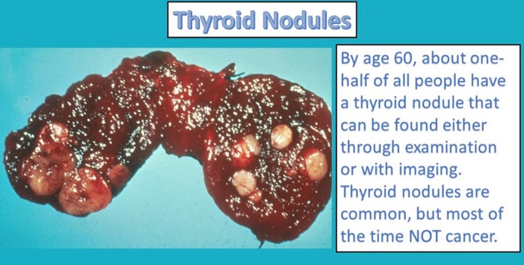
Thyroid Nodules

👉Thyroid nodules are a major health problem worldwide.

👉The prevalence of palpable thyroid nodules in the general population is approximately 5% in women and 1% in men living in parts of the world with sufficient iodine.

👉High resolution neck and thyroid ultrasound can detect thyroid nodules in a signif- icant proportion of randomly selected individuals, with higher frequencies in women and the elderly population.

👉The importance of thyroid nodules lies in the need to rule out cancer. The majority of thyroid nodules are benign, clinically irrelevant, and can be safely managed with a good surveillance program.

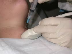
👉The detection and diagnosis of differentiated thyroid cancer have evolved over the years with increased use of high resolution cervical and thyroid ultrasound, fine needle aspiration biopsy (FNAB), molecular testing, and thyroglobulin as a serum tumor marker.

👉An algorithm that utilizes high resolution ultrasound and, when indicated, FNAB, and molecular testing for the diagnosis of thyroid nodules, facilitates a personalized, risk-based protocol that promotes high-quality care and minimizes cost and unnecessary testing.

Click to access 63f85b77cd57d842f1dd19ec8e4abf06e99e.pdf

#Arrangoiz

#ThyroidSurgeon

Parathyroid Awareness

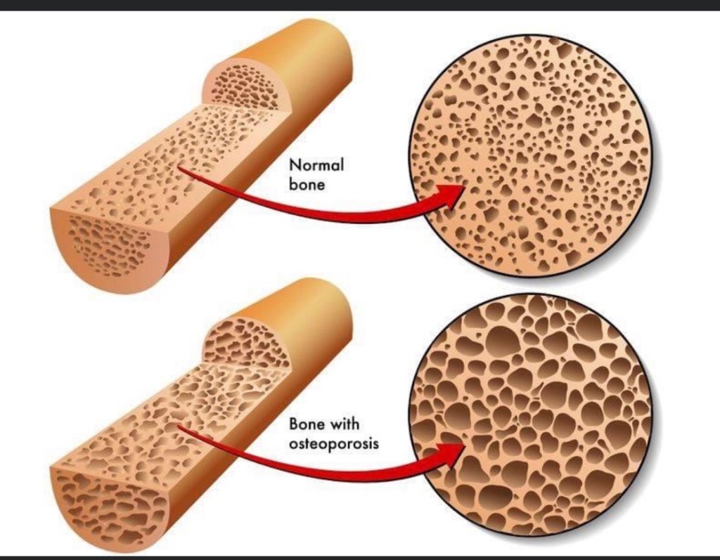
👉The majority of patients with both hyperparathyroidism and osteoporosis who undergo parathyroidectomy benefit from surgery with improvement in their bone mineral density.

👉https://pubmed.ncbi.nlm.nih.gov/23040710/

#CheckYourCalcium #Arrangoiz #ParathyroidSurgeon #ParathyroidExpert #HeadandNeckSurgeon

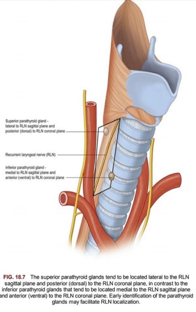
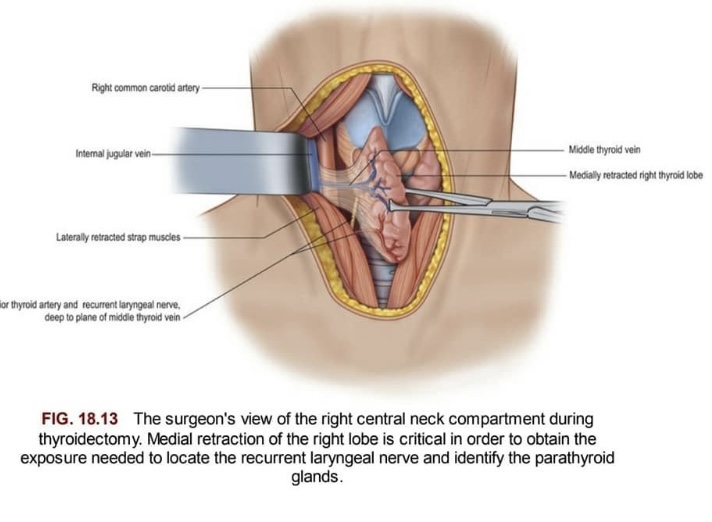
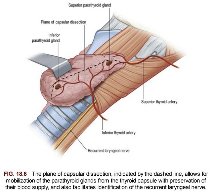
Parathyroid Gland Identification During Thyroid Surgery

#Arrangoiz #ParathyroidSurgeon #ParathyroidExpert #Hyperparathyroidism #CASO #CenterforAdvancedSurgicalOncology
Combination Pembrolizumab Plus Lenvatinib May Be Option in Anaplastic and Poorly Differentiated Thyroid Cancers

  • Clin Thyroidol 2021;33:131–133.
  • Background
    • Anaplastic thyroid carcinoma (ATC) and metas‐ tasized poorly differentiated thyroid carcinoma (PDTC):
      • Are rare aggressive malignancies with low overall survival despite the availability of extensive multimodal therapies
    • Tumors are highly proliferative:
      • With frequently increased tumor mutational burden as compared with differentiated thyroid carcinomas and elevated programmed cell death ligand 1 (PD-L1) levels
  • Methods
    • In a retrospective study, the authors analyzed six patients with metastasized ATC and two with PDTC:
      • Who received a combination therapy of lenvatinib and pembrolizumab
    • Lenvatinib was started at 14 to 24 mg daily and combined with pembrolizumab at a fixed dose of 200 mg every 3 weeks
    • Maximum treatment duration with this combination:
      • Was 40 months:
        • Three of the six patients with ATC were still on therapy
    • Patients’ tumors were characterized by whole-exome sequencing and PD-L1 expression levels (tumor proportion score [TPS], 1–90%)
  • Results
    • The best overall response within ATCs was:
      • 66% (4 of 6) with complete remission
      • 16% (1 of 6) with stable disease
      • 16% (1 of 6) with progressive disease
    • The best overall response in PDTCs was:
      • Partial remission (in 2 of 2)
    • Median progression-free survival was:
      • 17.75 months for all patients
      • 16.5 months for ATCs:
        • With treatment durations of 1, 4, 11, 15, 19, 25, 27, and 40 months
    • Grade III of IV toxicities developed in 4 of 8 patients and required dose reduction or discontinuation of lenvatinib
    • The median overall survival was 18.5 months:
      • With three ATC patients still alive without relapse (at 40, 27, and 19 months) despite metastatic disease at the start of treatment (International Union against Cancer [UICC] stage 4C)
    • All patients with long-term (greater than 2 years) or complete responses had either:
      • An increased tumor mutational burden or a PD-L1 TPS greater than 50%
  • Conclusions
    • These results suggest that the combination of lenvatinib and pembrolizumab might be safe and effective in patients with ATC or PDTC, leading to complete and long-term remissions
    • The combination treatment is now being systematically examined in a phase two clinical trial (Anaplastic Thyroid Carcinoma Lenvatinib Pembrolizumab – ATLEP; NCT04171622) in patients with ATC and PDTC
  • This small retrospective study demonstrates a rather provocative and high response rate in patients with ATC using the:
    • Multi–tyrosine kinase inhibitor lenvatinib combined with the immune checkpoint inhibitor pembrolizumab
  • While there is little doubt that pembrolizumab is active in ATC with activity that is likely comparable to that of spartalizumab (a similar PD-1 inhibitor that was tested in a robust phase two study of ATC and demonstrated a radiographic response rate of 19%), questions remain about the use of lenvatinib in this disease
  • Following initial reports from Japan with similarly remarkable response rates and a subsequent phase two study that seemed to confirm some efficacy, an international follow-up study failed to reproduce these results and was terminated after finding only one positive response in 33 patients (3) (NCT02657369)
  • Why there was such a discrepancy between those results?
    • At least one possible explanation is that in the Japanese study not all cases might have been anaplastic thyroid cancer, but could have been a mix between anaplastic and poorly differentiated thyroid cancer due to a lack of central pathology review
    • In PDTC, the high response rate to lenvatinib has been well described
    • One could argue that the combination of a checkpoint inhibitor plus lenvatinib could result in synergy:
      • With one drug enhancing the other’s activity perhaps through exposure of a broader range of neoantigens to the immune system
    • This hypothesis was the basis for a recently presented phase two study in radioiodine-refractory differentiated thyroid cancer conducted by the International Thyroid Oncology Group, which also combined pembrolizumab plus lenvatinib and which used the rate of complete responses as the primary end point
    • Unfortunately, there was no signal of synergistic activity, and the results were very much in line with those observed with lenvatinib alone
    • With that in mind, it is somewhat difficult to anticipate that the current data will be reproducible in the planned phase two study, but given the generally bad prognosis in this disease, every possible lead should be explored

#Arrangoiz #ThyroidSurgeon #ThyroidExpert #CancerSurgeon #ThyroidCancer #HeadandNeckSurgeon #CASO #CenterforAdvancedSurgicalOncology
Parathyroid Hormone Physiology

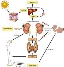
Parathyroid hormone (PTH) acts to raise blood calcium levels via its action on 3 organs. 1. The kidneys by Increased calcium reabsorption, Decreased phosphate reabsorption, Increased conversion of vitamin D to its final form – 1,25(OH) vitamin D; 2) Bones (increased resorption); 3) Intestines (increased absorption largely due to increased vitamin D in its final form).

Find out more at https://www.scirp.org/pdf/IJOHNS_2017072615211601.pdf

#Arrangoiz #ParathyroidSurgery #PararhyroidSurgeon #CASO #CenterforAdvancedSurgicalOncology
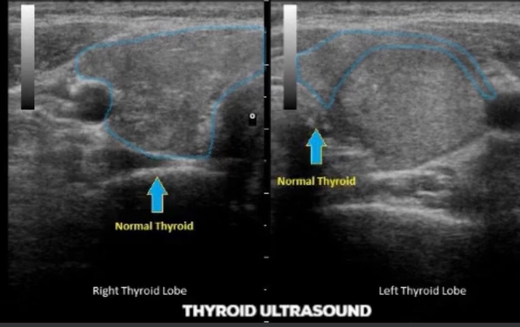
The Usefulness of Contrast-Enhanced Ultrasound to Evaluate Small Solid Thyroid Nodules Compared to TI-RADS

  • Clin Thyroidol 2021;33:114–116.
  • Background
    • Thyroid nodules are very common and frequently incidentally detected with the increasing use of imaging
    • The primary concern is how to identify those with suspicious features in order to avoid unnecessary diagnostic exams like fine-needle aspiration biopsy (FNAB) and overtreatment
    • Even though the Thyroid Imaging Reporting and Data System (TI-RADS):
      • Created by the American College of Radiologists:
        • Is widely accepted as an easy and useful tool to avoid unnecessary FNAB and close follow-up it still suffers from some biases, including experience and heterogeneity among sonographers
    • Contrast-enhanced ultrasound (CEUS) is a novel technology:
      • That can help differentiate between benign and malignant thyroid nodules
    • TI-RADS does not recommend FNAB for nodules up to 1 cm:
      • Even though such nodules have mild, moderate, or high (TI-RADS 3, 4, and 5, respectively) sonographic risk for malignancy
    • However, CEUS represents a new tool in the evaluation of thyroid nodules
    • The aim of this study was to compare the use of CEUS versus conventional ultrasound (as TI-RADS) in the risk stratification of thyroid nodules
  • Methods
    • In this study, 185 solid thyroid nodules between 0.5 and 1 cm in 154 patients were evaluated by conventional ultrasound and CEUS at the same time and by the same investigator
    • In conventional ultrasound, each target nodule was scored based on the components of:
      • Echogenicity, shape, edge, and strong echogenic focus, according to the 2017 edition of ACR TI-RADS in order to ascertain a TI-RADS classification
    • Qualitative indicators of CEUS analysis in the targeted thyroid nodules included:
      • Enhancement intensity, patterns of enhancement, internal homogeneity, the presence of perfusion defect, clearness of boundary, morphology, and size at enhance‐ ment peak
    • The CEUS characteristics of benign and malignant thyroid nodules with the significant differential diagnosis were scored, and the total scores of each thyroid nodule were calculated
  • Results
    • Of 185 thyroid nodules, the diagnosis was confirmed in 133 by surgical pathology, with 90 nodules (67.67%) showing papillary thyroid cancer (PTC) and 43 (32.33%) having benign results
    • In 52 thyroid nodules, cytology from the FNAB showed PTC in 11 nodules (21.15%) and nonmalignancy in 41 (78.85%)
    • Overall, there were 101 malignant thyroid nodules
    • When comparing the risk assessment scores:
      • CEUS outperformed TI-RADS using conventional ultrasound for benign diseases, but this was not the case in malignant nodules
    • Comparing the diagnostic performance between TI-RADS TR5 nodules and CEUS nodules that scored 5 points:
      • Sensitivity was 90.10% versus 86.13%, specificity 55.95% versus 89.29%, accuracy 74.59% versus 87.57%, positive predictive value 72.22% versus 90.63%, negative predictive value 82.46% versus 84.27%, and the area under the receiver-operating-characteristic curve 0.738 (95% CI, 0.663–0813) versus 0.916 (95% CI, 0.871– 0.961).
  • Conclusions
    • This study found that CEUS:
      • When evaluating small solid thyroid nodules, had higher diagnostic performance and a higher specificity than TI-RADS using conventional ultrasound
      • CEUS may be a valuable imaging tool to select patients with thyroid nodules for FNAB or surgery
    • Conventional ultrasound is currently the initial diagnostic tool for the risk stratification of thyroid nodules
    • To increase the diagnostic confidence and avoid unnecessary FNABs, many societies have proposed systems to stratify malignancy risks by ultrasound and guide physicians in when to perform FNAB:
      • One of the most popular and practical methods is TI-RADS
    • However, how to accurately differentiate malignant from benign nodules remains a significant challenge in many situations:
      • Since at least a half of all biopsied nodules are benign and up one third of FNABs are inconclusive
    • The new imaging method of CEUS can show tumor perfusion and vascular distribution after intravenous injection of a microbubble contrast agent:
      • Since vascular structures in the nodule differ from those of normal tissues
  • The specific characteristics of CEUS, like enhancement intensity, patterns of enhancement, and internal homogeneity, seem to be valuable in determining which nodules should undergo an FNAB and which may be kept under observation
  • In this study evaluating small nodules:
    • CEUS had significantly higher performance than TI-RADS in differentiating malignant from benign nodules:
      • Suggesting that CEUS qualitative analysis could be more effective in excluding malignant nodules and avoiding unnecessary biopsies
    • The use of CEUS in clinical practice has some crucial limitations, including the:
      • Cost of the exam, especially if the patient has more than one nodule to be analyzed:
        • Each nodule requires injection of a contrast agent
      • In addition, the small numbers of radiologists trained to perform this imaging study, as well as the absence of well-estab‐ lished criteria and consensus regarding the patterns of enhancement and classification of thyroid nodules, make its broad use in clinical practice controversial at present

#Arrangoiz #CancerSurgeon #ThyroidSurgeon #ThyroidExpert #HeadandNeckSurgeon #SurgicalOncologist #EndocrineSurgery #CASO #CenterforAdvancedSurgicalOncology