Inflammatory Breast Cancer IBC

  • Inflammatory breast cancer (IBC):
    • Is a clinical diagnosis characterized by:
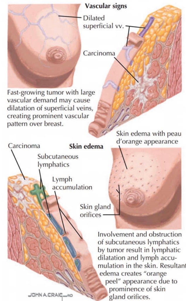
      • The rapid progression of an enlarged breast with skin changes including:
        • Redness
        • Edema
        • Peau d’orange
    • As mentioned previously IBC is a clinical diagnosis defined by the American Joint Committee on Cancer as a:
      •  “Diffuse erythema and edema involving approximately a third or more of the skin of the breast” and is staged cT4d
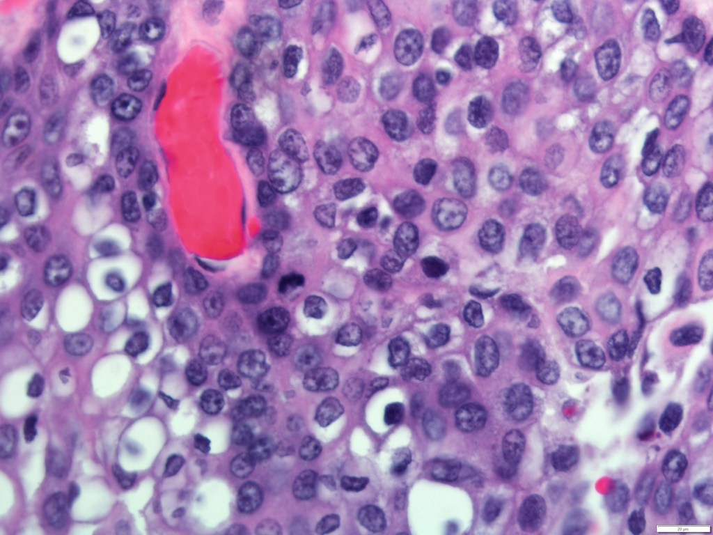
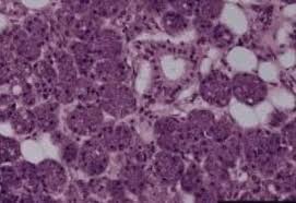
    • A punch biopsy of the skin:
      • Demonstrates tumor emboli within dermal lymphatics:
        • Approximately 75% of the time
      • A negative skin biopsy does NOT preclude the diagnosis, as it is clinical
    • The appearance may lead to:
      • Misdiagnosis of mastitis or breast cellulitis
    • The rapid evolution of symptoms (within 3 to 6 months):
      • Distinguishes IBC from a locally advanced breast cancer with associated edema
    • IBC is rare:
      • It presents in 2% to 4% of breast cancer patients:
        • Although the reported annual incidence has been increasing.
    • The tumor biology is disproportionately:
      • ER negative and HER2 amplified, compared with non-IBC
    • Patients should be evaluated in a multidisciplinary setting for trimodal therapy
    • Treatment should be initiated with:
      • Neoadjuvant chemotherapy, followed by aggressive local therapy
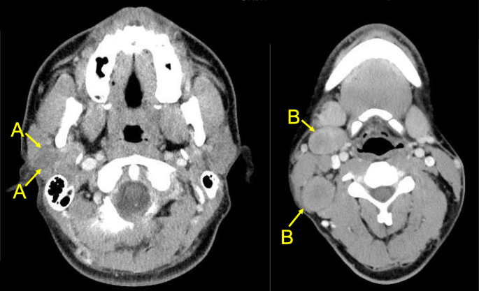
    • The majority of patients with IBC present with clinical lymph node involvement:
      • Sentinel lymph node biopsy is not reliable in IBC:
        • Due to blockage of dermal lymphatics:
          • Thus axillary dissection should be performed
    • Following neoadjuvant chemotherapy:
      • Modified radical mastectomy is the appropriate surgery:
        • Skin should not be spared so as not to leave behind residual disease
      • Immediate reconstruction should be avoided
    • Patients should receive post-mastectomy radiation:
      • To the skin, chest wall, and regional lymph nodes following surgery:
        • To optimize local control
    • Survival in IBC has improved with trimodal therapy:
      • A recent analysis of Surveillance, Epidemiology, and End Results data evaluated 10,197 patients with non-metastatic IBC between 1998 and 2010:
        • Patients who underwent trimodal therapy had improved 5- and 10-year survival (55.4% and 37.3%) over those that did not receive all three modalities
        • Survival was lowest at 10 years (16.5%) for patients who underwent surgery alone
    • Staging scans, including a CT chest / abdomen/ pelvis, PET scan, and / or bone scan:
      • Should be completed prior to initiating treatment
    • Inflammatory breast cancer is a clinical stage T4d:
      • And the most fatal form of breast cancer:
        • Accounting for 7% of all breast cancer deaths:
          • Real-world observational data have demonstrated that inflammatory breast cancer has significantly worse survival compared to other non-metastatic locally advanced and metastatic non-inflammatory breast cancers
        • Despite this, 5-year survival of IBC patients has increased from:
          • 40% to 50% in the 1990’s to almost 70% in 2008
    • Recent national and international guidelines for IBC recommend:
      • Full staging (PET / CT preferred over CT chest / abdomen / pelvis + bone scan) and bilateral breast and axillary nodal imaging, followed by neoadjuvant systemic therapy, modified radical mastectomy (including level I and II lymph node dissection), and radiation
      • Adjuvant targeted therapy and hormonal therapy should be considered in appropriate cases
      • Notably, lumpectomy is contraindicated
      • Breast reconstruction should be delayed
      • Multi-modal therapy for IBC has resulted in the best overall survival rates
    • For HER2-negative breast cancers:
      • Preoperative chemotherapy regimens should include:
        • Sequential doxorubicin and cyclophosphamide followed by a taxane:
          • To achieve the highest pathologic complete response rate
    • For HER2-positive breast cancers:
      • Chemotherapy should be used with dual anti-HER2-directed therapy with pertuzumab and trastuzumab:
        • To achieve the best pathologic complete response rate
  • References
    • National Comprehensive Cancer Network. NCCN Clinical Practice Guidelines in Oncology. Breast Cancer. Available with login at: https://subscriptions.nccn.org.
    • Fouad TM, Barrera AMG, Reuben JM, Lucci A, Woodward WA, Stauder MC, et al. Inflammatory breast cancer: a proposed conceptual shift in the UICC-AJCC TNM staging system. Lancet Oncol. 2017;18(4):e228-e232.
    • Ueno NT, Espinosa Fernandez JR, Cristofanilli M, Overmoyer B, Rea D, Berdichevski F, et al. International consensus on the clinical management of inflammatory breast cancer from the Morgan Welch Inflammatory Breast Cancer Research Program 10th Anniversary Conference. J Cancer. 2018;9(8):1437-1447.
    • Rueth NM, Lin HY, Bedrosian I, Shaitelman SF, Ueno NT, Shen Y, et al. Underuse of trimodality treatment affects survival for patients with inflammatory breast cancer: an analysis of treatment and survival trends from the National Cancer Database. J Clin Oncol. 2014;32(19):2018-2024.
    • Amin MB, Edge S, Greene F, et al., eds. AJCC Cancer Staging Manual. 8th ed. New York, NY: Springer; 2017
    • Hance KW, Anderson WF, Devesa SS, Young HA, Levine PH. Trends in inflammatory breast carcinoma incidence and survival: the surveillance, epidemiology, and end results program at the National Cancer Institute. J Natl Cancer Inst 2005;97(13):966-975.
    • Menta A, Fouad TM, Lucci A, et al. Inflammatory breast cancer: what to know about this unique, aggressive breast cancer. Surg Clin North Am. 2018;98(4):787-800.
    • Rueth NM, Lin HY, Bedrosian I, et al: Underuse of trimodality treatment affects survival for patients with inflammatory breast cancer: an analysis of treatment and survival trends from the National Cancer Database. J Clin Oncol. 2014;32(19):2018-2024.

#Arrangoiz #BreastSurgeon #CancerSurgeon #SurgicalOncologist #BreastCancer #InflammatoryBreastCancer #CASO #CenterforAdvancedSurgicalOncology

Recurrent Laryngeal Nerve Monitoring

👉Many surgeons use intraoperative recurrent laryngeal nerve monitoring to evaluate nerve function throughout surgery.

👉This is done by using a special breathing tube with sensors near your vocal cords.

Impact of Contralateral Prophylactic Mastectomy (CPM) on Psychosocial Outcomes

  • The decision to undergo CPM:
    • Is intensely personal
    • Frequently driven by:
      • A shifting balance between perceived future breast cancer risk
      • Anxiety over annual screening and potential future diagnostic procedures
      • The unknown physical, emotional, and cosmetic outcomes of the surgery
  • Long-term outcomes for women who have undergone CPM:
    • Report that 86% to 90% of respondents:
      • Were satisfied with the decision to undergo prophylactic surgery
    • With 20 years of follow-up:
      • More than 90 % of women definitely or probably would choose to undergo CPM again:
        • However, many of these same women report dissatisfaction with areas such as:
          • Body image, chronic pain, problems with implants, and sexual changes even though they noted overall satisfaction with their decision making
    • In a study of 296 women who participated in the National Prophylactic Mastectomy Registry and provided detailed responses to a survey evaluating their outcomes with CPM:
      • Only 6 % expressed regrets with the decision:
        • But of these women 39 % reported poor cosmetic outcomes and 22 % reported a reduced sense of sexuality:
          • Studies with longer follow-up had outcome data only on a proportion of the initial cohort, introducing possible bias between responders and nonresponders, limiting the strength of the evidence.
  • Few studies have examined quality of life between CPM and non-CPM patients:
    • One study, approximately 10 years ago:
      • Showed no difference in quality of life between patients undergoing CPM and those undergoing unilateral mastectomy or lumpectomy
    • In a study from Sweden:
      • No differences in overall health-related quality of life were identified up to two years post surgery in 60 women undergoing (delayed) CPM
  • Summary:
    • While 80% to 90 % of women report satisfaction with their decision to undergo CPM:
      • 20% to 30 % of these women report postsurgical dissatisfaction with cosmesis, body image, and sexuality
    • Studies show that CPM does not affect overall quality of life parameters
    • Women should be counseled on the potential long-term outcomes of CPM on body image and sexuality
  • References:
    • Roberts A, Habibi M, Frick KD. Cost-effectiveness of contralateral prophylactic mastectomy for prevention of contralateral breast cancer. Ann Surg Oncol. 2014;21:2209–2217. doi: 10.1245/s10434-014-3588-7.
    • Frost MH, Slezak JM, Tran NV, et al. Satisfaction after contralateral prophylactic mastectomy: the significance of mastectomy type, reconstructive complications, and body appearance. J Clin Oncol. 2005;23:7849–7856. doi: 10.1200/JCO.2005.09.233
    • Rosenberg SM, Sepucha K, Ruddy KJ, et al. Local therapy decision-making and contralateral prophylactic mastectomy in young women with early-stage breast cancer. Ann Surg Oncol. 2015;22:3809–3815. doi: 10.1245/s10434-015-4572-6
    • Geiger AM, West CN, Nekhlyudov L, et al. Contentment with quality of life among breast cancer survivors with and without contralateral prophylactic mastectomy. J Clin Oncol. 2006;24:1350–1356. doi: 10.1200/JCO.2005.01.9901
    • Frost MH, Hoskin TL, Hartmann LC, Degnim AC, Johnson JL, Boughey JC. Contralateral prophylactic mastectomy: long-term consistency of satisfaction and adverse effects and the significance of informed decision-making, quality of life, and personality traits. Ann Surg Oncol. 2011;18:3110–3116. doi: 10.1245/s10434-011-1917-7
    • Altschuler A, Nekhlyudov L, Rolnick SJ, et al. Positive, negative, and disparate–women’s differing long-term psychosocial experiences of bilateral or contralateral prophylactic mastectomy. Breast J. 2008;14:25–32. doi: 10.1111/j.1524-4741.2007.00521.x

#Arrangoiz #BreastSurgeon #CancerSurgeon #SurgicalOncologist #BreastCancer #ProphylacticMastectomy #CASO #Miami #CenterforAdvancedSurgicalOncology

Nipple Discharge Part 1

  • Nipple discharge is a relatively frequent event in females:
    • Being the third most common breast symptom prompting medical care:
      • After breast pain and breast palpable mass
  • Over 80% of females will develop an episode of nipple discharge during their fertile life:
    • Which can be categorized as:
      • Lactational
      • Physiological
      • Pathological 
        • According to the clinical history and the characteristics of the discharge
  • Lactational nipple discharge:
    • Is considered as a normal milk production
    • It is expected during pregnancy and lactation:
      • May persist for up to one-year post-partum or after cessation of breastfeeding
  • When a milky nipple discharge:
    • Occurs in females without recent history of pregnancy or lactation:
      • It is called galactorrhea:
        • Commonly involves bilateral multiple ducts
        • This is the result of an inappropriate increase in prolactin release:
          • Usually supported by a prolactinoma:
            • A prolactin-producing benign tumor of pituitary gland
  • Physiological nipple discharge:
    • Is a benign entity:
      • Usually bilateral
      • White, green, or yellow in color
      • It involves multiple ducts
      • Is associated with nipple squeezing
    • Some causes of physiological nipple discharge are:
      • Hypothyroidism
      • Medication side-effects
  • Pathologic nipple discharge (PND):
    • Is defined as a clear, serous, or bloody nipple secretion (not green or milky):
      • It is spontaneous
      • Discharging from a single duct and unilateral
    • It is frequently caused by:
      • A benign lesion, such as:
        • Intraductal papilloma(s):
          • 35% to 56% of the cases
        • Ductal ectasia:
          • 6% to 59% of the cases
      • An underlying malignancy can be present in a percentage of cases:
        • Reported to be variable from 5% to 33%
    • Because to differentiate between a benign from a malignant etiology of a PND based on clinical and diagnostic assessment is not easy:
      • Surgical excision has been considered the main way for getting both definitive diagnosis and eliminating the symptom
  • Clinical History and Physical Examination:
    • Clinical history plays an important role:
      • For evaluating the probability of malignancy
    • Predicting factors for malignancy in the presence of PND are:
      • BRCA 1 / mutations
      • History of ipsilateral cancer
      • Previous breast biopsy with diagnosis of atypia
      • Age over 50 years:
        • In a study including 318 patients with nipple discharge (any fluid from the nipple, spontaneous discharge or observed during breast examination):
          • Seltzer has reported a higher incidence of breast cancer:
            • Equal to 9% in females over 50 (95 patients and 9 cancers):
              • While the incidence was of only 1.3% in younger patients (223 patients and 3 cancers)
    • Physical examination:
      • Has the aim of distinguishing between benign and pathological discharge and of verifying the presence of palpable mass or other associated findings
      • It usually includes:
        • complete breast evaluation:
          • With inspection and palpation
          • Followed by a focused inspection of the nipple area:
            • Using a magnifying lamp
      • The physical examination is essential to investigate the:
        • Color of discharge
        • The number of ducts involved
        • The frequency of discharge (persistent or intermittent)
        • If it is unilateral or bilateral
      • spontaneous single-pore bloody and clear discharge:
        • Is suspect for pathological discharge
  • Mammography:
    • Represents the first conventional imaging technique to investigate nipple discharge:
      • At least after 39 years old
    • For patients with PND, aged between 30 and 40 years old with high-family risk:
      • Mammography could be appropriated in order to exclude the presence of microcalcifications
    • As well as for females younger than 30 of age:
      • When initial ultrasound shows suspicious findings
    • The protocol includes:
      • The standard cranio-caudal and mediolateral oblique views
    • Mammography findings that are suspect to be associated to an occult malignancy can range from:
      • Microcalcifications
      • Masses
      • Focal density asymmetry
      • Architectural distortion or ductal ectasia
      • Otherwise no abnormality can be identified
    • Mammography has low sensitivity and limited accuracy:
      • In the detection of retroareolar lesions that are often small, intraductal, and without calcifications
    • Ductal ectasia:
      • May occur as a general increase in density of the retroareolar region and in order to better visualize the area:
        • Spot compression views could be performed
    • In order to improve spatial resolution:
      • Magnification mammography can be performed:
        • To identify microcalcifications and to distinguish between benign or malignant duct disease
    • Microcalcifications with:
      • Branching or linear pattern, variable density, or distributed in a segmental way:
        • Are all highly suspicious of malignancy
      • Whereas round or rod-like calcifications:
        • Suggest for benign disease
    • Bahl et al studied 252 patients with at least one pathological feature of nipple discharge (unilateral, clear or bloody, or spontaneous discharge) who underwent surgical excision or a 2-year follow-up:
      • Of 20 cancers diagnosed:
        • Only three were revealed by mammography:
          • With a 15% (3/20) sensitivity
    • In other studies, the sensitivity of mammography:
      • Ranged from 7% to 26%. 
  • Ultrasound:
    • Offers a better performance than mammography:
      • For detecting intraductal lesions
    • Ductal ectasia:
      • Defined by a duct caliber greater than 3 mm
        • Is one of the most common findings seen on ultrasound:
          • It appears as dilated retroareolar ducts containing anechoid fluid or hypoecoic debris
    • An intraductal papilloma appears as:
      • hypoechoic nodule with a central vascular pedicle on color Doppler:
        • Doppler ultrasound is helpful in differentiating:
          • Intraductal viscous secretion versus intraductal nodule with vascular sign
    • Ultrasound malignant features are:
      • Irregular duct margins
      • Wall thickening
      • Hypoechoic intraductal mass with acoustic shadowing 
    • In a study by Park et al:
      • The detection rate of malignant lesions occult on mammography and ultrasound-detected:
        • Was reported to be 8 of 53 females with PND examined (15%)
    • Yoon et al:
      • Have also reported that adding ultrasound to mammography in the pre-operative setting of PND:
        • Led to the detection of malignancies in 26% of patients (ultrasound detected fivebreast cancers in addition to the 19 breast cancers found by mammography)
    • The role of ultrasound elastography is disputable in predicting malignancy in patients with PND:
      • Guo et al have evaluated the diagnostic accuracy of elastography in patients with PND:
        • Affirming that it is a useful tool for predicting malignancy:
          • With sensitivity for malignancy of 90% and that it could be used as a helpful test before more invasive examination (such as ductoscopy or duct excision):
            • However, it is only a preliminary study and further studies are needed to verify the diagnostic perfor- mance of elastography
  • Nipple discharge cytology:
    • Is performed by squeezing the nipple with a gentle compression of the areola area and spreading the secretion onto a glass slide:
      • After smearing, the slides are immediately fixed by spray fixation or by immersion in 95% ethyl alcohol:
        • Then stained with the Papanicolaou stain
    • It is a simple and fast examination, easy to perform and painless:
      • But strongly limited by a low sensitivity for cancer:
        • With a false negative rate over 50%
      • Moreover, it can be technically impossible when discharge is not present on the moment of the examination
      • According to the American College of Radiology:
        • This examination has not proven to be effective in differentiating benign from malignant lesions:
          • Therefore, discharge cytology is not routinely recommended
    • Nipple discharge smears are classified as abnormal if they contained:
      • Papillary, atypical, suspicious, or malignant cells:
        • Malignant nipple discharge cytology is correlated with higher specificity values

#Arrangoiz #BreastSurgeon #CancerSurgeon #SurgicalOncologist #Teacher #BreastCancer #NippleDischarge #PathologicNippleDischarge #PhysiologicNippleDischarge #CASO #Miami #CenterforAdvancedSurgicalOncology #IntraductalPapilloma #DuctalEctasia

Age as a Prognostic Factor in Thyroid Cancer

👉Thyroid cancer is the only malignancy that has age as a prognostic factor in TNM staging.

👉Due to the favorable prognosis in younger patients the highest possible stage for patients less than 55 is Stage II

AJCC TNM Staging for Thyroid Cancer

#Arrangoiz #ThyroidSurgeon #ThyroidExpert #Teacher #CancerSurgeon #HeadandNeckSurgeon #SurgicalOncologist #CASO #Miami #CenterforAdvancedSurgicalOncology

Papillary Thyroid Cancer Recurrence

👉Up to 20% of patients with papillary thyroid cancer will experience recurrent disease after surgery throughout their lifetime.

👉Certain mutations (e.g. BRAFV600E) are independent risk factors for recurrence.

#Arrangoiz #ThyroidSurgeon #ThyroidExpert #Teacher #CancerSurgeon #HeadandNeckSurgeon #SurgicalOncologist #CASO #Miami #CenterforAdvancedSurgicalOncology

Thyroid Cancer – Lymph Node Recurrences

👉Not all thyroid cancer recurrences require surgery.

👉Small lymph node recurrences in the neck can often be observed, however recurrent nodes greater than 0.8 cm in the central neck and greater than 1 cm in the lateral neck are typically removed.

#Arrangoiz #ThyroidSurgeon #ThyroidExpert #Teacher #CancerSurgeon #HeadandNeckSurgeon #SurgicalOncologist #CASO #Miami #CenterforAdvancedSurgicalOncology

Anatomy of the Nasolacrimal System

  • The purpose of the nasolacrimal system is to:
    • Drain tears from the ocular surface to the lacrimal sac and, ultimately, the nasal cavity
    • Blockage of the nasolacrimal system:
      • Can cause tears to flow over the eyelid and down the cheek:
        • This condition is epiphora
  • Structure and Function:
    • Both the upper eyelid and the lower eyelid have a small opening on the surface of the eyelid margin near the medial canthus:
      • These are called puncta:
        • Each puncta leads to a drainage canal that eventually flows into the lacrimal sac and then the nasal cavity
    • The drainage canal connecting the ocular surface to the nasal cavity consists of multiple parts:
      • Within the lower eyelid:
        • The punctum leads to a 2 mm long ampulla:
          • Which runs perpendicular to the eyelid margin
        • The ampulla turns 90 degrees medially:
          • Becoming the inferior canaliculus and travels 8 to 10 mm before reaching the common canaliculus
        • The upper canaliculus travels 2 mm superiorly in the eyelid before turning 90 degrees medially and moving 8 to 10 mm before connecting to the common canaliculus
      • The common canaliculus:
        • Drains into the lacrimal sac
      • Within the junction between the common canaliculus and the lacrimal sac:
        • Is the valve of Rosenmuller:
          • This apparatus is a one-way valve that prevents reflux from the lacrimal sac to the puncta
    • The lacrimal sac drains:
      • Inferiorly to the nasolacrimal duct:
        • Which is bordered:
          • Medially by:
            • Palatine bone and the inferior turbinate in the nose
          • Laterally by:
            • Maxillary bone
      • The nasolacrimal duct:
        • Opens at the inferior meatus:
          • Located underneath the inferior nasal turbinate
      • The lacrimal sac is:
        • Approximately 10 to 15 mm in axial length and 13 to 20 mm in corneal length
      • The nasolacrimal duct is:
        • 12 to 18 mm long
      • The inferior nasal meatus is partially covered by a mucosal fold:
        • Known as the valve of Hasner
  • Embryology:
    • The nasolacrimal duct:
      • Starts forming around five weeks of gestation
      • It starts out as a linear thickening of ectoderm:
        • Located in a groove between the nasal and maxillary prominences
      • This thickening:
        • Eventually separates into a solid cord and sinks into the surrounding mesenchyme
        • Over time the cord canalizes:
          • Forming the lacrimal sac and the beginning of the nasolacrimal duct
      • The nasolacrimal duct extends:
        • Intranasally until it exits under the inferior turbinate
      • The lacrimal sac extends caudally:
        • To complete the canalicular system
      • The inside of the canal breaks down and forms a lumen:
        • So that the nasolacrimal system is patent:
          • This process is generally complete by the time of birth
  • Blood Supply and Lymphatics:
    • Blood supply to the nasolacrimal area of the face:
      • Is generally from the angular artery:
        • The angular artery is considered a branch of the facial artery:
          • However, some studies have shown that it can originate from the ophthalmic artery in some individuals
        • It terminates in anastomosis with the dorsal nasal branch of the ophthalmic artery
        • The angular artery and vein:
          • Appear alongside the nose near the medial orbit
        • A correlating angular vein drains this region
    • The medial and lateral portions of the eyelids have different lymphatic drainage systems:
      • The medial one-third of the upper eyelid and the medial two-thirds of the lower eyelid:
        • Drain to the submandibular lymph nodes
      • The lateral two-thirds of the upper eyelid and the lateral one-third of the lower eyelid:
        • Drain to the pre-auricular lymph nodes
  • Nerves:
    • Cranial nerve VII:
      • Supplies the motor innervation to the muscles of the face
    • The movement of these muscles:
      • Aid in proper drainage of the tears through the nasolacrimal system:
        • By what is known as the lacrimal pump mechanism
    • Cranial nerve III and cranial nerve VII:
      • Innervate the muscles that control the blinking of the eyelids:
        • This action is the primary driver of the lacrimal pump mechanism
    • Irritation of the ocular surface:
      • Stimulates the ophthalmic branch of cranial nerve five:
        • Which begins the reflex tear arc pathway:
          • The efferent pathway involves cranial nerve VII and parasympathetic fibers
        • The role of the sympathetic nervous system in tear production:
          • Is not well understood
  • Muscles:
    • The action of the orbicularis muscle and surrounding tissues:
      • Helps propel the flow of tears from the canaliculi to the nasolacrimal duct:
        • Via the lacrimal pump mechanism
  • References:
    • Computed tomography dimensions of the lacrimal gland in normal Caucasian orbits., Tamboli DA,Harris MA,Hogg JP,Realini T,Sivak-Callcott JA,, Ophthalmic plastic and reconstructive surgery, 2011 Nov-Dec.
    • An Unusual Case of Nasolacrimal Obstruction Caused by Foodstuffs., Matsumoto H,Matsumoto A,, Case reports in ophthalmology, 2015 Sep-Dec.
    • Lacrimal Gland Volume Changes in Unilateral Primary Acquired Nasolacrimal Obstruction., Yazici A,Bulbul E,Yazici H,Sari E,Tiskaoglu N,Yanik B,Ermis S,, Investigative ophthalmology & visual science, 2015 Jul.
    • Incidence of neoplasia in patients with unilateral epiphora., Bewes T,Sacks R,Sacks PL,Chin D,Mrad N,Wilcsek G,Tumuluri K,Harvey R,, The Journal of laryngology and otology, 2015 Jul.
    • Ducasse A,Arndt C,Brugniart C,Larre I, [Lacrimal traumatology]. Journal francais d’ophtalmologie. 2016 Feb.
    • Modified External Dacryocystorhinostomy in Primary Acquired Nasolacrimal Duct Obstruction., Sharma HR,Sharma AK,Sharma R,, Journal of clinical and diagnostic research : JCDR, 2015 Oct.

#Arrangoiz #CancerSurgeon #HeadandNeckSurgeon #SurgicalOncologist #CASO #Miami #CenterforAdvancedSurgicalOncology

Mucoepidermoid carcinoma (MEC)

  • Mucoepidermoid carcinoma (MEC):
    • Is the most common malignant neoplasm of the:
      • Mayor and minor salivary glands
  • Epidemiology:
    • They encompass between 2.8% to 15.5% of all salivary gland tumors
    • Among 12% to 35% of malignant salivary gland tumors
    • Among 6.5% to 41% of all minor salivary gland tumors:
      • Representing the most common type of malignant minor salivary gland tumor in most series
    • Approximately half the cases occur in the major salivary glands:
      • 65% to 80% of these occur in the parotid
      • 8% to 13% occur in the submandibular gland
      • 2% to 4% involve the sublingual gland
    • MEC of the minor salivary glands:
      • Ordinarily arises on the palate:
        • But a number may also be found in the:
          • Retro molar area
          • Floor of the mouth
          • Buccal mucosa
          • Lip
          • Tongue
    • Its prevalence is highest in:
      • The fourth to fifth decade of life (35 to 65 years of age):
        • With a female preponderance as high as 4:1
  • Grossly:
    • The tumor is poorly circumscribed and measures from 3 to 5 cm
  • Histologically:
    • They are characterized by a mixed population of cells, including:
      • Mucin-producing cells
      • Epidermoid cells with squamoid differentiation
      • Clear cells
      • Intermediate cells:
        • That may predominate in numbers
        • Are believed to be the progenitor of the other types of cells
      • No myoepithelial cells are present
  • The clinical behavior of MEC has proved to be difficult to predict:
    • But correlations to tumor grade and stage have been reported
    • The histologic features that are most useful in predicting the aggressive nature of these tumors are:
      • A minor cystic component (less than 20%)
      • Tumor necrosis
      • Neural invasion
      • Cellular anaplasia
      • Brisk mitotic activity
    • Based on the presence or absence of these features and the clinical behavior, MEC are classified as:
      • Low grade
      • Intermediate grade
      • High grade
    • Low-grade MEC are:
      • Well circumscribed, with pushing margins and dilated cystic areas containing mucin
      • Mucin producing, intermediate, or epidermoid cells make up the lining of these cystic structures
    • Intermediate-grade MEC:
      • As the grade worsens:
        • The tumors become more infiltrative, poorly circumscribed
        • Cystic formations are lost
        • Nests of tumor become more solid and irregular with intermediate or epidermoid cells dominating
    • High-grade MEC are characterized by:
      • The invasion of adjacent structures
      • Atypical mitoses
      • Necrosis
      • Perineural invasion
      • Lymph node metastasis:
        • 40% to 50%
      • Distant metastases
    • Differential diagnosis of these high-grade lesions are:
      • Primary of metastatic squamous cell carcinoma:
        • MEC is differentiated from metastatic SCC by:
          • The presence of intracellular mucin
      • Sebaceous carcinomas
      • Clear cell carcinomas
  • Histologic grade and tumor stage:
    • Appear to have profound effects on survival
    • Aro et al:
      • Found a statistically significant difference in disease free survival (DFS) by grade:
        • Between low-grade MEC and intermediate / high-grade MEC (P = 0.001)

#Arrangoiz #CancerSurgeon #HeadandNeckSurgeon #SurgicalOncologist #SalivaryGlandCancer #MucoepidermoidCarcinoma #ParotidTumors #MEC #CASO #CenterforAdvancedSurgicalOncology #Miami

Cost of Contralateral Prophylactic Mastectomy (CPM) Versus Surveillance

  • There is robust literature to support the use of CPM as a cost-effective strategy:
    • In patients with hereditary breast cancer syndromes
  • Anderson et al. demonstrated that the most cost-effective strategy:
    • With and without quality adjustment:
      • For women with BRCA1 or BRCA2 mutations was:
        • Prophylactic bilateral salpingo-oophorectomy with bilateral mastectomy
  • Simulation models analyzing costs for CPM versus surveillance in patients with sporadic breast cancer reveal disparate findings:
    • An initial Markov model study found that CPM was cost effective compared with surveillance for:
      • Patients younger than 70 years:
        • But this finding was highly dependent on the quality of life assumptions
    • A second study that included operative complications and breast reconstruction costs used a decision-tree model and concluded that:
      • Although CPM resulted in a cost savings over surveillance for women younger than 50 years:
        • It also reduced quality of life years
      • When MRI was inserted in the model as the primary method of screening:
        • The cost-effectiveness of CPM increased
      • Loss of quality of life years was largely attributed to complications from reconstructive procedures
    • The two models differ in the assumptions regarding quality of life:
      • If we assume an improvement in quality of life after CPM:
        • Then CPM could be cost effective
      • Alternatively, if quality of life is decreased,:
        • CPM would not be a cost-effective strategy
  • The available data on cost effectiveness for CPM is limited
  • Summary:
    • CPM is a cost-effective strategy for women with BRCA mutations
    • At this time, there is insufficient evidence to support the concept of superior cost effectiveness for CPM in women with sporadic breast cancer and the cost effectiveness is highly dependent on the quality of life assumptions
  • Reference:
    • Contralateral Prophylactic Mastectomy Consensus Statement from the American Society of Breast Surgeons: Additional Considerations and a Framework for Shared Decision Making. Ann Surg Oncol (2016) 23:3106–3111

#Arrangoiz #BreastSurgeon #BreastCancer #CancerSurgeon #SurgicalOncologist #ContralateralProphylacticMastectomy #Cost-effectiveness #Miami #CASO #CenterforAdvancedSurgicalOncology