
- Lobular neoplasia (LN):
- Is an umbrella term for a spectrum of non-invasive, dyscohesive epithelial proliferations arising in the terminal duct-lobular unit (TDLU)
- It encompasses:
- Atypical lobular hyperplasia (ALH)
- Lobular carcinoma in situ (LCIS)
- Subdivided into:
- Classic (C-LCIS)
- Florid (F-LCIS)
- Pleomorphic (P-LCIS)
- Nakhlis et al., JAMA Surgery, 2026; Tjendra and Susnik, Seminars in Diagnostic Pathology, 2025
- Subdivided into:
- Its defining molecular feature is:
- Loss of E-cadherin membrane expression:
- A CDH1-encoded cell-adhesion protein
- Loss of E-cadherin membrane expression:
- LN is both a:
- Risk marker for—and a non-obligate precursor of—invasive breast cancer in either breast:
- With most subsequent cancers being invasive ductal rather than lobular (Morrow et al., Nature Reviews Clinical Oncology, 2015)
- Risk marker for—and a non-obligate precursor of—invasive breast cancer in either breast:
- Definition and Histopathologic Classification:
- The WHO classifies non-invasive LN by:
- Nuclear atypia and architecture into:
- ALH and LCIS (classic, florid, pleomorphic)
- Nuclear atypia and architecture into:
- The common cytology is:
- A non-cohesive, non-polarized proliferation of small monotonous cells with scant cytoplasm and low-grade nuclei:
- Frequently with intracytoplasmic vacuoles producing a “fried egg” or signet-ring appearance:
- Pagetoid extension up adjacent ducts is common (Brogi, Virchows Archiv, 2022; Kuba and Brogi, Histopathology, 2023)
- Frequently with intracytoplasmic vacuoles producing a “fried egg” or signet-ring appearance:
- A non-cohesive, non-polarized proliferation of small monotonous cells with scant cytoplasm and low-grade nuclei:
- ALH vs. C-LCIS (quantitative distinction):
- The threshold is the proportion of acini that are both filled and distended (> 8 to 10 cells across):
- LCIS:
- Is diagnosed when > 50% of acini within a TDLU are filled and distended
- ALH:
- Is diagnosed when ≤ 50% are involved with only minimal expansion
- LCIS:
- The cells are cytologically indistinguishable:
- ALH and C-LCIS are often grouped as classic LN and represent a morphologic continuum (Nakhlis et al., JAMA Surgery, 2026; Jani et al., The Breast Journal, 2022)
- Florid LCIS (F-LCIS):
- Cells resemble C-LCIS but there is marked acinar distention (~ 40 to 50 cells across) with little to no intervening stroma (often mass-forming) and frequent comedo-type necrosis / calcification (Brogi, Virchows Archiv, 2022)
- Pleomorphic LCIS (P-LCIS):
- High-grade pleomorphic nuclei > 4× the size of a lymphocyte (similar to high-grade DCIS):
- Sometimes with apocrine features and necrosis (Kuba and Brogi, Histopathology, 2023)
- High-grade pleomorphic nuclei > 4× the size of a lymphocyte (similar to high-grade DCIS):
- The threshold is the proportion of acini that are both filled and distended (> 8 to 10 cells across):
- Immunophenotype:
- Classic LN is:
- Typically ER / PR-positive and HER2-negative
- F-LCIS and P-LCIS:
- Can show less favorable phenotypes
- P-LCIS:
- In particular may be ER-negative and carries recurrent ERBB2 alterations
- Classic LN is:
- E-cadherin loss:
- Distinguishes LN from DCIS (Jani et al., The Breast Journal, 2022; Chung et al., Breast Cancer Research and Treatment, 2024)
- The WHO classifies non-invasive LN by:
- Incidence:
- True population incidence is unknown:
- Because classic LN is clinically and mammographically occult
- Historically it is reported in 1% to 4% of benign breast biopsies
- True population incidence is unknown:
- Incidence of C-LCIS:
- Has risen an estimated two- to fourfold since the 1980s:
- Attributed to increased biopsy volume, improved imaging and immuno-histochemistry, and population aging
- C-LCIS is most common in premenopausal women (median age 51 to 55)
- Historically C-LCIS:
- Is multicentric in ~ 80% of the cases
- Bilateral in ~ 40% of the cases (Brogi, Virchows Archiv, 2022)
- Has risen an estimated two- to fourfold since the 1980s:
- Incidence of P-LCIS is rare:
- A SEER analysis (ICD-O-3 code 8519/2) found an age-adjusted incidence of 0.08 per 100,000 woman-years versus 4.68 for classic disease:
- Comprising 2.6% of LCIS and peaking at ages 65 to 69 (Zihni and Sabuncuoğlu, Clinical Breast Cancer, 2026)
- A SEER analysis (ICD-O-3 code 8519/2) found an age-adjusted incidence of 0.08 per 100,000 woman-years versus 4.68 for classic disease:
- Breast Cancer Risk:
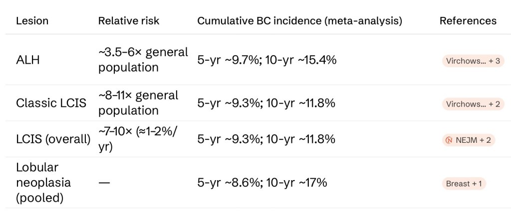
- LN confers a bilateral, lifelong elevation in breast cancer risk (Harris et al., New England Journal of Medicine, 1992; Morrow et al., Nature Reviews Clinical Oncology, 2015)
- See table 1
- The annual risk for LCIS is a steady ~1% to 2% per year:
- Translating to a lifetime risk often cited as 30% to 40% and inversely related to age at diagnosis:
- Example: ~40% at age 50 vs. ~30% at age 60)
- In a cohort of 1,060 women with C-LCIS:
- The cumulative cancer rate was 7% at 5 years and 21% at 10 years without chemoprevention
- Risk is distributed to both breasts:
- Though contemporary data suggest an ipsilateral predilection:
- One series found 90.9% of subsequent cancers ipsilateral (63.6% at the LCIS site), supporting a precursor role
- Though contemporary data suggest an ipsilateral predilection:
- Most subsequent cancers are:
- Ductal, early- stage, ER-positive, HER2-negative, and low / intermediate grade:
- With breast cancer–specific survival exceeding 95% at 10 years
- Ductal, early- stage, ER-positive, HER2-negative, and low / intermediate grade:
- Importantly, standard risk models (Gail, Tyrer-Cuzick) do not accurately estimate risk in patients with ALH / LCIS (Nakhlis et al., JAMA Surgery, 2026; Chung et al., Breast Cancer Research and Treatment, 2024)
- Translating to a lifetime risk often cited as 30% to 40% and inversely related to age at diagnosis:
- LN confers a bilateral, lifelong elevation in breast cancer risk (Harris et al., New England Journal of Medicine, 1992; Morrow et al., Nature Reviews Clinical Oncology, 2015)
- Diagnostic Imaging Findings:
- Classic LN (ALH, C-LCIS) is usually mammographically and clinically occult and discovered incidentally when biopsying another target:
- Mammography misses > 30% of lobular lesions owing to the subtle, infiltrative growth pattern
- When findings are present:
- Mammography:
- Grouped amorphous calcifications are the most common finding:
- Often associated with adjacent columnar cell change rather than the LN itself
- P-LCIS and F-LCIS are more often the actual imaging target, typically as:
- Pleomorphic / suspicious calcifications
- Grouped amorphous calcifications are the most common finding:
- Ultrasound:
- Low, operator-dependent sensitivity; when a correlate exists it is usually:
- An irregular, hypoechoic, avascular, shadowing mass
- Low, operator-dependent sensitivity; when a correlate exists it is usually:
- MRI:
- Most sensitive (> 90%), best for defining extent and detecting multifocality / multicentricity; typical appearance is heterogeneous non-mass enhancement with persistent kinetics
- Contrast-enhanced mammography (CEM):
- Preliminary data show performance comparable to MRI for detection, extent, and multifocality, predominantly as non-mass enhancement
(Scoggins et al., Academic Radiology, 2013; Nicosia et al., Breast Cancer Research and Treatment, 2024; Amitai et al., Breast Cancer Research and Treatment, 2020)
- Preliminary data show performance comparable to MRI for detection, extent, and multifocality, predominantly as non-mass enhancement
- Mammography:
- Classic LN (ALH, C-LCIS) is usually mammographically and clinically occult and discovered incidentally when biopsying another target:
- Management:
- Management hinges on the specific lesion and on radiologic-pathologic concordance (Nakhlis et al., JAMA Surgery, 2026; NCCN Breast Cancer Screening and Diagnosis, 2026)
- ALH and classic LCIS on core needle biopsy:
- Routine surgical excision is not required in the presence of radiographic-pathologic concordance:
- Because of exceedingly low upgrade rates:
- Select patients may be suitable for monitoring in lieu of excision
- Because of exceedingly low upgrade rates:
- Excision should be considered case-by-case when there is:
- Radiologic-pathologic discordance
- Concerning histologic features:
- Marked atypia or necrosis
- Inadequate sampling
- Routine surgical excision is not required in the presence of radiographic-pathologic concordance:
- Surveillance typically includes:
- Clinical examination and / or imaging at 6 to 12 months before returning to routine screening, with supplemental MRI considered on the basis of overall risk
- Counseling on risk reduction should be offered, including endocrine chemoprevention (e.g., tamoxifen in premenopausal, and tamoxifen or an aromatase inhibitor / raloxifene in postmenopausal women
- Pleomorphic LCIS and florid LCIS on core needle biopsy:
- Complete surgical excision with negative margins is recommended:
- Given upgrade rates approaching 40%
- Outcomes data for these non-classic variants remain limited
- Complete surgical excision with negative margins is recommended:
- Bilateral risk-reducing mastectomy:
- Is reserved for a minority of patients with additional risk factors (e.g., strong family history, deleterious germline mutation) and requires individualized multidisciplinary discussion
(Nakhlis et al., JAMA Surgery, 2026; NCCN Breast Cancer Screening and Diagnosis, 2026; Jani et al., The Breast Journal, 2022; Elfgen et al., Virchows Archiv, 2023)
- Is reserved for a minority of patients with additional risk factors (e.g., strong family history, deleterious germline mutation) and requires individualized multidisciplinary discussion
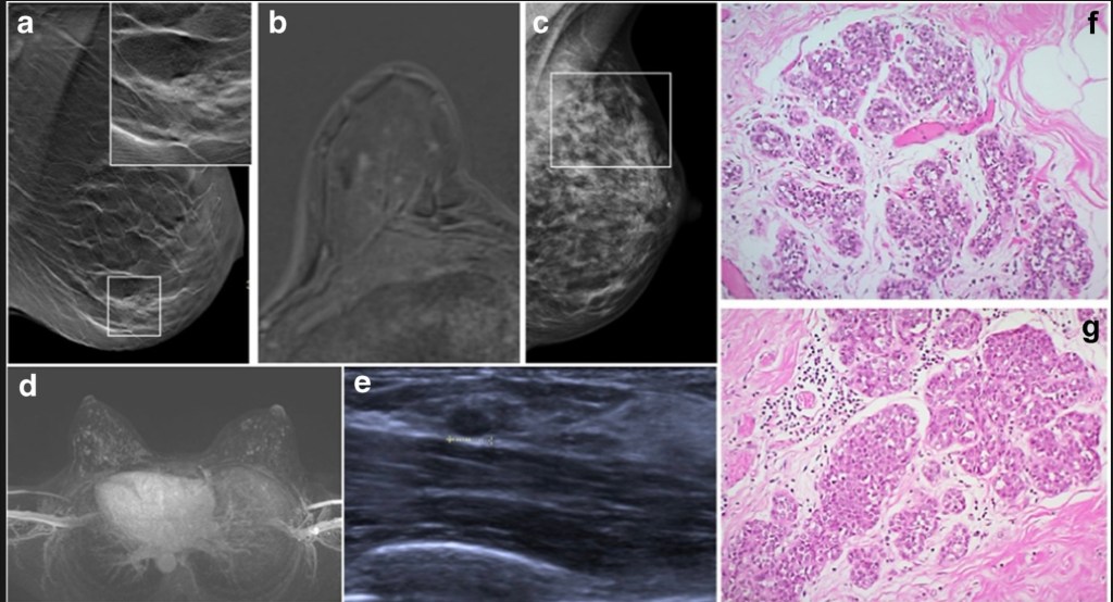
Classical lobular neoplasia (LN). a Screen detected calcification (in square) in the breast on mammography. Inset shows clustered calcifications, which were associated to LCIS and adenosis on the subsequent stereotactic vacuum biopsy. b Foci corresponding to small areas of LCIS on MRI. c Mammography shows dense fibroglandular tissue with diffuse calcifications (in square); the consecutive MRI-guided biopsy confirmed LCIS. d Screening MRI shows bilateral strongly enhancing foci within bilateral diffuse non-mass enhancement. e The target ultrasound (from the patient in d) reveals a small oval mass in the left breast, which was biopsied and histologically confirmed as invasive lobular carcinoma. d Morphology of classical LN, type ALH consisting of monotonous cells, subtotally filling the ductular units. f Morphology of classical LN, type LCIS, consisting of the same monotonous cells as in g, however, almost completely occupying the ductulo-lobular unit


