
- The National Comprehensive Cancer Network (NCCN) recommends:
- That every patient with suspected oropharyngeal squamous cell carcinoma (OPSCC) undergo a structured workup anchored by:
- Mandatory tumor HPV testing by p16 immunohistochemistry (IHC), tissue confirmation, cross-sectional imaging, and clinical staging by AJCC 8th edition:
- With p16 status determining which staging table and treatment algorithm applies
- Mandatory tumor HPV testing by p16 immunohistochemistry (IHC), tissue confirmation, cross-sectional imaging, and clinical staging by AJCC 8th edition:
- That every patient with suspected oropharyngeal squamous cell carcinoma (OPSCC) undergo a structured workup anchored by:
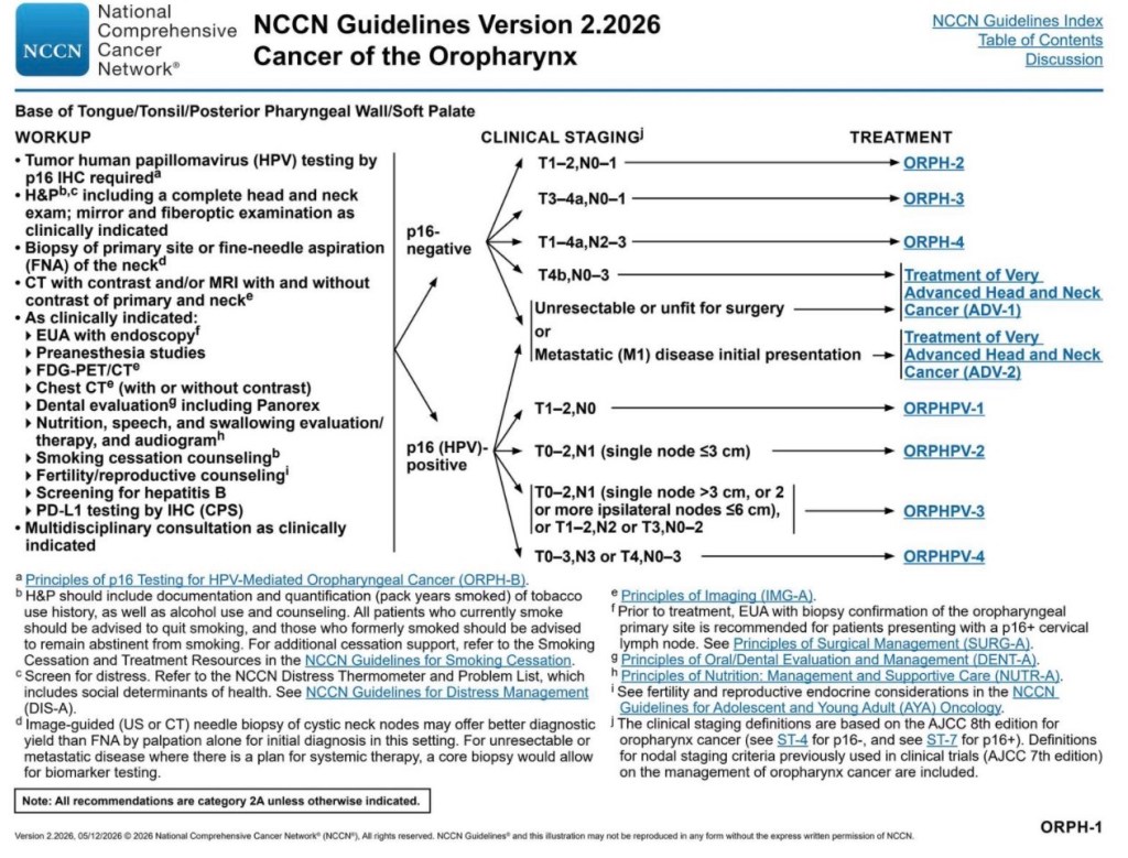
- Required workup elements (base of tongue / tonsil / posterior pharyngeal wall / soft palate):
- Tumor HPV testing by p16 IHC is required:
- The 70% cutoff with nuclear and cytoplasmic expression of at least moderate-to-strong intensity is used:
- Direct HPV confirmatory testing (PCR or RNA ISH) is recommended, especially for clinical trials and when p16 is used as a surrogate
- The 70% cutoff with nuclear and cytoplasmic expression of at least moderate-to-strong intensity is used:
- History and physical including a complete head and neck exam, with mirror and fiberoptic examination as clinically indicated:
- H&P should document / quantify tobacco (pack-years) and alcohol use with counseling, and screen for distress
- Biopsy of the primary site or FNA of the neck:
- Image-guided (US or CT) needle biopsy of cystic neck nodes offers better yield than palpation-guided FNA:
- A core biopsy is preferred when biomarker testing is planned for unresectable / metastatic disease
- Image-guided (US or CT) needle biopsy of cystic neck nodes offers better yield than palpation-guided FNA:
- CT with contrast and / or MRI with and without contrast of the primary and neck
- Additional studies as clinically indicated
EUA with endoscopy:- Prior to treatment, EUA with biopsy confirmation of the oropharyngeal primary is recommended for patients presenting with a p16+ cervical node
- FDG-PET/CT; chest CT (with or without contrast)
- Dental evaluation:
- Including Panorex
- Nutrition, speech, and swallowing evaluation / therapy, and audiogram
- Smoking cessation counseling
- Fertility / reproductive counseling
- Hepatitis B screening
- PD-L1 testing by IHC (CPS)
- Multidisciplinary consultation.
- Tumor HPV testing by p16 IHC is required:
- Imaging principles:
- Assess the primary with CT (with contrast) or MRI (with and without contrast) of the neck:
- Imaging from skull base to thoracic inlet:
- CT is complementary for cortical bone erosion
- MRI is preferred for bone marrow invasion, skull base / intracranial / orbital invasion, and perineural spread, and in patients with extensive dental amalgam
- Evaluate nodal disease with the same modality
- Consider FDG-PET / CT for its higher sensitivity:
- Particularly for midline tumors approaching the contralateral neck or when definitive RT is planned
- For locoregionally advanced disease (T3 to T4 or ≥ N1), FDG-PET/CT is preferred to evaluate for distant / thoracic metastases:
- If PET / CT is not done, obtain chest CT
- FDG-PET/CT cannot exclude brain metastasis
- If imaging does not reveal an obvious primary:
- PET / CT should be obtained before EUA, biopsies, and tonsillectomy to identify potential primary sites before intervention
- Assess the primary with CT (with contrast) or MRI (with and without contrast) of the neck:
- Search for the occult primary (neck mass presentation):
- Because the first sign of OPSCC is often a neck mass with a small, asymptomatic, radiographically occult primary:
- The NCCN emphasizes diligent identification and pathologic confirmation of the primary:
- Usually in the base of tongue or tonsil
- The NCCN emphasizes diligent identification and pathologic confirmation of the primary:
- Cross-sectional imaging:
- Should precede direct examination and confirmatory biopsy
- EUA may entail unilateral or bilateral biopsies of suspicious oropharyngeal areas:
- Palatine tonsillectomy may reveal a small primary, and lingual tonsillectomy may be considered if palatine tonsils and biopsies are negative:
- Bilateral palatine and lingual tonsillectomies are ill-advised due to swallowing morbidity
- Palatine tonsillectomy may reveal a small primary, and lingual tonsillectomy may be considered if palatine tonsils and biopsies are negative:
- FNA of the neck mass (often US-guided) usually establishes metastatic carcinoma:
- p16 immunostaining supports HPV-associated OPSCC when an oropharyngeal primary is present
- In occult-primary cases with p16-positive nodal metastasis, confirmation with HPV ISH / PCR is recommended:
- Open excisional node biopsy is rarely needed:
- If performed, the surgeon should be prepared for neck dissection if frozen section confirms SCC
- Open excisional node biopsy is rarely needed:
- The occult-primary pathway similarly directs HPV / EBV testing on nodal SCC:
- If HPV-positive with a T0 primary, the patient is treated as oropharyngeal cancer (ORPH-1)
- Because the first sign of OPSCC is often a neck mass with a small, asymptomatic, radiographically occult primary:
- Clinical staging (AJCC 8th edition):
- p16 status splits OPSCC into two separate staging systems:
- p16-negative OPSCC is staged with the oropharynx (p16-) / hypopharynx TNM (ST-6):
- Which incorporates extranodal extension (ENE) into N categories
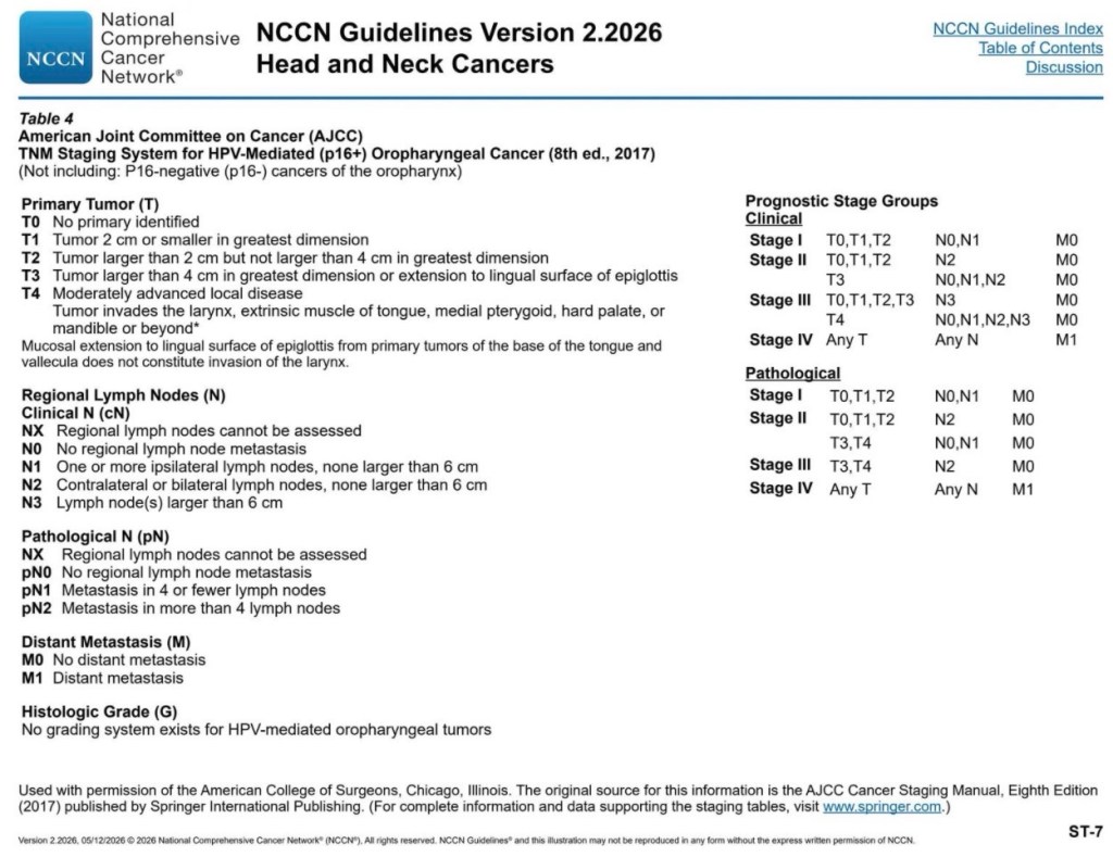
- p16-positive (HPV-mediated) OPSCC uses a distinct TNM (ST-7):
- With different clinical N categories:
- N1: ipsilateral nodes ≤ 6 cm
- N2: contralateral / bilateral ≤ 6 cm
- N3: > 6 cm
- A separate pathologic N classification:
- pN1 ≤ 4 nodes
- pN2 > 4 nodes
- Stage groups that markedly downstage nodal disease relative to p16-negative cancer
- There is no Tis or T4b category, and no histologic grading system, for HPV-mediated tumors
- With different clinical N categories:
- p16-negative OPSCC is staged with the oropharynx (p16-) / hypopharynx TNM (ST-6):
- p16 status splits OPSCC into two separate staging systems:
- The following NCCN algorithm summarizes the oropharynx workup and the p16-based staging / treatment branch point
- HPV / p16 testing nuances that affect prognosis and staging:
- While p16 IHC is the preferred surrogate for AJCC 8th edition staging:
- Roughly 9% to 20% of p16-positive OPSCC lack detectable HPV DNA / mRNA, and these p16+ / HPV− (and p16−/HPV+) discordant tumors:
- Carry a worse prognosis than double-positive tumors
- The 2025 College of American Pathologists update supports p16 IHC alone in high-prevalence regions (US, Canada, Northern Europe) when clinicopathologic surrogates of HPV disease are present:
- But recommends adding HPV-specific testing (RNA-ISH or DNA PCR) when p16 is equivocal, when morphology and p16 are discordant, for large multisite tumors, for nontonsillar / non–base-of-tongue oropharyngeal sites, in low-prevalence regions, and for clinical trials
- Roughly 9% to 20% of p16-positive OPSCC lack detectable HPV DNA / mRNA, and these p16+ / HPV− (and p16−/HPV+) discordant tumors:
- While p16 IHC is the preferred surrogate for AJCC 8th edition staging:
- Examination under anesthesia and tonsillectomy for the occult primary :
- The AAO-HNS neck mass guideline advises that when a persistent neck mass evades diagnosis after FNA, imaging, and exam, endoscopy under anesthesia with directed biopsies should precede open neck biopsy to avoid tumor seeding and its associated complications:
- A meta-analysis found palatine tonsillectomy has ~ 10-fold higher diagnostic yield than blind tonsil biopsy for detecting occult primaries:
- Because tonsillar tumors often lie deep within crypts or submucosa
- A meta-analysis found palatine tonsillectomy has ~ 10-fold higher diagnostic yield than blind tonsil biopsy for detecting occult primaries:
- The AAO-HNS neck mass guideline advises that when a persistent neck mass evades diagnosis after FNA, imaging, and exam, endoscopy under anesthesia with directed biopsies should precede open neck biopsy to avoid tumor seeding and its associated complications:
- Imaging modality performance:
- For detecting nodal metastases, high-resolution CT has ~ 82% sensitivity / 85% specificity, whereas PET/CT reaches ~ 90% sensitivity / 94% specificity
- Dedicated brain imaging is reserved for neurologic symptoms
- The ACR Appropriateness Criteria list CT neck with contrast, MRI without / with contrast, and FDG-PET / CT as the recommended studies for initial staging of oral cavity / oropharyngeal cancer, with FDG-PET / CT complementary for mapping systemic disease and detecting synchronous second primaries
- Special populations:
- A diagnosis of HNSCC in an adolescent or young adult without risk factors warrants evaluation for Fanconi anemia


 Rodrigo Arrangoiz MS, MD, FACS, FSSO