- Diagnosis of precancerous lesions or early cancer can be difficult]
- Leukoplakia and erythroplakia:
- Are precancerous lesions that have a varying risk of progression to malignancy
- Conversion from leukoplakia to carcinoma:
- Is reported in up to 5% to 7% of patients observed over several years
- Leukoplakia develops as a result of:
- Chronic irritation of the mucous membranes by carcinogens:
- This irritation stimulates proliferation of epithelial and connective tissue
- Histopathologic examination reveals:
- Underlying hyperkeratosis associated with epithelial hyperplasia
- Chronic irritation of the mucous membranes by carcinogens:
- In the absence of underlying dysplasia:
- Leukoplakia rarely (less than 5 % to 7%) is associated with progression to malignancy
- Keratoses of a variety of degrees:
- Manifest as leukoplakia
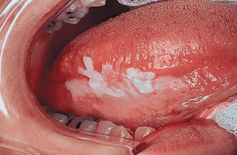

of the tongue
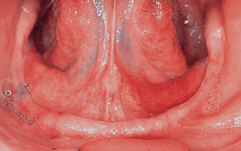
- Red spots, friable adjacent normal mucosa, characterize erythroplakia:
- It is associated with underlying epithelial dysplasia:
- Has a much greater potential for malignancy than leukoplakia:
- Carcinoma is found in nearly 30% to 40 % of cases of erythroplakia
- Has a much greater potential for malignancy than leukoplakia:
- Erythroplakia usually manifests as a pinkish, velvety flat
- It is associated with underlying epithelial dysplasia:
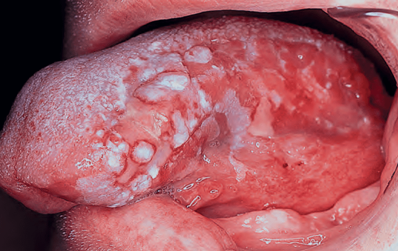
- Speckled leukoplakia:
- Has a particularly high incidence of malignant transformation:
- Similar to erythroplakia
- Has a particularly high incidence of malignant transformation:
#Arrangoiz #CancerSurgeon #HeadandNeckSurgeon #SurgicalOncologist #ThyroidSurgeon #ParathyroidSurgeon #MountSinaiMedicalCenter #MSMC #Miami #Mexico
