Ultrasound is safe and painless. It produces pictures of the inside of the body using sound waves. Ultrasound imaging is also called ultrasound scanning or sonography. It uses a small probe called a transducer and gel placed directly on the skin. High-frequency sound waves travel from the probe through the gel into the body. The probe collects the sounds that bounce back. A computer uses those sound waves to create an image. Ultrasound exams do not use radiation (as used in x-rays). Because images are captured in real-time, they can show the structure and movement of the body’s internal organs. They can also show blood flowing through blood vessels.

Ultrasound imaging is a noninvasive medical test that helps physicians diagnose and treat medical conditions.
Doppler ultrasound is a special ultrasound technique that evaluates movement of materials in the body. It allows the doctor to see and evaluate blood flow through arteries and veins in the body.
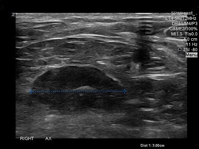
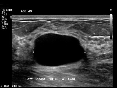
Ultrasound imaging of the breast produces a picture of the internal structures of the breast.
During a breast ultrasound examination, the sonographer or physician performing the test may use Doppler techniques to evaluate blood flow or lack of flow in any breast mass. In some cases, this may provide additional information as to the cause of the mass.
What are some common uses of the procedure?
- Determining the Nature of a Breast Abnormality
The primary use of breast ultrasound is to help diagnose breast abnormalities detected by a physician during a physical exam (such as a lump) and to characterize potential abnormalities seen on mammography or breast magnetic resonance imaging (MRI).
Ultrasound imaging can help to determine if an abnormality is solid (which may be a non-cancerous lump of tissue or a cancerous tumor), fluid-filled (such as a benign cyst) or both cystic and solid.
Doppler ultrasound is used to assess blood supply in breast lesions. - Supplemental Breast Cancer Screening
Mammography is the only screening tool for breast cancer that is known to reduce deaths due to breast cancer through early detection. Even so, mammograms do not detect all breast cancers. Some breast lesions and abnormalities are not visible or are difficult to interpret on mammograms. Breasts that are considered dense have a lot of glandular and connective tissues and not much fatty tissue, and that makes cancer harder to detect.
Many studies have shown that ultrasound and magnetic resonance imaging (MRI) can help supplement mammography by detecting breast cancers that may not be visible with mammography. Your doctor can help you determine if either of these tests is appropriate for you. MRI is more sensitive than ultrasound in depicting breast cancer, but MRI may not be available to all women. If screening MRI is performed, then screening ultrasound is not needed, though ultrasound may be used to characterize and biopsy abnormalities seen on MRI. When ultrasound is used for screening, abnormalities not visible with mammography may be identified, including some that may require biopsy. Many of the abnormalities found with screening breast ultrasound are not cancer (false positives).
Ultrasound can be offered as a screening tool for women who:
- are at high risk for breast cancer and unable to undergo an MRI examination.
- are pregnant or should not be exposed to x-rays (which are necessary for a mammogram).
- have increased breast density — when the breasts have a lot of glandular and connective tissue and not much fatty tissue (see the Dense Breasts page for more information).
- Ultrasound-guided Breast Biopsy
When an ultrasound examination reveals a suspicious breast abnormality, a physician may choose to perform an ultrasound-guided biopsy. Because ultrasound provides real-time images, it is often used to guide biopsy procedures. An ultrasound exam will usually need to be performed before the biopsy in order to plan the procedure and to determine if this method of biopsy can be used
#Arrangoiz #CancerSurgeon #BreastSurgeon #CASO #CenterAdvancedforSurgicalOncology #PalmettoGeneralHospital #BreastCancer