Introduction
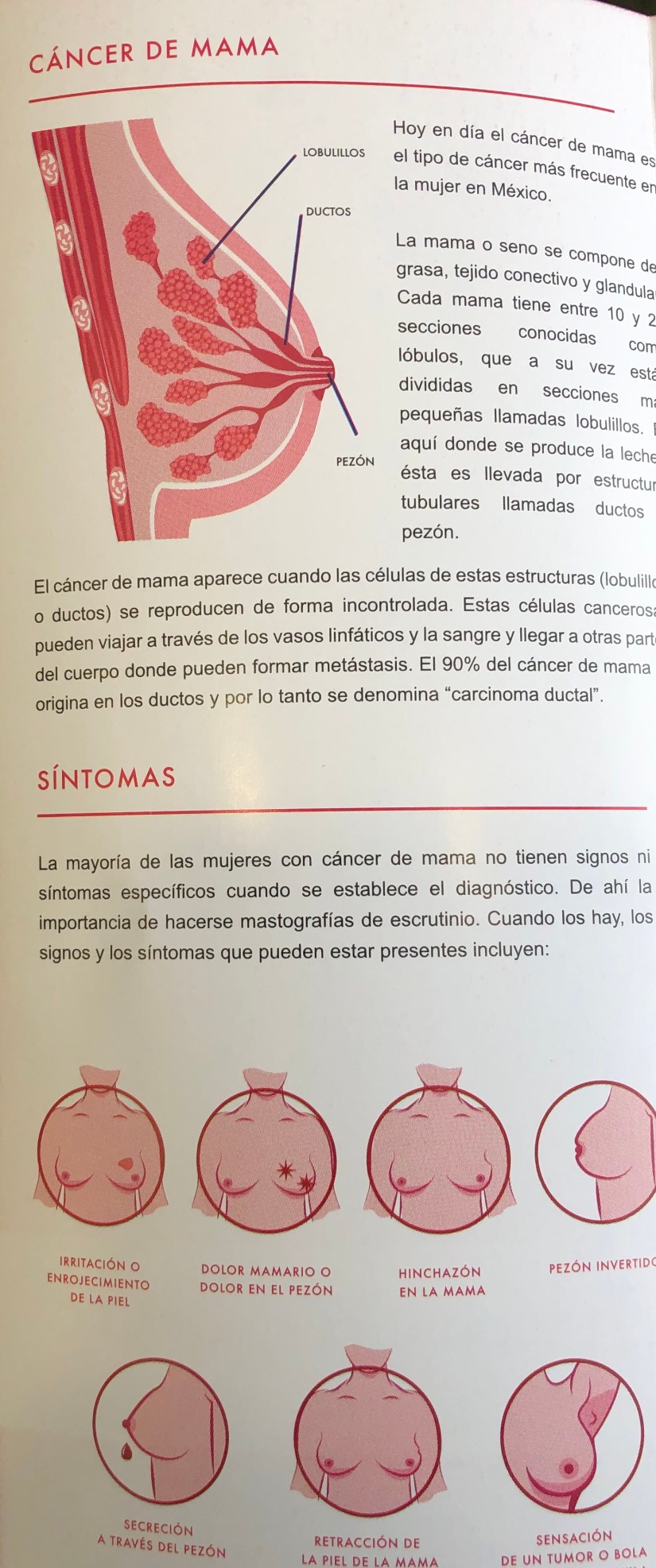
- In the breast, intraductal papilloma (IDP) is a benign lesion that consists of branching fibrovascular cores occurring within a cystic cavity with overlying layers of epithelial and myoepithelial cells:
- The classic pathologic features of intraductal papilloma (IDP) include an encysted solid mass with a branching fibrovascular pattern.


- The large/central subtype (L/C ST) specifically refers to an IDP arising from a large duct.
- IDP (L/C ST) is generally grossly apparent, solitary, and centrally located in the breast; it has accordingly been referred to as “solitary papilloma” and “central papilloma.”
- IDP (L/C ST) stands in contrast to IDP small/peripheral subtype (S/P ST), which originates at the terminal duct lobular unit (TDLU) and is usually located peripherally in the breast, is not grossly apparent, and generally occurs in multiples.

Epidemiology
- IDP (L/C ST) is primarily found in middle-aged women.
- In a study of 179 women with solitary / central papilloma:
- The mean age at diagnosis was 48 years, and occurrence substantially decreased after age 75 years.
- Younger women were also identified in this series:
- The youngest was aged 18 years.
- In a study of 179 women with solitary / central papilloma:
- IDPs are relatively common and are found in 1% to 5% of breast biopsies.
- Current evidence suggests that IDP (L/C ST) is more common that IDP (S/P ST).
Etiology
- Molecular evidence has shown that IDPs frequently demonstrate loss of heterozygosity (LOH):
- Involving specific loci on chromosome 16:
- This suggests they are clonal neoplasms.
- However, the studies showing this do not distinguish between IDP (L/C ST) and IDP (S/P ST):
- Therefore, they are unable to evaluate genetic differences between these lesions.
- However, the studies showing this do not distinguish between IDP (L/C ST) and IDP (S/P ST):
- This suggests they are clonal neoplasms.
- Involving specific loci on chromosome 16:
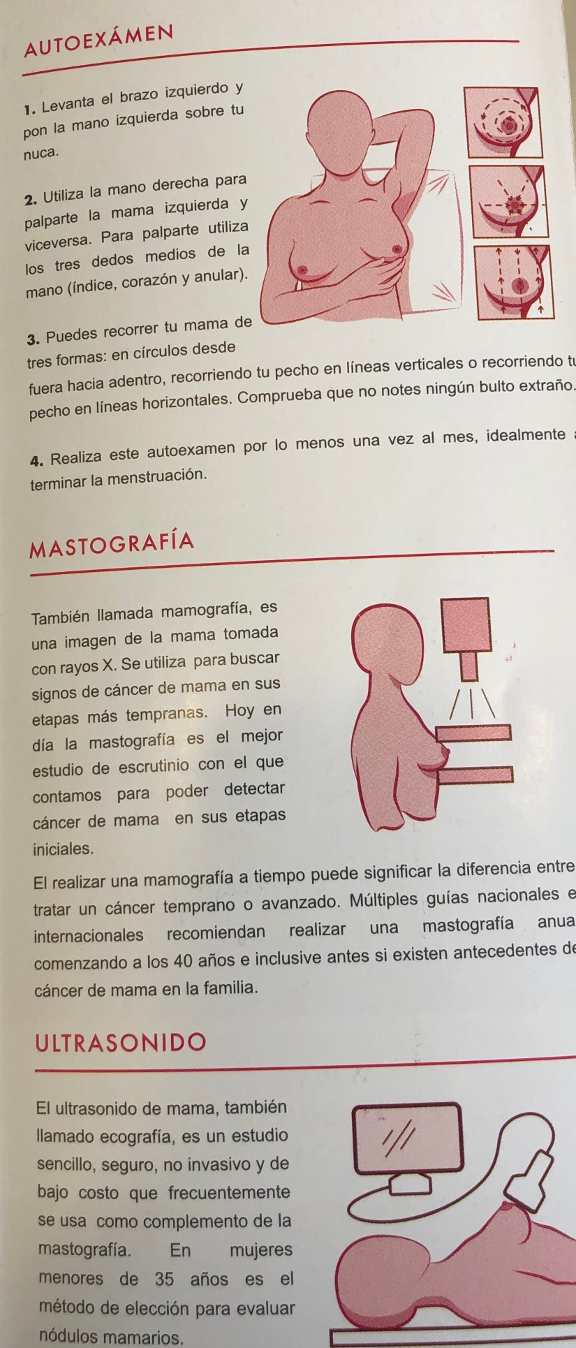
Presentation
- IDP (L/C ST) frequently presents as a unilateral serous or bloody nipple discharge:
- It may also present as a palpable breast mass and may on occasion present as breast pain.
- Mammography reveals no abnormality in most cases but may show duct ectasia, microcalcifications, or a mass.
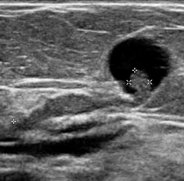
- Ultrasonography may be more sensitive than mammography for detecting IDP (L/C ST) and can reveal duct ectasia, nodules, or a cyst with or without a polyp.
- On MRI, IDP (L/C ST) appears as dilated ducts with an associated enhancing, well-circumscribed mass:
- MRI is currently the most sensitive imaging modality for detecting IDP (L/C ST).


Rodrigo Arrangoiz MS, MD, FACS a surgical oncologist and is a member of Sociedad Quirúrgica S.C at the America British Cowdray Medical Center in Mexico City:
-
He is an expert in the management of breast cancer:
-
If you have any questions about intraductal papilloma and bloody nipple discharge please fill free to ask Dr. Arrangoiz
-
Training:
• General surgery:
• Michigan State University:
• 2004 al 2010

• Surgical Oncology / Head and Neck Surgery / Endocrine Surgery:
• Fox Chase Cancer Center (Filadelfia):
• 2010 al 2012

• Masters in Science (Clinical research for health professionals):
• Drexel University (Filadelfia):
• 2010 al 2012

• Surgical Oncology / Head and Neck Surgery / Endocrine Surgery:
• IFHNOS / Memorial Sloan Kettering Cancer Center:
• 2014 al 2016

Sociedad Quirúrgica S.C. multidisciplinary breast cancer clinic: