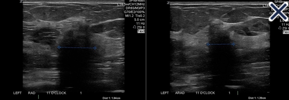
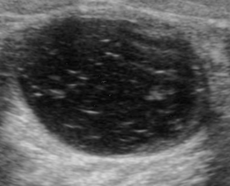
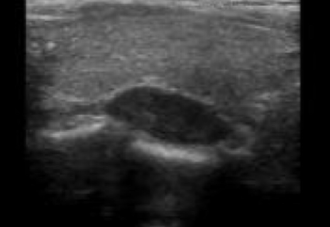
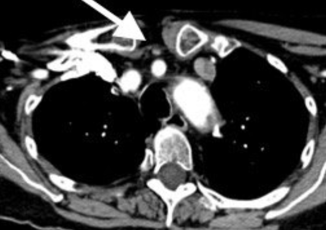
Severe eccentric compression and displacement of the mediastinum to the edge of the node
Convex indentations of the hilum:
Favors metastatic diseased
Severe compression of the hilum to slit-like configuration:
Can occur in metastatic or severely inflamed lymph node
Severe eccentric compression and displacement of the hilum to the edge of the node:
Favors metastatic disease
Complete obliteration of the hilum and rounding of the node:
Favors metastatic disease (can also occur in severe necrotizing lymphadenitis)
Perinodal invasion by metastasis:
The outer thin echogenic capsule cannot be identified
Angular margins
References
Stavros AT. Evaluation of regional lymph nodes in breast cancer patients. In: Stavros AT. Breast Ultrasound. Philadelphia, PA: Lippincott Williams & Wilkins; 2004:858-859.
Creates images derived solely from higher frequencies
The ultrasound beam:
Is transmitted centered at 1 frequency, e.g., 6 MHz, and received centered at a multiple of the transmitted frequency, e.g., 12 MHz
Different techniques can be used to process the received signals so that only the returning high-frequency harmonic signal is used to produce the image:
Whereas echoes from the fundamental / lower frequencies are rejected
THI increases signal-to-noise ratio:
Resulting in better tissue contrast
THI:
Reducesreverberation, clutter, and speckle artifacts:
Improving contrast resolution
It accentuates real echoes in addition to suppressing artifactual echoes
The suppression of speckle artifact by coded harmonics makes solid nodules more hypoechoic and conspicuous than they are with fundamental imaging (Images)
It makes the thin, echogenic capsule that surrounds most benign lesions appear to be thinner, more echogenic and more complete than with fundamental imaging
THI cannot be combined with simultaneous color Doppler imaging because the resulting frame rate would be unacceptable:
When Doppler is required, the image that is interlaced with Doppler must be constructed at fundamental rather than harmonic frequencies
THI is of limited value in differentiating benign from malignant lesions
Another method of reducing artifactual echoes, improving contrast, and making the thin, echogenic capsule more conspicuous is real time spatial compounding of images
In conventional imaging, each frame is created by a single sweep of the beam at a 90 degree angle to the long axis of the transducer:
In compound imaging, there are multiple sweeps of the beam from different angles, creating a spatially and temporally compounded image from multiple angles over time
Among other things, the lateral borders of lesions can be seen better with compound imaging
Infiltrating ductal carcinoma with fundamental imaging.The same infiltrating ductal carcinoma as above with coded harmonic imaging
References:
Mesurolle B, Helou T, El-Khoury M, Edwardes M, Sutton EJ, Kao E. Tissue harmonic imaging, frequency compound imaging, and conventional imaging use and benefit in breast sonography. J Ultrasound Med. 2007;26(8):1041-1051.
Cha JH, Moon WK, Cho N, Kim SM, Park SH, Han BK, et al. Characterization of benign and malignant solid breast masses: comparison of conventional US and tissue harmonic imaging. Radiology. 2007;242(1):63-69.
Stavros AT. Breast ultrasound equipment requirements. In: Stavros AT. Breast Ultrasound. Philadelphia, PA: Lippincott Williams & Wilkins; 2004:16-41.
Is important in identifying breast cancer at an early stage
Calcifications have many forms:
The characteristics of the calcifications:
Help identify whether they are associated with a benign or malignant process
Smooth, round, large and layering calcium:
Are generally associated with benign findings
Round and punctate calcifications. Lobular. This group of calcifications is characterized by relatively monomorphic round and punctate calcifications having some variation in density. No linear forms or linear orientation is seen. BI-RADS 2: Benign finding
Fine, irregular, punctate, linear and branching, and pleomorphic calcifications:
Are characteristics generally associated with malignant findings
Fine Linear or Fine Linear Branching These are thin, linear or curvilinear irregular calcifications. They may be discontinuous. Their appearance suggests filling of the lumen of a duct, i.e. ‘casting’ calcifications. These calcifications are classified as Bi-RADS 4C. On the left calcifications in a segmental distribution. Some have a linear distribution and some have a branching morphology. This is highly suggestive of malignancy (Bi-RADS 4C)
Magnification views of the calcifications are essential in helping to evaluate the calcifications to determine the appearance and to be able to accurately interpret the findings
References:
Brant W, Helms C. Fundamentals of Diagnostic Radiology, 5th Edition. Lippincott, Williams & Wilkins; 2019
Baldwin P. Breast calcification imaging. Radiol Technol. 2013;84(4):383M-404M.
At first glance, the rounded, almost anechoic sonographic mass could be mistaken for a simple cyst:
However, closer inspection shows some small spiculations, angular margins, and a suggestion of an echogenic halo
These findings alone make it a BIRADS 4 lesion
Even if they were not present:
The lesion is rounded and, therefore, does not meet any of the 3 benign definitions:
That would allow it to be classified BIRADS 3
Although the lesion is small, homogeneous, and does not cause worrisome posterior acoustical artifacts:
Cancers this small frequently have no shadowing or enhancement
It could be benign:
But it has the appearance that a high-grade carcinoma would have when it is less than a centimeter in diameter
When a nodule seen on mammogram and ultrasound are in the same location, have the same size and shape, and the same surrounding tissue:
They can be confidently assumed to be the same
If it were going to be followed, mammograms would expose this young woman to ionizing radiation and thus a small but real risk of developing a radiation-induced malignancy:
So a 6-month follow-up ultrasound would be the modality of choice
Because the lesion cannot be confidently considered to be less than BIRADS 4a:
Evaluation with a needle is required:
It is possible aspiration would collapse a small cyst:
If not, percutaneous biopsy should be done with a spring-loaded, vacuum-assisted, or intact-removal device, depending on the experience and expertise of the surgeon
Steps in the evaluation of breast nodules by ultrasound:
You first evaluate the lesion for any of the 10 malignant signs:
Shadowing
Hypoechoic echotexture
Spiculation
Angular margins
Thick echogenic halo
Microlobulation
Taller than wide
Duct extension
Branching pattern
Calcifications)
Finding none:
You move on to the second step in the evaluation process and specifically look for one of the three strictly defined benign signs:
If any of them are found:
The lesion can be considered BIRADS 3:
The three benign findings defined by Stavros are:
A purely hyperechoic lesion with no hypoechoic area larger than a normal duct or lobule
Elliptical, wider than tall, well-circumscribed and thin echogenic capsule
Gently lobulated, wider than tall, well-circumscribed and thin echogenic capsule
Combining the elliptical or gently lobulated shapes with the presence of a complete, thin echogenic capsule:
Is necessary because many circumscribed carcinomas and most ductal carcinoma in situ are encompassed in a thin, echogenic capsule:
However, the shape of circumscribed invasive carcinoma or pure ductal carcinoma in situ:
Is rarely elliptical or gently lobulated
Because this lesion is elliptical, wider-than-tall, well-circumscribed, and has a thin, echogenic capsule:
It is BIRADS 3:
A 6-month follow-up ultrasound would be appropriate unless the anxiety of the patient makes core biopsy a better option
References
D’Orsi CJ, Sickles EA, Mendelson EB, Morris EA. ACR BI-RADS® Atlas, Breast Imaging Reporting and Data System, 5th ed. Reston, VA, American College of Radiology, 2013.
Madjar H, Mendelson EB. The Practice of Breast Ultrasound. 2nd ed. Thieme; 2008;141-144.
Stavros AT. Breast Ultrasound. Philadelphia, PA: Lippincott Williams & Wilkins; 2004.
Sonographic mass with a benign mammographic imaging:
Is managed based on ultrasound features
References
Evaluation and imaging features of malignant breast masses. In: Cardenosa G. Clinical Breast Imaging: The Essentials. Philadelphia, PA: Wolters Kluwer; 2015:234-282.
Stavros AT. Breast Ultrasound. Philadelphia, PA: Lippincott Williams & Wilkins; 2004.
The mammogram shows trabecular thickening and an increased thickness of the skin:
Trabecular thickening is an increased prominence of linear structures in the breast:
Primarily corresponding to Cooper’s ligaments
It is almost always seen in conjunction with skin thickening:
It indicates significant breast edema
It has a number of different causes, including:
Congestive heart failure
Renal failure
Venous or lymphatic obstruction
Diffusely infiltrating carcinoma
Radiation
When the cause is lymphatic obstruction:
The obstruction can be the result of surgery, radiation, or lymphatic invasion by cancer
Unilateral breast edema with increased trabecular and skin thickening:
Sometimes occurs with congestive heart failure:
The pedal edema that develops during the daytime decreases at night:
And the fluid accumulates in the dependent breast in patients who sleep on their side
References
Berg, WA, Birdwell RL, Kennedy A. Diagnostic Imaging: Breast. Salt Lake City, UT: Amirsys; 2006.
Verbelen H, Gebruers N, Beyers T, De Monie AC, Tjalma W. Breast edema in breast cancer patients following breast-conserving surgery and radiotherapy: a systematic review. Breast Cancer Res Treat. 2014;147(3):463-471.
Wratten CR, O’Brien PC, Hamilton CS, Bill D, Kilmurray J, Denham JW. Breast edema in patients undergoing breast-conserving treatment for breast cancer: assessment via high frequency ultrasound. Breast J. 2007;13(3):266-273.
Menta A, Fouad TM, Lucci A, Le-Petross H, Stauder MC, Woodward WA, et al. Inflammatory breast cancer: what to know about this unique, aggressive breast cancer. Surg Clin North Am. 2018;98(4):787-800.
Is recommended for most ADH lesions diagnosed on core needle breast biopsy (CNB):
The chance of upgrade at excision to ductal carcinoma in situ (DCIS) or invasive carcinoma:
Is generally in the 12% to 22% range in the literature
The need for routine excision of pure flat epithelial atypia (FEA) has been less clear:
Some authors have reported an upgrade rate of 9.6% following excision of lesions that show pure FEA without ADH:
When the vast majority of biopsies were done with a 14-gauge spring-loaded core biopsy device
It is not clear that biopsy with a vacuum-assisted device would yield the same results:
In fact, in one study reporting biopsy of low-risk calcifications with a vacuum-assisted device:
Pure FEA never resulted in an upgrade to malignancy
An article from the Mayo Clinic:
Showed that FEA does not seem to convey an independent risk of breast cancer beyond that of associated proliferative disease without atypia or associated ADH
The risk of upgrade at surgical excision for ADH:
Has been reported to correlate with the number of ducts or terminal duct lobular units involved on vacuum-assisted core biopsy:
With 2 or fewer foci of involvement:
There was no upgrade on excision
With 4 or more foci of involvement:
There was a strong probability of upgrade to ductal carcinoma in situ or invasive carcinoma at excision
Work continues to try to define a low-risk group who could potentially avoid excisional biopsy:
Particularly those with small areas of calcifications completely removed with core needle biopsy and only focal ADH on pathology
Apocrine metaplasia, florid epithelial hyperplasia of the usual variety, and columnar cell change without atypia:
Do not confer a significant risk of upgrade and do not require excision
References
Eby PR, Ochsner JE, DeMartini WB, Allison KH, Peacock S, Lehman CD. Is surgical excision necessary for focal atypical ductal hyperplasia found at stereotactic vacuum-assisted breast biopsy? Ann Surg Oncol. 2008;15(11):3232-3238.
Ely KA, Carter BA, Jensen RA, Simpson JF, Page DL. Core biopsy of the breast with atypical ductal hyperplasia: a probabilistic approach to reporting. Am J Surg Pathol. 2001;25(8):1017-1021.
Khoumais NA, Scaranelo AM, Moshonov H, Kulkarni SR, Miller N, McCready DR, et al. Incidence of breast cancer in patients with pure flat epithelial atypia diagnosed at core-needle biopsy of the breast. Ann Surg Oncol. 2013;20(1):133-138.
Said SM, Visscher DW, Nassar A, Frank RD, Vierkant RA, Frost MH, et al. Flat epithelial atypia and risk of breast cancer: a Mayo cohort study. Cancer. 2015;121(10):1548-1555.
McGhan LJ, Pockaj BA, Wasif N, Giurescu ME, McCullough AE, Gray RJ. Atypical ductal hyperplasia on core biopsy: an automatic trigger for excisional biopsy? Ann Surg Oncol. 2012;19(10):3264-3269.
Patients who are candidates for surgery for primary hyperparathyroidism (PHPT) but have negative or discordant imaging findings by ultrasound or Sestamibi scan:
Should still undergo surgical evaluation according to American Association of Endocrine Surgeons guideilnes:
Debate on adjunct screening in women with dense breasts has resulted from legislation mandating that women be informed if their mammograms show dense breast tissue, including:
Informing them that other screening modalities are available
In addition to MRI and molecular imaging:
Both tomosynthesis and breast ultrasound are additional techniques for enhanced screening in patients with dense breasts
Multiple studies show significant benefits from the addition of tomosynthesis to conventional digital mammography alone in screening programs:
Ciatto et al:
Found an increase in detection rate of invasive breast cancer from 5.3 / 1000 to 8.1 / 1000:
While also decreasing the recall rate by 17%
Skaane et al:
Found a 40% increase in the detection of invasive cancers:
With a 15% reduction in false negatives
Rose et al. and Haas et al:
Showed statistically significant relative reductions in recall rates of:
37% and 30%, respectively
A recent retrospective review of 454,850 examinations in 13 screening centers in the United States:
Demonstrated a 41% increase in invasive cancer detection, a 15% reduction in call backs, and a 49% increase in the positive predictive value for recall
Implementation of tomosynthesis did not lead to a significant reduction in biopsy rates as compared to digital mammography screening:
As yet, there are no data that show a reduction in mortality with enhanced screening in dense breasts
A prospective multicenter study compared tomosynthesis with bilateral physician hand-held ultrasound screening in 3,231 asymptomatic patients with mammography-negative dense breasts:
In all, 24 additional cancers were detected, 23 of which were invasive
Tomosynthesis detected 13 cancers, and ultrasound detected 23
These data suggest that even though tomosysthesis significantly increases the number of cancers found in dense breasts, in the hands of a skilled breast radiologist:
Ultrasound may be even better
References
Ciatto S, Houssami N, Bernardi D, Caumo F, Pellegrino M, Brunelli S, et al. Integration of 3D digital mammography with tomosynthesis for population breast-cancer screening (STORM): a prospective comparison study. Lancet Oncol. 2013;14(7):583-589.
Skaane P, Bandos AI, Gullien R, Eben EB, Ekseth U, Haakenaasen U, et al. Comparison of digital mammography alone and digital mammography plus tomosynthesis in a population-based screening program. Radiology. 2013;267(1):47-56.
Haas BM, Kalra V, Geisel J, Raghu M, Durand M, Philpotts LE. Comparison of tomosynthesis plus digital mammography and digital mammography alone for breast cancer screening. Radiology. 2013;269(3):694-700.
Rose SL, Tidwell AL, Bujnoch LJ, Kushwaha AC, Nordmann AS, Sexton R Jr. Implementation of breast tomosynthesis in a routine screening practice: an observational study. AJR Am J Roentgenol. 2013;200(6):1401-1408.
Friedewald SM, Rafferty EA, Rose SL, Durand MA, Plecha DM, Greenberg JS, et al. Breast cancer screening using tomosynthesis in combination with digital mammography. JAMA. 2014;311(24):2499-2507.
Tagliafico AS, Calabrese M, Mariscotti G, Durando M, Tosto S, Monetti F, et al. Adjunct screening with tomosynthesis or ultrasound in women with mammography-negative dense breasts: interim report of a prospective comparative trial. J Clin Oncol. 2016;34(16):1882-1888.
Ductal calcifications have a wide variety of presentations:
Depending upon the underlying process that created them
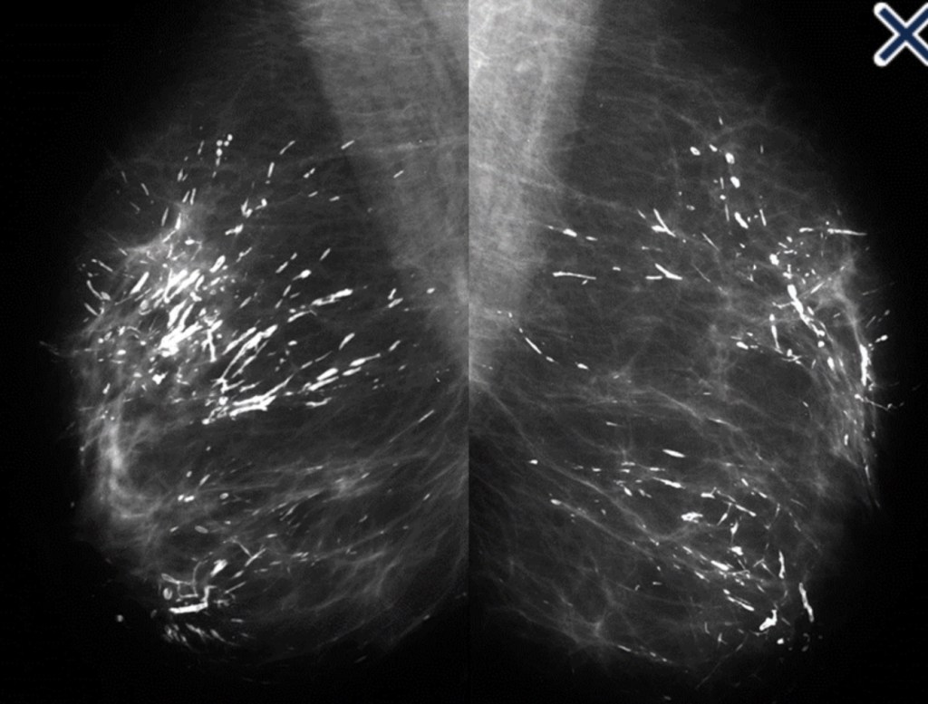
When coarse rod-like ductal calcifications:
Are diffuse, bilateral, and not confined to a single lobe:
They can be confidently assumed to result from plasma cell mastitis:
They do not require further evaluation or biopsy (Image)
The process is called secretory disease:
Because there is a stagnant, viscous fluid that eventually petrifies and results in the smooth contoured calcifications
Some of them are branching and look like malignant casting type calcifications:
But the key distinguishing feature:
Is the diffuse, multilobe, bilateral nature of the process:
Calcifications become much more worrisome when they are confined to a single lobe
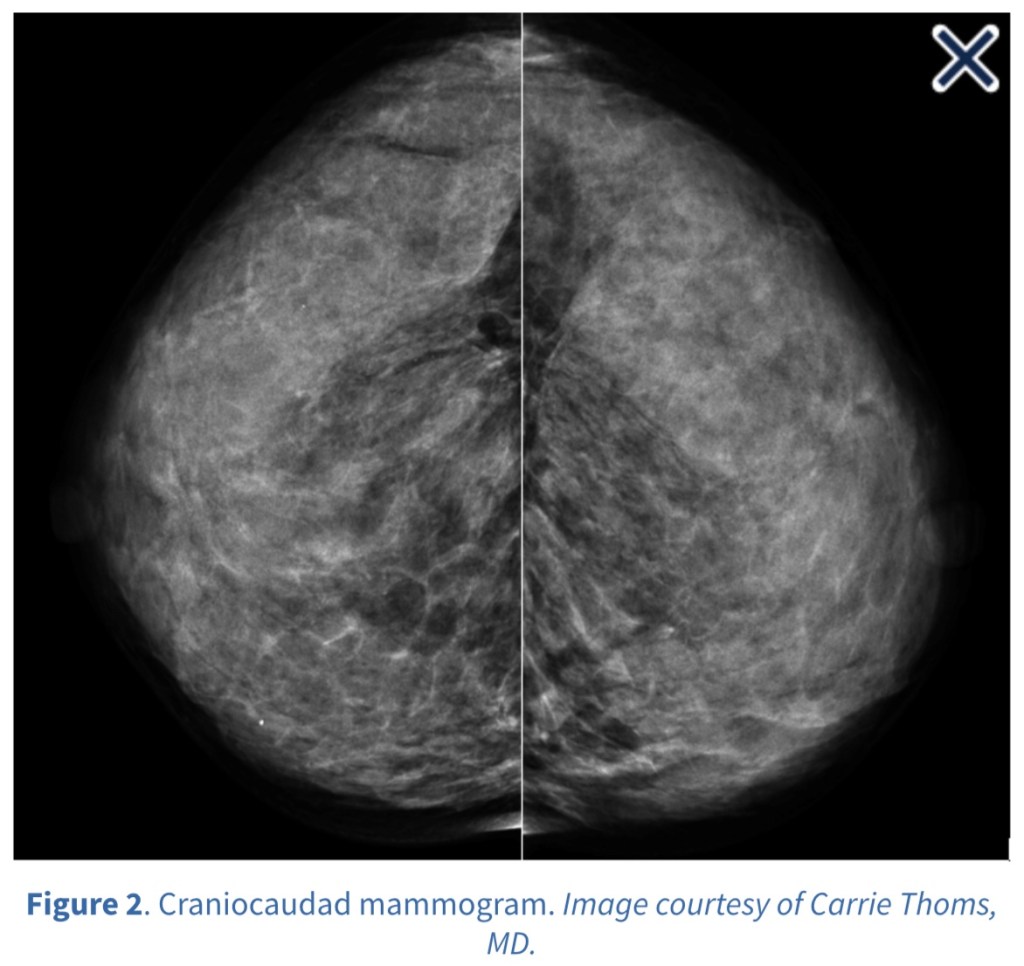
Coarse rod-like ductal calcifications: Plasma Cell Mastitis Casting type calcification: fragmented, linear, and branching
The most frequent malignant, ductal “casting type” calcifications:
Are fragmented, linear, and branching:
They are the most reliable mammographic sign of malignancy (Image)
The presence of fragmented and / or dotted casting type calcifications on the mammogram:
Restricted to one lobe:
Is a pathognomonic sign of a diffuse, grade 3 breast cancer subtype that originates in the major ducts and usually has a solid or micropapillary pattern:
Traditionally, this subtype has been called “comedo carcinoma”:
The cancer cells either produce a viscous, proteinaceous fluid which gradually concentrates and eventually calcifies, or they undergo necrosis (apoptosis) followed by calcification
In both instances, the intraluminal pressure increases, distending the ducts considerably
Dotted casting type calcifications:
Have been referred to as “snakeskin-like calcifications”:
They accumulate in the fluid:
Produced by either micropapillary or solid cancer cell growth patterns (Image)
Snakeskin type calcifications.
The tips of the micropapillary growths may become detached and eventually calcify, contributing to the intraluminal calcifications (Image).
The micropapillary growths break off and calcify in the lumen, resulting in the individual dots of calcification
Occasionally, malignant ductal calcifications present in a manner that can be easily mistaken for a benign process:
It occurs when fluid production:
Rather than necrosis, dominates the picture
The intraductal carcinoma can be grades 1, 2, or 3 and a micropapillary and / or cribriform architecture is present
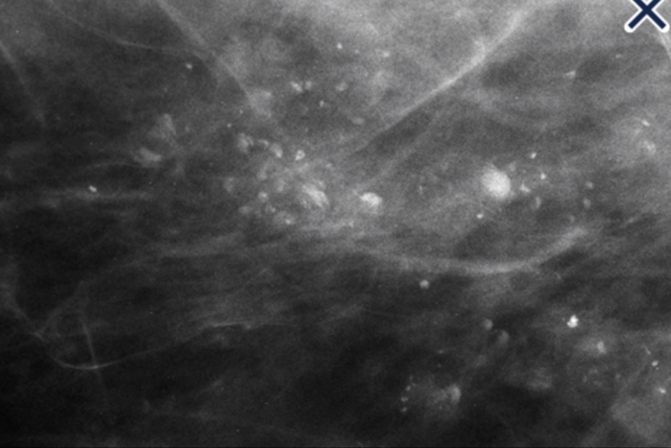
Tabár has done extensive work comparing the mammographic imageswith 3D subgross sections and whole mount histology slides in order to determine that the calcification occurs in large lakes of fluid that calcify, forming round or oval, discoid calcifications:
He credits Stavros with suggesting the name “skipping stone” calcifications because they are reminiscent of stones that children skip across a pond (Images 5a, 5b, 6, and 7)
Magnification view of “skipping stone” type calcificationsMagnification view of “skipping stone” type calcifications
A variation of this type of calcification occurs in grades 1 and 2 fluid-producing intraductal carcinomas when large psammoma body-like calcifications develop in the ducts in a pattern suggestive of a “string of pearls”