Blog

Oncoplastic Breast Surgery (OBS)

  • Oncoplastic breast surgery (OBS):
    • Combines principles of oncology and plastic surgery:
      • Toward achieving sound oncological and aesthetically pleasant results for breast tumors amenable to segmental mastectomies in patients with a favorable tumor to breast volume
  • OBS expands the indications for breast conservation:
    • Allowing the resection of much larger tumors, and is now an option for the surgical treatment of tumors larger than 4 cm and locally advanced cancers especially in large-breasted patients
    • When compared to conventional breast-conserving surgery (BCS):
      • OBS commonly results in higher specimen resection volumes and lower re-excision rates 
    • OBS does not eliminate or change the need for adjuvant therapies; most, if not all, patients undergoing segmental mastectomy for invasive breast cancers will benefit from adjuvant radiation therapy
    • OBS training is widely accepted as a prerequisite for performing these surgeries and should be represented in any multidisciplinary team, treating breast cancer and offering OBS: 
      • While no definitive test exists to assess aesthetic results and compare these between OBS and BCS for matched defects, most studies report favorable aesthetic results following OPS, in the majority of patients
  • References
    • Behluli I, Le Renard PE, Rozwag K, Oppelt P, Kaufmann A, Schneider A. Oncoplastic breast surgery versus conventional breast-conserving surgery: a comparative retrospective study. ANZ J Surg. 2019;89(10):1236-1241.
    • Strach MC, Prasanna T, Kirova YM, Alran S, O’Toole S, Beith JM, et al. Optimise not compromise: the importance of a multidisciplinary breast cancer patient pathway in the era of oncoplastic and reconstructive surgery. Crit Rev Oncol Hematol.2019;134:10-21.
    • Papanikolaou IG, Dimitrakakis C, Zagouri F, Marinopoulos S, Giannos A, Zografos E, et al. Paving the way for changing perceptions in breast surgery: a systematic literature review focused on oncological and aesthetic outcomes of oncoplastic surgery for breast cancer. Breast Cancer. 2019;26(4):416-427.

Work-up of Breast Implant-Associated Anaplastic Large Cel Lymphoma

  • Breast implant-associated anaplastic large cell lymphoma (BIA-ALCL):
    • Is an uncommon lymphoma that has only been reported in patients with a history of a textured breast implant device
  • Suggested theories of the cause of BIA-ALCL include:
    • Textured implant particulate
    • Chronic allergic inflammation, and / or response to a biofilm
  • Following National Comprehensive Cancer Network (NCCN) guidelines:
    • A swollen breast can be evaluated with ultrasound for either a:
      • Fluid collection
      • Capsular mass
      • Lymph node swelling
    • Fluid collections should be aspirated percutaneously:
      • A minimum of 20 ml but ideally as much fluid available should be sent for:
        • CD30 immunohistochemistry
        • Cell block cytology and flow cytometry evaluation and labeled to “rule out BIA-ALCL
      • CD30 testing is critical to direct pathologists
      • Efforts should be made to establish a diagnosis:
        • Prior to any surgical intervention
  • References
  • Brody GS, Deapen D, Taylor CR, Pinter-Brown L, House-Lightner SR, Andersen JS, et al. Anaplastic large cell lymphoma occurring in women with breast implants: analysis of 173 cases. Plast Reconstr Surg. 2015;135(3);695-705.

Fat Grafting in After Breast Cancer Surgery

  • Autologous fat grafting:
    • Is a procedure to transfer fat from one part of the patient’s body to another without requiring microvascular anastomosis
    • The graft initially takes similar to a skin graft, depending on the surrounding tissue bed before new blood vessels form
    • The grafted tissue is at risk for ischemia or fat necrosis:
      • As a result, there may be both palpable changes (e.g., cyst) or radiographic changes (e.g., calcifications) following fat grafting:
        • These have benign appearance on mammogram and are generally distinguishable from malignancy but may prompt additional imaging
  • Retrospective data suggest fat grafting does not interfere with cancer screening or detection
  • Fat grafting can be performed for all types of reconstruction, including primary defects as in lumpectomy, asymmetry, contour changes after radiation, or contour irregularity over an implant or tissue flap:
    • To date, there have been no clinical studies that demonstrate an increase in local regional recurrence in patients who have undergone fat grafting after breast cancer treatment
  • There is debate as to whether there is an ideal time to perform fat grafting:
    • But it is not routinely performed at the same time as lumpectomy or mastectomy:
      • Especially if radiation is indicated, as the tissue grafts will be susceptible to radiation injury
  • Finally, graft resorption is possible following the procedure, and therefore patients should be counseled that more than one operation may be necessary to achieve the desired result
  • References
    • Khouri RK Jr, Khouri RK. Current clinical applications of fat grafting. Plast Reconstr Surg 2017;140(3):466e-486e.
    • Agha RA, Fowler AJ, Herlin C, Goodacre TE, Orgill DP. Use of autologous fat grafting for breast reconstruction: a systematic review with meta-analysis of oncological outcomes. J Plast Reconstr Aesthet Surg. 2015;68(2):143-161.

Innervation of the Nipple Areolar Complex (NAC):

  • The innervation of the nipple areolar complex (NAC):
    • Is from the anterior and lateral branches of the 3rd, 4th, and 5th intercostal nerves:
      • Of which the lateral cutaneous branch of the 4th intercostal is dominant
  • This nerve has the greatest risk of injury during a resection of the central lower pole of the breast:
    • A superior pedicle, with resection of the lower pole:
      • Has the highest incidence of NAC sensory compromise
    • All patients should be advised that there is a risk of permanent loss of sensation
  • References
    • Riccio CA, Zeiderman MR, Chowdhry S, Brooks RM, Kelishadi SS, Tutela JP, et al. Plastic surgery of the breast: keeping the nipple sensitive. Eplasty. 2015;15:e28.
    • Chiummariello S, Angelisanti M, Arleo S, Alfano C. Evaluation of the sensitivity after reduction mammoplasty. Our experience and review of the literature. Ann Ital Chir.2013;84(4):385-388.
    • Schlenz I1, Rigel S, Schemper M, Kuzbari R. Alteration of nipple and areola sensitivity by reduction mammaplasty: a prospective comparison of five techniques. Plast Reconstr Surg. 2005;115(3):743-751.

Diagnosis of Breast Implant Associated Large Cell Lymphoma

  • Periprosthetic fluid collections:
    • Occurring more than 1 year after implantation:
      • Should be evaluated to rule out breast implant-associated anaplastic large cell lymphoma (BIA-ALCL)
  • Diagnosis of BIA-ALCL:
    • Is made by aspiration of the periprosthetic fluid:
      • Usually performed by fine needle aspiration:
        • Under ultrasound guidance
      • A sample of 50 ml or greater is sufficient to establish a diagnosis by CD30 immunohistochemistry:
        • CD30 is a cell surface protein expressed by roughly 5% of normal circulating T-cells:
          • However, BIA-ALCL demonstrates confluent staining of CD30
  • Open biopsy:
    • Is more invasive, and the capsule may be negative even in the presence of disease
  • Therefore, effusion FNA is preferred for diagnosis
  • Plastic surgery evaluation:
    • Is not appropriate or necessary
  • Mammogram evaluation is non-specific for a disease process and is not indicated with an obvious fluid collection
  • References
    • Clemens MW, Horwitz S. NCCN Consensus Guidelines for the diagnosis and management of breast implant-associated anaplastic large cell lymphoma. Aesth Surg J. 2017;37(3):285-289.
    • Brody GS, Deapen D, Taylor CR. Anaplastic large cell lymphoma occurring in women with breast implants: analysis of 173 cases. Plast Reconstr Surg. 2015;135(3):695-705.
    • Horwitz SM, Ansell SM, Ai WZ, Barnes J, Barta SK, Choi M, et al. NCCN guidelines insights: t-cell lymphomas, version 2.2018. J Natl Compr Canc Netw. 2018;16(2):123-135.

Can you have Nipple Sparing Mastectomy after Having Breast Reduction Surgery?

  • Several studies have concluded that a prior mammaplasty procedure (at least 3 months prior) has a negligible effect on the nipple necrosis rate following a nipple-sparing mastectomy
  • References
    • Alperovich M, Tanna N, Samra F, Blechman KM, Shapiro RL, Guth AA, et al. Nipple-sparing mastectomy in patients with a history of reduction mammaplasty or mastopexy: how safe is it? Plast Reconstr Surg. 2013;131(5):962-967.
    • Spear SL, Rottman SJ, Seiboth LA, Hannan CM. Breast reconstruction using a staged nipple-sparing mastectomy following mastopexy or reduction. Plast Reconstr Surg. 2012;129(3):572-581.
    • Frederick MJ, Lin AM, Neuman R, Smith BL, Austen WG Jr, Colwell AS. Nipple-sparing mastectomy in patients with previous breast surgery: comparative analysis of 775 immediate breast reconstructions. Plast Reconstr Surg. 2015;135(6):954e-962e.

Breast Oncoplastic Incisions

  • Disease located in the inferior hemisphere of the breast:
    • Can be easily incorporated into a Wise pattern reduction mammoplasty
Schematic representation of Wise pattern incision and various choices of pedicles. (A) Wise pattern incision. (B) Superior pedicle. (C) Superior-medial pedicle. (D) Inferior pedicle. (E) Lateral pedicle
  • Split reduction techniques:
    • Are best used for disease located in the upper inner or upper outer quadrants requiring skin excision due to the proximity of the tumor to the skin
    • With a split reduction technique:
      • The lateral (for upper outer quadrant cancers) or medial (for upper inner quadrant cancers) triangle of the Wise pattern is not positioned at the base of the breast:
        • But instead advanced up onto the breast where the area of disease was removed
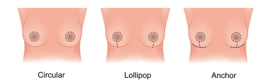
  • Donut mastopexy:
    • Is best used in patients with grade 1 ptosis with the area of disease is located close to the nipple areolar complex
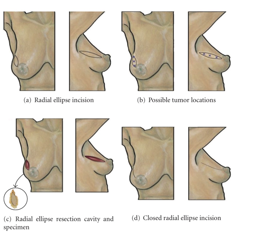
  • The radial ellipse lumpectomy:
    • Is useful for lateral tumors
    • With this technique a radial ellipse of skin and breast parenchyma is excised full thickness
    • This technique however does not improve significant ptosis
Radial ellipse segmentectomy. (a) Shows location of radial ellipse segmentectomy skin incision in upper outer quadrant. (b) Shows multiple “stars” indicating possible tumor locations suitable for this approach. (c) Shows resection cavity following excision of malignancy with excised specimen (inset). (d) Shows breast following closure of the skin incision.
  • References
    • Savalia NB, Silverstein MJ. Oncoplastic breast reconstruction: patient selection and surgical techniques. J Surg Oncol. 2016;113(8):875-882.
    • Silverstein MJ, Mai T, Savalia N, Vaince F, Guerra L. Oncoplastic breast conservation surgery: the new paradigm. J Surg Oncol. 2014;110(1):82-89.
    • Anderson BO, Masetti R, Silverstein MJ. Oncoplastic approaches to partial mastectomy: an overview of volume-displacement techniques. Lancet Oncol.2005;6(3):145–57.
    • Clough KB, Ihrai T, Oden S, Kaufman G, Massey E, Nos C. Oncoplastic surgery for breast cancer based on tumour location and a quadrant-per-quadrant atlas. Br J Surg. 2012;99(1):1389-1395

Donut Mastopexy in Oncoplastic Breast Surgery

  • Donut mastopexy:
    • Is best used to address small to medium cancers centrally located in any quadrant
  • Results are best in patients with:
    • Grade 1 ptosis with enlarged nipple areola complex
  • This approach in patients with significant ptosis:
    • Will result in flattening of the central breast and would be better treated with other onocoplastic techniques
  • Treatment of triple negative breast cancers:
    • Should consider neoadjuvant chemotherapy prior to surgical excision
  • Young patients with triple negative breast cancer:
    • Also should consider hereditary risk assessment:
      • As this may alter their decision regarding breast conservation
  • Patients with a history of prior breast cancer treated with adjuvant radiation therapy:
    • Should not consider breast conservation unless there are special circumstances
  • References
    • Savalia NB, Silverstein MJ. Oncoplastic breast reconstruction: patient selection and surgical techniques. J Surg Oncol. 2016;113(8):875-882.
    • Piper M, Peled A, Sbitany H. Oncoplastic breast surgery: current strategies. Gland surg. 2015;4(2):154-163.
    • Clough KB, Kaufman GJ, Nos C, Buccimazza I, Sarfati IM. Improving breast cancer surgery: a classification and quadrant per quadrant atlas for oncoplastic surgery. Ann Surg Oncol 2010;17(5):1375-1391.
    • Omarini C, Guaitoli G, Pipitone S, Moscetti L, Cortesi L, Cascinu S, et al. Neoadjuvant treatments in triple-negative breast cancer patients: where we are now and where we are going. Cancer Manag Res. 2018;10:91-103.

Oncoplastic Breast Incision in Large Breast with Ptosis

  • In general, patients with significant ptosis of the breast:
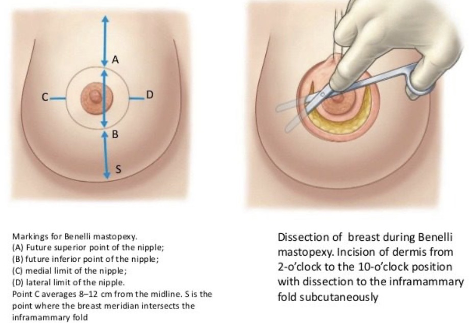
    • Obtain better cosmetic results with a reduction mammoplasty compared to donut or Benelli mastopexy
  • Lesions distant to the nipple areolar complex:
    • Can be difficult to access through a mastopexy incision
  • References
    • Lebovic GS. Oncoplastic surgery: a creative approach to breast cancer management. Surg Oncol Clin N Am. 2010;19(3): 567-580.
    • Piper M, Peled A, Sbitany H. Oncoplastic breast surgery: current strategies. Gland Surg. 2015;4(2):154-163.
    • Clough, KB, Kaufman, GJ, Nos, C, Buccimazza, I, Sarfati, IM. Improving breast cancer surgery: a classification and quadrant per quadrant atlas for oncoplastic surgery. Ann Surg Oncol. 2010;17(5):1375-1391.
    • Clough KB, Benyahi D, Nos C, Charles C, Sarfati I. Oncoplastic surgery: pushing the limits of breast-conserving surgery. BreastJ. 2015;21(2):140-146.

Risk of Finding High Risk Pathology in the Contralateral Breast in Patients Undergoing Oncoplastic Breast Conserving Surgery

  • A recent study by Liang et al:
    • Showed that in patients undergoing contralateral breast procedures at the time of oncoplastic breast-conserving surgery:
      • 16.5% were found to have high-risk pathology in the contralateral breast:
        • With almost 5% of these being cancer
    • Larger studies from Europe have shown the contralateral occult malignancy rate:
      • To be 1% to 5%:
        • This rate of occult disease is higher than the reported occult malignancy rate in patients who are undergoing cosmetic reduction mammoplasty
    • Orientation of the specimen(s) from symmetry procedure may be beneficial when performing these operations in the setting of cancer treatment
  • References
    • Liang Y, Muse-Fisher C, Rambukwella M, Naber SP, Chatterjee A. Malignant and high-risk lesions in the contralateral breast symmetry mastopexy and reduction specimens when performing large-volume displacement oncoplastic surgery. Ann Plast Surg. 2019;82(4S Suppl 3):S185-S191.
    • Sorin T, Fyad JP, Pujo J, Colson T, Bordes V, Leroux A, et al. Incidence of occult contralateral carcinomas of the breast following mastoplasty aimed at symmetrization. Ann Chir Plast Esthet. 2014;59(2):e21-e28.