In 2013 the SSO and ASTRO convened a multidisciplinary expert panel to review the available evidence regarding margin width and IBTR in patients with invasive cancer having breast conservation therapy
Meta-analysis and secondary data from prospective and retrospective trials led them to conclude that positive margins (defined as ink on invasive cancer):
Is associated with at least a 2-fold increase in IBTR
Among patients with negative margins:
A margin width of no ink on tumor represented the optimal margin width to minimize the risk of IBTR:
Notably the routine practice of obtaining wider negative margins than no ink on tumor is not indicated
While younger age:
Is associated with both increased IBTR after breast-conserving therapy as well as increased local chest wall relapse after mastectomy:
There is no evidence that increased margin width (over no ink on tumor) nullifies this increased risk of IBTR in younger patients
In 2016, margin guidelines related to the treatment of non-invasive breast cancer (DCIS) in the setting of breast conservation therapy were developed by the SSO, ASTRO, and ASCO in a similar manner
A consensus statement released by a multidisciplinary panel included the optimal margins for pure ductal carcinoma in situ (DCIS) and mixed tumors (invasive and non-invasive components within the same tumor) in the setting of breast conservation:
Results from the meta-analysis showed that a 2 mm margin decreases the risk of IBTR in pure DCIS compared to closer negative margins
This differs from the previous margin recommendation for invasive cancer, which remains no ink on tumor
However, in the setting of mixed tumors (invasive cancer with a DCIS component) the recommendation for negative margins remains no ink on tumor, as patients with mixed disease are treated as invasive cancer and therefore receive systemic therapy more often than pure DCIS patients:
In the setting of DCIS with micro-invasion (no focus of invasive disease larger than 0.1 cm):
The multidisciplinary panel recommends a 2 mm margin, as these lesions have similar rates of IBTR as pure DCIS
References
Moran MS, Schnitt SJ, Giuliano AE, Harris JR, Khan SA, Horton J, et al. Society of Surgical Oncology-American Society for Radiation Oncology consensus guideline on margins for breast-conserving surgery with whole-breast irradiation in stages I and II invasive breast cancer. Int J Radiat Oncol Biol Phys. 2014;88(3):553-564.
Morrow M, Van Zee KJ, Solin LJ, Houssami N, Chavez-MacGregor M. et al. Society of Surgical Oncology-American Society for Radiation Oncology-American Society of Clinical Oncology Consensus Guideline on margins for breast-conserving surgery with whole-breast irradiation in ductal carcinoma in situ. J Clin Oncol. 2016;34(33):4040-4046.
To investigate the risk of ipsilateral breast events (IBEs) in patients with DCIS treated with local excision without radiation:
The ECOG and North Central Cancer Treatment Group (NCCTG):
Conducted a prospective trial between 1997 and 2002 known as the E5194 study
Patients were stratified into two groups based on grade:
Low- and intermediate-grade DCIS measuring 2.5 cm or smaller
High-grade DCIS measuring 1 cm or smaller
Margin widths of 3 mm or wider were required along with no residual calcifications on postoperative mammograms
The low- and intermediate-grade DCIS group:
Had a 12-year IBE rate of 14.4%
The high-grade DCIS group:
Had a 12-year IBE rate of 24.6% (p=0.003)
Larger tumor size:
Was also found to be statistically significantly associated with developing an IBE (P = .01)
The risks of developing an IBE for either DCIS or invasive cancer:
Increased over time through 12 years of follow-up:
Without plateau
Current literature reports a 50% decrease in local recurrence with radiotherapy after surgical excision of DCIS:
However, we continue to look for low risk subsets of patients with DCIS:
In whom the absolute benefit of radiation therapy of lumpectomy may be small
The DCIS score (12-gene signature):
Was developed from tissue samples from the E5194 study:
With the goal of identifying low and high-risk subsets of patients with DCIS following lumpectomy alone
This assay utilized quantitative reverse transcriptase-polymerase chain reaction (RT-PCR):
From tumor specimens from 327 patients with DCIS treated with surgical excision without radiation from the E5194 study
The DCIS score of 0 to 100 (low less than 39, intermediate 39 to 54, high ≥ 55):
Was then designed to predict the recurrence of IBE overall, as well as DCIS or invasive cancer recurrence
The DCIS score correlated with 10-year IBE risk of:
10.6% in the low-risk group
26.7% in the intermediate-risk group
25.9% in the high-risk group
Young age and larger tumor size:
Also were found to be independent predictors of recurrence
So, this test may be most beneficial in post-menopausal women with small tumors
References:
Solin LJ, Gray R, Hughes LL, Wood WC, Lowen MA, Badve SS, et al. Surgical excision without radiation for ductal carcinoma in situ of the breast: 12-year results from the ECOG-ACRIN E5194 Study. J Clin Oncol. 2015;33(33):3938-3944.
Solin LJ, Gray R, Baehner FL, Butler SM, Hughes LL, Yoshizawa C, et al. A multigene expression assay to predict local recurrence risk for ductal carcinoma in situ of the breast. J Natl Cancer Inst. 2013;105(10):701-710.
Is a rare, benign, chronic inflammatory condition of the breast occurring mostly in non-white women of childbearing age
In the United States:
It’s more commonly seen in the Hispanic population
Idiopathic granulomatous mastitis often presents with:
A unilateral, hard, discrete tender mass
Nonetheless, it may also present with:
Multiple indurated inflammatory masses
A non-tender mass
With pain as a solitary symptom
In many cases, a central mass is accompanied by:
Multiple simultaneous areas of peripheral infection with abscesses
Erythema and ulceration of the overlying skin, and / or fistula formation
Pathologic features of IGM include:
Non-caseating granulomatous inflammation with necrosis and giant cell formation on a background of neutrophils all surrounding a breast lobule
Given the overlap between clinical and radiographic features of IGM and other disease processes:
Histopathologic evaluation has emerged as the gold standard for diagnosis of IGM
There is no formal consensus regarding the appropriate treatment for patients diagnosed with IGM:
The available treatment options include:
Expectant management
Steroid therapy
Immunomodulators such as:
Methotrexate and azathioprine
Limited or wide surgical excision
Antibiotic treatment in the setting of superimposed infection
References:
Mohammed S., Statz A, Lacross JS, Lassinger BK, Contreras A, Gutierrez C, et al. Granulomatous mastitis: a 10-year experience from a large inner city county hospital. J Surg Res. 2013;184(1):299-303.
Pandey TS, Mackinnon JC, Bressler L, Millar A, Marcus EE, Ganschow PS. Idiopathoic granulomatous mastitis – a prospective study of 49 women and treatment outcomes with steroid therapy. Breast J. 2014;20(3):258-266.
Oran ES, Gurdal SO, Yankol Y, Öznur M, Calay Z, Tunacı M, et al. Management of idiopathic granulomatous mastitis diagnosed by core biopsy: a retrospective multicenter study. Breast J. 2013;19(4):411-418.
Akbulut S, Yilmaz D, Bakir S. Methotrexate in the management of idiopathic granulomatous mastitis: review of 108 published cases and report of four cases. Breast J. 2011;17(6):661-668.
What is the short-term safety of an active monitoring approach vs guideline-concordant care (surgery with or without radiation therapy):
For hormone receptor–positive, grade 1 or grade 2 breast ductal carcinoma in situ?
Findings:
In this prospective randomized clinical trial of:
957 participants
The 2-year Kaplan-Meier cumulative rate of ipsilateral invasive cancer was 5.9% in the guideline-concordant care group vs 4.2% in the active monitoring group:
A difference of −1.7% (upper limit of the 95% CI, 0.95%):
Indicating that active monitoring is not inferior to guideline concordant care
Meaning:
These data support the short-term safety of active monitoring compared with guideline-concordant care in patients with low-risk ductal carcinoma in situ
Abstract:
Importance:
Active monitoring for low-risk ductal carcinoma in situ (DCIS) of the breast has been proposed as an alternative to guideline-concordant care, but the safety of this approach is unknown
Objective :
To compare rates of invasive cancer in patients with low-risk DCIS receiving active monitoring vs guideline-concordant care
Design, Setting, and Participants:
Prospective, randomized noninferiority trial enrolling 995 women aged 40 years or older with a new diagnosis of hormone receptor–positive grade 1 or grade 2 DCIS without invasive cancer at 100 US Alliance Cancer Cooperative Group clinical trial sites from 2017 to 2023
Interventions:
Participants were randomized to receive active monitoring (follow-up every 6 months with breast imaging and physical examination; n = 484) or guideline-concordant care (surgery with or without radiation therapy; n = 473)
Main Outcomes and Measures :
The primary outcome was 2-year cumulative risk of ipsilateral invasive cancer diagnosis, according to planned intention-to-treat and per-protocol analyses, with a noninferiority bound of 5%
Results:
The median age of the 957 participants analyzed was 63.6 (95% CI, 55.5-70.5) years in the guideline-concordant care group and 63.7 (95% CI, 60.0-71.6) years in the active monitoring group
Overall, 15.7% of participants were Black and 75.0% were White
In this prespecified primary analysis, median follow-up was 36.9 months; 346 patients had surgery for DCIS, 264 in the guideline-concordant care group and 82 in the active monitoring group
Forty-six women were diagnosed with invasive cancer, 19 in the active monitoring group and 27 in the guideline-concordant care group
The 2-year Kaplan-Meier cumulative rate of ipsilateral invasive cancer was 4.2% in the active monitoring group vs 5.9% in the guideline-concordant care group, a difference of −1.7% (upper limit of the 95% CI, 0.95%):
Indicating that active monitoring is not inferior to guideline-concordant care
Invasive tumor characteristics did not differ significantly between groups
Conclusions and Relevance:
Women with low-risk DCIS randomized to active monitoring did not have a higher rate of invasive cancer in the same breast at 2 years compared with those randomized to guideline-concordant care
Is a self-limiting superficial thrombophlebitis of the breast
The etiology is not always clear:
But it has been reported to be associated with:
Trauma
Core biopsy
Breast surgery
Cancer
Radiation treatment
Underwire bras
Oral contraceptives
It typically presents as a:
Vertically oriented, tender cord on the breast:
Which becomes more prominent when the arm is raised and can be associated with skin retraction
It can be treated with:
Warm compresses
Aspirin, or non-steroidal anti-inflammatory drugs:
It usually resolves within 4 to 6 weeks
Axillary web syndrome:
Can also present as a palpable cord, but is often found in the axilla:
Can extend to involve the medial aspect of the ipsilateral arm down to the antecubital fossa
It is associated with limitations in shoulder mobility
Fat necrosis:
Can cause a palpable mass with skin retraction:
But it does not generally conform to the distribution of a superficial vein
Zuska’s disease:
Is a condition consisting of:
Chronic subareolar abscesses and fistulae
Caused by squamous metaplasia and keratin plugging of the breast ducts, and it is not related to Mondor’s disease
References
Pasta V, D’Orazi V, Sottile D, Del Vecchio L, Panunzi A, Urciuoli P. Breast Mondor’s disease: diagnosis and management of six new cases of this underestimated pathology. Phlebology. 2015;30(8):564-568.
Salemis NS, Vasilara G, Lagoudianakis E. Mondor’s disease of the breast as a complication of ultrasound-guided core needle biopsy: management and review of the literature. Breast Dis. 2015;35(1):73-76.
Shetty MK, Watson AB. Mondor’s disease of the breast: sonographic and mammographic findings. AJR Am J Roentgenol. 2001;177(4):893-896.
Tilley A, Thomas-MacLean R, Kwan W. Lymphatic cording or axillary web syndrome after breast cancer surgery. Can J Surg. 2009;52(4):E105-E106.
Lannin DR. Twenty-two year experience with recurring subareolar abscess and lactiferous duct fistula treated by a single breast surgeon. Am J Surg. 2004;188(4):407-410.
Demonstrated that 95% of women with breast cancer and strong indications for post-lumpectomy radiation:
Went on to receive it:
But rates of post-lumpectomy radiation therapy use have been shown to vary:
Depending on the region of the country that the patient lives in
The age of the patient
The disease being treated (DCIS vs Invasive Disease)
Among patients who undergo BCT for DCIS:
Only 50% are estimated to receive adjuvant radiation
Many patients choose mastectomy over breast-conserving surgery for DCIS:
Because they are not able or willing to complete 6 weeks of daily radiation therapy:
Secondary to social or health considerations
Other patients who are candidates for breast-conserving surgery:
Choose to undergo a mastectomy:
Because of concerns about postradiation complications
Breast-conserving surgery alone (i.e., without radiation therapy):
May be sufficient in a select subgroup of patients with DCIS
Initial data that supported the use of breast-conserving surgery alone in the treatment of DCIS came from a study by Lagios et al. (1989):
In which 79 patients with mammographically detected DCIS underwent margin-negative excision alone
After a follow-up time of 124 months:
The local recurrence rate was 16% overall, specifically:
33% for the subgroup of patients with high-grade lesions and comedo necrosis versus only 2% for the patients with low- or intermediate-grade lesions
The USC / VNPI score can be a helpful tool in clinical decision making:
But even though margin width is an independent prognostic factor for recurrence using the USC / VNPI score:
It is unlikely that margin width alone can identify the patients with DCIS treated with breast conservation for whom radiation therapy can be safely omitted
In a retrospective analysis of 469 patients with DCIS who underwent breast conservation with margins that were at least 10 mm, Silverstein et al. (1999):
Did not detect a lower recurrence rate:
When postoperative radiation therapy was employed
In contrast, even on reanalysis of the NSABP B-17 data:
All patient cohorts benefited from radiation therapy:
Regardless of the clinical or mammographic tumor characteristics
Furthermore, Wong et al. (2003, 2014):
Reported the early termination of a prospective single-arm trial conducted at the Dana-Farber / Harvard Cancer Center:
In which radiation therapy was omitted in patients with grade 1 to 2 DCIS that was no more than 25 mm and excised with 10 mm or greater margins:
At a median follow-up of 3.3 years:
The number of local recurrences observed was 2.5% per patient-year:
Corresponding to a 5-year rate of 12.5%
In 2010, Rudloff and colleagues at Memorial Sloan Kettering Cancer Center:
Published a multivariable nomogram:
To estimate risk for local recurrence in women with DCIS treated with breast-conserving surgery
The nomogram incorporates commonly available factors that have previously been shown to affect risk of ipsilateral breast tumor recurrence, these include:
Age at diagnosis
Family history
Type of patient presentation:
Radiologic or clinical
Nuclear grade
Necrosis, margins
Number of excisions
Receipt of radiation and / or adjuvant endocrine therapy
The nomogram calculates an actual, individualized estimate of absolute risk of ipsilateral breast tumor recurrence at 5 or 10 years:
Which can be weighed against the use of available adjuvant treatment options
There are two large, prospective, observational studies:
Designed to investigate the role of observation versus radiation therapy after breast-conserving therapy in patients with DCIS
As mentioned earlier, Wong and colleagues at Harvard:
Conducted a single-arm, phase III observational study examining long-term outcomes in women with small (≤ 2.5 cm), low- and intermediate-grade DCIS who were treated with lumpectomy and margins ≥ 1 cm and did not receive adjuvant tamoxifen or radiation:
With a median follow-up of 11 years:
13% (19 of 143) of patients experienced local recurrence:
Approximately two-third of which were DCIS
In the Eastern Cooperative Oncology Group–American College of Radiology Imaging Network (ECOG-ACRIN; formerly known as the Eastern Cooperative Oncology Group) Cancer Research Group E5194 study:
Patients with low- or intermediate-grade DCIS smaller than 25 mm (cohort 1), or high-grade DCIS smaller than 10 mm (cohort 2), with excisional margins of at least 3 mm, underwent breast-conserving surgery without radiation therapy:
30% of patients received tamoxifen
At 12 years:
14.4% of the participants in cohort 1 experienced an in-breast even while 24.6% of those in cohort 2 experienced an in-breast event (p = 0.0003), and this difference was driven by a statistically significant difference (p = 0.02) in noninvasive recurrence
In addition, membership in cohort 2 and larger tumor size were both found to be associated with increased likelihood of recurrence (Solin, 2015)
Finally, in patients over 70:
There is increasing evidence that radiation therapy:
Depending on the aggressiveness of the DCIS and the expected life expectancy of the patient:
Prognostic role of estrogen receptor (ER) and HER2 in DCIS:
In observational studies:
ER status – 5 of 26 studies found a statistically significant lower risk of ipsilateral breast tumor recurrence (IBTR) in ER positive cases
HER2 status – 10 out of 27 studies reported a significant increasein the risk of recurrences to be associated with HER2 expression
Limitations of these observational studies were:
Small sample size (events) in the majority of the studies
Selection bias
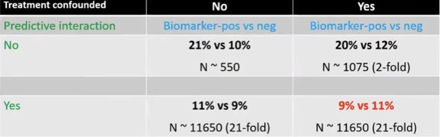
Treatment-related confounding:
ER expression is inversely associated and HER2 expression is positively associated with:
Adverse histologic features in DCIS
Therefore, ER negative or HER2 amplification in DCIS:
Is more likely to receive adjuvant treatment that ER positive or HER2 negative DCIS when ER or HER2 status is not known:
Potentially masking the true association
The probability of masking of the true association:
Increases greatly if the biomarker also has predictive characteristics
How to eliminate treatment-related confounding:
The study population should have random treatment allocation:
Cohorts from randomized controlled trials
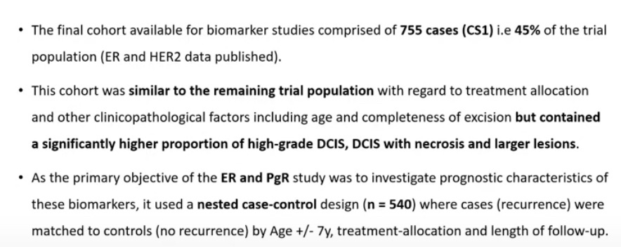
Case-control studies matching by treatment:
Does not permit investigation of predictive characteristics of the biomarker
Multivariable / adjusted analysis:
Power remains an issue
Biomarker cohort study:
UK, Australia and New Zealand DCIS trial Cuzick J et al Lancet Oncol. 2011; Houghton J eta al Lancet 2003):
2X2 randomized trial comparing the effectiveness of radiotherapy and tamoxifen in reducing recurrences in patients with complete locally excised DCIS
# of patients 1694
The 2X2 factorial design permits investigation pertaining to both adjuvant treatments in DCIS
After a median follow-up of 12.7 years, there have been 162 invasive and 197 DCIS events in these patients:
17 unknown
Total 376
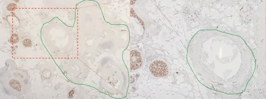
In the study they observed that in patient with ER positive DCIS:
They identified areas within the ducts that were ER negative in the same lesion
Multi-clonal ER Expression: On the right side panel you can see an ER negative duct with an ER positive duct adjacent to it. Multi-clonal ER Expression
In these study 11% of patients were identified to have multi-clonal DCIS:
Clonal method:
Estrogen receptor (ER) expression and recurrence:
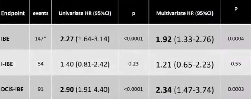
ER negative (multi-clonal) DCIS is associated with:
A five fold increase of in situ ipsilateral breast event
A three fold increase in overall ipsilateral breast event
Invasive ipsilateral breast event is not statistically increased
mOR; Matched Odds Ratio, IBE: Ipsilateral breast event, I-IBE: Invasive IBE, In situ IBE
The results show that the clonal method:
Is superior to the standard method in predicting IBE and DCIS-IBE
Progesterone receptor (PgR):
Was not significantly associated with recurrence in ER positive DCIS
It was not an independent predictor in multivariable models
Inclusion of PGR did not significantly improve multivariable models
HER2 expression and recurrence:
HER2 positively was identified in 55% of the cases of DCIS:
Compared to invasive breast cancer which is around 15% to 20%
The expression of HER2 was associated with a two fold increase in IBE and in situ IBE
Comparison is HER2 3+ vs. 0, 1+, 2+ Univariable Analysis N = 713; Multivariable Analysis N = 612 IBE: Ipsilateral breast event, I-IBE: Invasive IBE, In situ IBE
HER2 status (post-ERBB2-reflex test) and recurrence:
HER2 status (ERBB2 reflexes) as a predictor of recurrence:
Is associated with nearly a three fold increase in IBE and in situ IBE
Is associated with an increase risk of I-IBE but it did not reach statistical significance
HER2 status (ERBB2 reflexes) is assigned after ERBB 2 mRNA expression a reflex test. Comparison of HER2 positive (3+ of IHC 2+ and ERBB2 mRNA expression > 1.1007 vs. HER2 negative (0, 1+ or 2+ with ERBB2 mRNA expression </= 1.1007
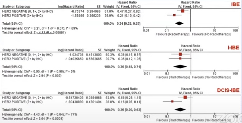
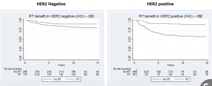
Radiation therapy benefit:
In HER2 positive disease was much larger as compared to HER 2 negative disease (statistically significant difference)
In HER2 negative DCIS radiation therapy reduced events by 53% compared to 80% in HER2 positive disease
Kaplan Meir curves showing the difference in benefit of radiation therapy in HER2 positive disease
In conclusion:
ER is a strong prognostic factor:
IBE mOR 3.33 for clinical method
HER2 is a strong prognostic factor:
IBE HR 2.84 for ERBB2-reflexes status
Radiotherapy benefit greater in HER2 positive DCIS:
HR 0.21 vs 0.47:
En by greater benefit in reducing DCIS-IBE
ER and HER2 evaluation should be routinely carried out?
Endocrine therapy considerations:
NSABP-B24:
ER is a predictor of tamoxifen benefit
Ipsilateral ER status is not a predictor of contralateral breast cancer (CBC) risk or tamoxifen benefit in preventing CBC
Combining the data from the RTOG 9804 trial and the UK/ANZ DCIS trial in the low risk DCIS (< 10 mm):
Can we recommend radiation therapy only to ER – negative or HER2 – positive DCIS?
The effect size and predictive benefit are an excellent fit to the RTOG 9804 results if 15% to 20% of patients in the trial were HER2 positive (proportion similar to the UK/ANZ DCIS trial)
With that proportion of HER2 expression:
15 year cumulative IBE rates (15.1% overall) in the RT arm of the trial would be 9% in HER2 negative (0.6% per years, same as CBC risk) and 26% in HER2 positive disease
Increased local recurrence and decreased long-term prognosis
Indications for postoperative radiotherapy (PORT) include:
Positive or close margins:
However despite PORT:
Local recurrence rates do not approach those in which adequate clearance is achieved at the primary operation
Increasing resection margins in the region of the head and neck:
Potentially results in increased functional and cosmetic deficit
Resection margins of up to 2 cm have been advocated:
However such margins result in significant functional deficit following the resection of even the smallest of tumors
Three-dimensional, 1 cm resection margins:
Have been demonstrated as acceptable when dealing with oral and oropharyngeal tumor:
Adopting 1 cm surgical margins:
Account is taken of the shrinkage that occurs post-resection:
So ensuring greater than 5 mm pathological margins
It should be remembered that the use of 5 mm as a cut-off point for ‘clear’ margins is arbitrary and purely represents a margin that is considered acceptable
It is vitally important to continually reassess margins visually and by palpation during tumor resection
If approaching the resection of a tumor with curative intent:
Then reconstructive considerations should not influence the tumor resection
Comparison of published data regarding the incidence of positive margins and their influence on survival or local recurrence is complicated by the variable definition of a positive margin:
The definition of a positive margin ranges from:
Invasive tumor at the margin, tumor within 1 mm and tumor within 5 mm
The UK Royal College of Pathologists have issued guidelines:
Suggesting clear margins if the histological clearance is > 5 mm
Close margins if 1 mm to 5 mm
Positive margins if less than 1mm
The incidence of positive margins for tumors of the oral cavity:
Has been demonstrated as being higher than other head and neck sites:
Potentially due to its complex anatomy and three-dimensional shape
Large tumors, perineural spread, vascular permeation, a noncohesive invasive front or cervical metastasis:
Are all associated with a greater risk of failing to achieve clear margins:
These features suggest that close or involved margins:
Potentially reflect a more aggressive tumor biology
The incidence of close or involved margins following tumor resection may be greater than 60% depending on tumor site and size:
Invariably, it is the deep margin that is close or positive:
However close deep margins do not necessarily require adjunctive treatment:
The use of ultrasonography to aid in determining deep margin resection has been described
Frozen sections are not routinely used by many surgeons:
Reasons cited being potential cost
Inability to reliably prevent positive final margins
Poor relocation of biopsy site should the result be positive
Ninety-nine percent of American head and neck surgeons:
Routinely use frozen section intraoperatively:
However overreliance on frozen section may result in undertreatment of tumors
When conducting a bony resection:
A 1 cm margin should be achieved:
It has been demonstrated that it is unusual for extension of tumor in bone to exceed the overlying soft tissue extension
The risk of underlying invasion in patients with DCIS:
Is roughly 25% (Bundred et al, BMJ. 2013) in this era of core biopsies
Risk of SLN metastasis in pure DCIS:
Is 0.2% to 0.7% (Zetterfund et al; BJS. 2014; Nicholson et al, EJSO. 2015):
Risk may rise to 9% if known micro-invasion (Meretoja et al, Ann Surg Onc. 2009)
Vacuum assisted devices (VAB):
Can lower the risk of invasion to 11% in retrospective data (Sumian et al, EJSO, 2016)
In prospective data (Cinnamome Study):
The upgrade was 39% (Tunon-De-Lara, Ann Surg Onco. 2015)
Routine MRI does not improve surgical outcomes:
Upgrade, size, or re-excision rates
(Fancellu et al, BJS. 2015; Chou et al ECON-AGRIN E4112, Radiology. 2021; Roque et al, NPJ breast cancer. 2022)
Dedicated breast PET? (Grana-Lopez et l Eur J Rad. 2020; Sasada et al, EJSO. 2021):
Up to know have not shown improve outcomes
There are approximately 50 retrospective studies evaluating nomograms and there predictive value on the risk of finding invasion in DCIS:
The usual risk factors are:
Size, grade 2 and 3, comedo necrosis, mass effect, micro-invasion:
Among “high-risk” DCIS:
Axillary evaluation in DCIS affects treatment but not survival (Coromilas et al, Ann Surg Onc 2016)
Axillary evaluation in DCIS increases complications and long-term morbidity (relative increase up to 6 to 8 times (Kilelea et al, Ann Surg Onc. 2018)
SLNB can be omitted in patients with DCIS planned for breast conserving surgery (BCS):
Detection rate at reoperation is 85.5% (GATA Study, Breast 2015)
NO data on feasibility after oncoplastic surgery
NO data on procedure accuracy
DCIS is noninvasive:
By definition, is unable to metastasize:
However, some studies have shown that up to 15% of patients with pure DCIS have isolated tumor cells (ITCs) or micrometastasis on nodal evaluation compared to others that show a 0.2% to 0.7% risk of nodal metastasis:
However, these small tumor deposits likely have little prognostic significance and may be cell clusters displaced by biopsy
In patients with DCIS detected by core biopsy:
There is a 15% to 25% associated risk of an invasive component when excised
Patients undergoing mastectomy for DCIS:
Should be offered SLNB since it would not be feasible to perform following mastectomy if invasive carcinoma is subsequently identified
ASCO consensus guidelines recommend that patients with DCIS who undergo breast-conserving operation should not routinely have SLNB:
However, SLNB could be discussed with patients undergoing breast conservation:
Who have a core biopsy diagnosis of DCIS and:
A large area of DCIS on imaging (2 to 5 cm)
High-grade DCIS
Comedonecrosis
When a physical examination or imaging shows a discrete mass
These findings have been associated with an increased risk of invasive cancer, and SLNB at the time of the initial operation could avoid a second operation
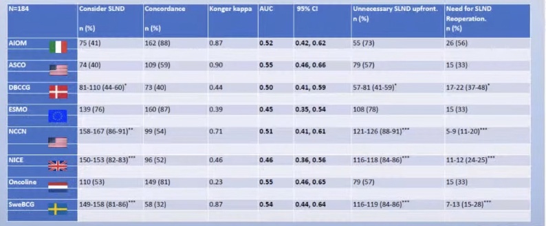
All of the guidelines that try and help us decide when to perform of SLNB in DCIS show a predictive value of a coin toss.