Blog

Pendrin Transporter Protein in Thyroid Hormone Synthesis

  • Pendrin, a transporter protein:
    • Is crucial for iodide efflux in thyroid follicular cells:
      • Playing a key role in thyroid hormone synthesis
    • It’s specifically located on the apical membrane of the thyroid follicular cells:
      • Where it mediates the movement of iodide:
        • From inside the cell to the colloid space within the follicle 
  • Function:
    • Pendrin acts as a transporter:
      • Facilitating the movement of iodide and chloride ions across the cell membrane
      • In the thyroid, it specifically transports iodide from the cytoplasm of thyroid follicular cells into the follicular lumen (colloid) 
  • Location:
    • Pendrin is located on the apical membrane of thyroid follicular cells:
      • Which is the membrane facing the colloid within the thyroid follicle. 
  • Significance:
    • This transport is essential for the synthesis of thyroid hormones (T4 and T3)
    • Iodide is first transported into the cell by the:
      •  Sodium-iodide symporter (NIS:
        • Which is located on the basolateral membrane of the thyroid follicular cell
      • Then, pendrin facilitates the efflux of iodide into the colloid:
        • Where it can be used in the synthesis of thyroglobulin (TG)
          • The precursor to thyroid hormones
  • Pendred Syndrome:
    • Mutations in the PDS gene, which codes for pendrin:
      • Can lead to Pendred syndrome:
        • A condition characterized by goiter (enlarged thyroid), and hearing loss:
          • Due to impaired iodide transport in the thyroid and inner ear
Thyroid Hormone Synthesis in the Thyroid Follicular Cell.
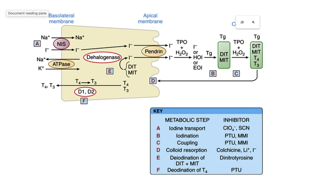
Thyroid hormone synthesis and secretion are activated when Thyroid stimulating hormone (TSH) binds to the TSH receptor on the basolateral membrane. Iodide is transported into the cell via the Na+/I− (NIS) symporter and flows down an electrical gradient, maintained by the sodium-potassium ATPase. Iodide becomes covalently attached to the tyrosyl residues of the precursor thyroid hormone glycoprotein, thyroglobulin, by thyroperoxidase (TPO) to form monoiodotyrosine (MIT) and diiodotyrosine (DIT). These are subsequently coupled by the action of TPO to form the iodothyronine hormones, tetraiodothyronine (T4) and triiodothyronine (T3). In the process of thyroid hormone secretion, Tg enters the cell by pinocytosis, forming colloid droplets. These fuse with lysosomes, forming phagolysosomes in which Tg is broken down by proteolysis, and then T4 and T3 are released and diffuse into circulation. MIT and DIT are formed by the iodination of tyrosyl amino acids on the thyroglobulin molecule. In a subsequent step, two DITs are coupled to form T4, or one DIT and one MIT are coupled to form T3. (From Brent GA, Koenig RJ. Thyroid and antithyroid drugs. In: Brunton L [ed]. Goodman and Gilman’s The Pharmacological Basis of Therapeutics. 13th ed. New York: McGraw-Hill; 2017.)

Axillary Staging in Ductal Carcinoma In Situ (DCIS)

👉DCIS is noninvasive and, by definition, is unable to metastasize.

👉However, studies have shown that up to 15% of patients with pure DCIS have isolated tumor cells (ITCs) or micrometastasis on nodal evaluation.

However, these small tumor deposits likely have little prognostic significance and may be cell clusters displaced by biopsy.

👉In patients with DCIS detected by core biopsy, there is a 15% to 20% associated risk of an invasive component when excised.

👉Patients undergoing mastectomy for DCIS should be offered sentinel lymph node biopsy (SLNB) since it would not be feasible to perform following a mastectomy if invasive carcinoma is subsequently identified.

👉ASCO consensus guidelines recommend that patients with DCIS who undergo breast-conserving operation should not routinely have SLNB.

👉However, SLNB could be discussed with patients undergoing breast conservation who have a core biopsy diagnosis of DCIS and a large area of DCIS on imaging (2 to 5 cm), high-grade DCIS, or comedonecrosis or when a physical examination or imaging shows a discrete mass.

👉These findings have been associated with an increased risk of invasive cancer, and SLNB at the time of the initial operation could avoid a second operation.

👉

Training:

• General surgery:

• Michigan State University:

• 2004 al 2010

• Surgical Oncology / Head and Neck Surgery / Endocrine Surgery:

• Fox Chase Cancer Center (Filadelfia):

• 2010 al 2012

• Masters in Science (Clinical research for health professionals):

• Drexel University (Filadelfia):

• 2010 al 2012

• Surgical Oncology / Head and Neck Surgery / Endocrine Surgery:

• IFHNOS / Memorial Sloan Kettering Cancer Center:

• 2014 al 2016

#Arrangoiz

#Surgeon

#Cirujano

#SurgicalOncologist

#CirujanoOncologo

#BreastSurgeon

#CirujanodeMama

#CancerSurgeon

#CirujanodeCancer

National Comprehensive Cancer Network (NCCN) Guidelines Recommend Consideration of Sentinel Lymph Node Biopsy (SLNB) for Ductal Carcinoma In Situ (DCIS)

  • The likelihood of finding invasive cancer on final pathologic evaluation following a core needle biopsy showing DCIS:
    • Has been reported to vary between 10% to 20%:
      • Therefore, a second operation will be required in this subset of patients who are later found to have invasive cancer
  • NCCN guidelines recommend:
    • Simultaneous performance of a sentinel lymph node biopsy when operating on patients with DCIS on core biopsy if:
      • The index operation may compromise subsequent lymphatic drainage:
        • Such as a mastectomy or a lumpectomy performed on a cancer in the axillary tail of the breast
  • While pathologic subtypes of DCIS may be associated with higher risks of local recurrence:
    • The presence of grade 3 disease and comedonecrosis:
      • Are not associated with higher probabilities of nodal involvement
  • Lastly, a history of prior invasive cancer is not associated with a higher probability of invasive cancer when the core biopsy demonstrated DCIS alone
  • References
  • Jakub JW, Murphy BL, Gonzalez AB, Conners AL, Henrichsen TL, Maimone S 4th, et al. A validated nomogram to predict upstaging of ductal carcinoma in situ to invasive disease. Ann Surg Oncol. 2017;24(10):2915-2924.
  • NCCN guidelines Breast Cancer (version 1.2018). National Comprehensive Cancer Network website. https://www.nccn.org/professionals/physician_gls/pdf/breast.pdf. Accessed October 18, 2019.

#Arrangoiz #BreastSurgeon #CancerSurgeon #SurgicalOncologist #BreastCancer #DCIS #SLNB #Miami #Mexico #Surgeon #Teacher

Lactational or Puerperal Breast Abscesses

  • Lactational or puerperal breast abscesses:
    • Occur in 0.4% to 11% of breastfeeding women
  • The objectives of management should include:
    • Resolution of the abscess
    • Relief of pain
    • Continuation of breastfeeding
  • Appropriate antibiotic coverage should include:
    • Coverage for Gram-positive organisms until cultures are available to guide therapy
  • The most common organisms isolated from cultures include:
    • Staphylococcus aureus
    • Staphylococcus epidermidis
    • Streptococci
  • Choice of interventional strategy is somewhat controversial:
    • As both needle aspiration and surgical I&D are highly effective in successfully treating the abscess and have similar recurrence rates
  • Needle aspiration:
    • Is associated with reduction of healing time, higher continuation of breastfeeding, and higher patient satisfaction, but the appropriate choice of management depends on the clinical situation
  • Aggressive pumping or overfeeding to empty the breast:
    • Can lead to hyperlactation and may contribute to further tissue damage and inflammation:
      • The patient should continue to breastfeed, if possible, or pump physiologic volumes of milk and empty the unaffected breast first
  • While mammary fistula (“milk fistula”) is the most common complication after breast abscess:
    • It is relatively rare, occurring in 1% to 3% of patients
    • They can develop spontaneously:
      • But more commonly occur after instrumentation of the breast (eg, needle biopsies, aspirations, surgery)
      • Not surprisingly, incidence of milk fistula after needle aspiration is significantly lower than surgical I&D for women with lactational breast abscesses (RR=0.21, p=0.013)
    • For patients undergoing surgery:
      • Incisions should be as small and as far from the nipple-areolar complex as possible
      • Placement of closed-suction drains or vacuum-assisted dressings should be avoided:
        • As they may promote fistula development due to granulation tissue formation:
          • Penrose drain or another non-suction drain should be used instead
      • While dopamine agonists have been shown to effectively cease lactation:
        • They are no longer routinely recommended
      • Continued breastfeeding is encouraged and can divert milk flow through the nipple, minimizing flow through the cavity
  • References:
    • Zhou F, Li Z, Liu L, et al. The effectiveness of needle aspiration versus traditional incision and drainage in the treatment of breast abscess: a meta-analysis. Ann Med. 2023;55(1):2224045. Published online June 23, 2023. doi: 10.1080/07853890.2023.2224045
    • Mitchell K, Johnson H, Rodriguez J, et al; Academy of Breastfeeding Medicine.  Academy of Breastfeeding Medicine Clinical Protocol #36: The Mastitis Spectrum, Revised 2022.  Breastfeed Med. 2022;17(5):360-376. doi: 10.1089/bfm.2022.29207.kbm
    • Johnson HM, Mitchell KB.  Low incidence of milk fistula with continued breastfeeding following radiologic and surgical interventions on the lactating breast. Breast Dis. 2021;40(3):183-189. doi: 10.3233/BD-201000

Lactational Breast Abscesses

  • Lactational breast abscesses:
    • Develop in up to a quarter of all breastfeeding women:
      • Typically within the first 12 weeks after birth or during the weaning process
  • Breast abscesses:
    • Rarely resolve with antibiotics alone
  • Abscesses generally require:
    • Drainage in conjunction with antibiotics
  • Depending on the prevalence of community-acquired MRSA in the region in which the patient presents:
    • MRSA coverage may be empirically initiated
  • A recent Cochrane review:
    • Found insufficient evidence to determine whether:
      • Needle aspiration is a more effective option to incision and drainage (I&D) for lactational breast abscesses, or whether an antibiotic should be routinely added to women undergoing I&D for lactational breast abscesses
      • The evidence for the primary outcome of treatment failure is low quality, with downgrading based on including small studies with few events and unclear risk of bias
  • The American Society of Breast Surgeons:
    • Has recommended an attempt at needle aspiration as first-line treatment for a breast abscess
  • Compared to operative incision and drainage:
    • Fine needle aspiration offers decreased morbidity
  • References:
    • Rao R, Ludwig K, Bailey L, Berry TS, Buras R, Degnim A, et al. Select choices in benign breast disease: an initiative of the American Society of Breast Surgeons for the American Board of Internal Medicine Choosing Wisely® Campaign. Ann Surg Oncol. 2018;25(1):2795-2800.
    • Boakes E, Woods A, Johnson N, Kadoglou N. Breast infection: a review of diagnosis and management practices. Eur J Breast Health. 2018;14(3):136-143.
    • Irusen H, Rohwer AC, Steyn DW, Young T. Treatments for breast abscesses in breastfeeding women. Cochrane Database Syst Rev. 2015(8):CD010490.

#Arrangoiz #BreastSurgeon #CancerSurgeon #SurgicalOncologist #LactationalBreastAbcesses #Miami #Mexico #Teacher

Axillary Staging after Neoadjuvant Chemotherapy in Breast Cancer

  • In accordance with National Comprehensive Cancer Network and American Society of Clinical Oncology guidelines:
    • Patients that converted from clinically node positive to negative on both physical exam and imaging after neoadjuvant chemotherapy:
      • Are candidates for targeted axillary dissection (TAD)
  • In order to minimize the false negative rate of TAD:
    • It is important to retrieve the:
      • Clipped malignant axillary lymph node and also perform a sentinel lymph node biopsy (SLNB)
  • Data from ACOSOG Z1071 and MD Anderson Cancer Center:
    • Demonstrated false negative rates greater than approximately 10% to 20% with SLNB alone
    • These rates decreased to nearly 1% to 7%:
      • With the addition of a directed retrieval of the initially malignant axillary lymph node
  • Despite the excellent response to neoadjuvant chemotherapy (NACT) on exam and imaging:
    • There is no data to support non-operative management of the axilla in this patient who presented with regionally advanced invasive breast cancer
  • Unlike with an upfront surgery approach:
    • Any residual disease in the axillary lymph nodes after NACT:
      • Warrants completion level I / II axillary lymph node dissection:
        • In fact, studies have shown rates of additional axillary disease:
          • Ranging from about 30% to 60% in patients with residual micrometastases after NACT
          • These rates are greater than the residual axillary disease rate in ACOSOG Z0011 and also suggest disease that is resistant to systemic therapy and in need of regional control
  • There is no role for random sampling of additional level one axillary lymph nodes
  • Furthermore, while nomograms exist to predict the probability of detecting additional positive non-sentinel lymph nodes in this setting and occasionally special circumstances exist that warrant discussion of results with the patient and one’s colleagues before moving forward:
    • Completion level I / II ALND at the time of the index surgery remains the standard of care
  • The results of Alliance 11202:
    • A randomized phase III trial comparing axillary lymph node dissection to axillary radiation in breast cancer patients (cT1 to cT3, cN1) who have positive sentinel lymph node disease after receiving neoadjuvant chemotherapy will help address the question of whether regional node irradiation is equivalent to cALND plus regional node radiation with respect to recurrence-free survival
  • It should also be noted that the OPBC-04 /Eubreast-06 study:
    • Examined oncologic outcomes following omission of axillary lymph node dissection in node positive patients downstaging to node negative with neoadjuvant chemotherapy
    • Axillary recurrence after omission of ALND in patients who successfully downstage from N+ to ypN0 with NAC is a rare event following both SLNB or TAD, and was not significantly different between those who underwent a TAD versus SLNB
    • The 3-year axillary recurrence rate was less than 1% with TAD or SLNB, while locoregional recurrence at 3 and 5 years was 1.5% and 2.7%, respectively, in all patients and did not differ significantly with the two strategies of lymph node re-staging
  • Furthermore, when the SLNB procedure is optimized with dual tracer and retrieval of ≥ 3 SLNs, the clipped node is an SLN in the majority of cases:
    • Suggesting that failure to retrieve the clipped node should not be an indication for ALND
  • References

Timeliness for Surgery in Breast Cancer Care

  • The management of breast cancer:
    • Has become increasingly complex and multidisciplinary:
      • With increasing imaging studies, appointments, and often, second or third opinions patients seek for care
    • Together, many of these factors have led to lengthening time intervals between diagnosis and surgery
  • At the same time, time from diagnosis to surgical treatment of 60 days:
    • Is now a Commission on Cancer quality metric
  • Minimizing delays in treatment:
    • Is a sensible goal believed to lead to improved outcomes
  • The precise time frame that is considered reasonable and safe versus detrimental to breast cancer survival is not known:
    • Although a number of recent large retrospective studies have evaluated this
  • Bleicher et al:
    • In a 2016 study of nearly 100,000 women > age 65 in the SEER-Medicare database:
      • Showed that overall survival decreased by 9% after a 60-day delay from diagnosis to surgery
    • In addition, the association between overall survival and time to surgery:
      • Was significant for stage I (HR 1.13, p<0.001) and stage II (HR 1.06, p<0.01):
        • But not for stage III breast cancer patients
    • The association between breast cancer-specific survival and time to surgery (HR 1.84, p=0.02):
      • Persisted solely for stage I patients:
        • Likely attributable to the baseline mortality in this group being smaller than the relative impact imposed by a delay in treatment
  • A 2020 study of ~350,000 patients (of all ages) in the NCDB with stage I to III breast cancer treated with up front surgical therapy examined the relationship between overall survival, time to surgery, and biologic subtype of breast cancer (i.e. triple negative, ER+PR+, HER2+):
    • Prevailing opinion prior to this study was that delays would be more detrimental to those with more biologically aggressive tumors such as TN or HER2+ due to downstream delays in adjuvant systemic therapy resulting from delayed surgical treatment
    • This study found that overall survival was observed to decline with every month delay in surgical treatment (HR 1.1, p<0.001), and this did not vary by biologic subtype (p>0.33)
  • A more recent 2023 study of NCDB stage I to III breast cancer patients treated with up front surgery analyzed survival for every one-week interval after 30 days post-diagnosis:
    • Median time to surgery was 30 days:
      • 90% of patients underwent surgery within 60 days
    • Delays of 9 weeks or greater:
      • Were found to be more common in younger women and the uninsured
    • They found that there was no significant association between time to surgery and survival:
      • For any of the groups until after 9 weeks post-diagnosis
    • A surgical delay of 9 weeks or longer after diagnosis was associated with worse overall survival (HR 1.15, p < 0.001) compared with surgery within 4 weeks of diagnosis
    • Again, no significant interaction was found between tumor biologic subtype and time to surgery’s association with survival
    • Therefore, the conclusion was made that 8 weeks or shorter serve as a standard quality metric for timeliness of surgery
  • References:
    • Bleicher RJ et al. Preoperative delays in the US Medicare population with breast cancer. J Clin Oncol 2012; 30:4485-92
    • Bleicher RJ et al. Time to Surgery and Breast Cancer Survival in the United States. JAMA Surg 2016; 2:330-9
    • Mateo AM et al. Time to Surgery and the Impact of Delay in the Non-Neoadjuvant Setting on Triple-Negative Breast Cancers and Other Phenotypes. Ann Surg Oncol 2020; 27:1679-92
    • Wiener AA et al. Reexamining Time From Breast Cancer Diagnosis to Primary Breast Surgery. JAMA Surg 2023; 158:485-92

Timing of Surgical Management for Breast Cancer

  • The timing of surgical management for breast cancer patients:
    • Has been evaluated in multiple retrospective studies
  • Patients are increasingly more anxious regarding a breast cancer diagnosis and are wanting more immediate intervention despite the delays which are possible from increased use of reconstructive procedures
  • Multiple studies have aimed to determine the optimal timing of surgical intervention in breast cancer as it pertains to overall and survival outcomes
  • One of the most recent retrospective reviews performed in 2023 was conducted by Weiner et al:
    • This study evaluated whether there was a correlation between time from a breast cancer diagnosis to surgical intervention and the patient’s overall survival:
      • Data from the NCDB database was utilized from 2010 to 2014 and included female patients who were 18 and older with stage I to III breast cancer who underwent upfront surgery
      • This review found that patients undergoing surgery greater than 8 weeks from diagnosis:
        • Had a poorer overall survival when compared to patients who underwent surgical intervention less than 8 weeks from diagnosis
      • The review concluded that time to surgery of 8 weeks or less:
        • Should be used as the quality metric for breast cancer patients
      • Data analysis from this same study revealed that while time to surgery may portend a worse survival when greater than 8 weeks:
        • The most impactful driver of breast cancer survival outcomes still remains tumor characteristics to include:
          • Loco-regional spread and nodal status
          • Tumor size and receptor subtypes
  • The European Union of Breast Cancer Specialists published quality indicators for breast cancer patients in 2010 to include timeliness of surgery as a recommended but not mandatory quality metric:
    • This recommendation specifically endorsed that patients who were planned for upfront surgery:
      • Should receive surgical intervention within 6 weeks of biopsy confirmation of breast cancer:
        • With a minimum of 75% of patients falling within the 6-week timeframe and a goal that 90% or greater of patients with breast cancer will have surgical treatment within this timeframe
  • References

Breast Cancer-Related Lymphedema (BCRL)

  • Breast cancer-related lymphedema:
    • Has been a significant concern for breast cancer patients undergoing axillary surgery
  • The development of BCRL:
    • Is associated with significantly lower physical and psychosocial well-being and increased health care utilization
  • The risk of BCRL:
    • Is a function of the extent of axillary intervention:
      • Ranging from about 12% following a sentinel node biopsy to about 30% after an axillary lymph node dissection (ALND)
    • The highest risk of BCRL (51%):
      • Has been reported in patients with inflammatory breast cancer:
        • Who receive trimodality therapy (neoadjuvant taxane-containing chemotherapy, modified radical mastectomy, and adjuvant radiation)
    • Therefore, adjuvant radiotherapy:
      • Is associated with an increased risk of BCRL
  • The value of routine screening for BCRL in patients at risk:
    • Is controversial
  • There is growing evidence that subclinical lymphedema:
    • Defined as relative volume change of the affected arm of 5% to 10% compared to the baseline measurement:
      • Is strongly associated with the development of more symptomatic BCRL:
        • Which correlates with a relative volume change of > 10%
  • Consequently, identifying patients with subclinical lymphedema:
    • Is a potential opportunity for early intervention and long-term improvement in quality of life
  • Furlan et al:
    • Prospectively evaluated 85 breast cancer patients (n=40 had an ALND and n=45 had a sentinel node biopsy) by obtaining serial circumferential arm measurements preoperatively, then 1 month, 3, 6, 12, and 24 months after surgery
    • Study results showed that the earliest signs of subclinical lymphedema:
      • Were detected no sooner than the 6-month assessment:
        • Those with subclinical lymphedema were promptly referred for decongestive therapy
  • An international randomized trial comparing bioimpedance spectroscopy (BIS) and tape measurement to detect subclinical lymphedema:
    • Showed that BIS had a higher sensitivity and was associated with an earlier referral for decongestive therapy
    • In the same study, earlier administration of decongestive therapy:
      • Was associated with a lower risk of progression to symptomatic BCRL
  • The practical aspects of implementing BCRL screening with bioimpedance spectroscopy (BIS) versus tape measurements and other techniques warrant further study
  • References:
    • Coriddi M, Kim LN, Haglich K, et al. The impact of lymphedema on patient-reported outcomes after breast reconstruction: a preliminary propensity score-matched analysis. Ann Surg Oncol. 2023;30(5):3061-3071. doi: 10.1245/s10434-022-12994-z
    • Cheville A, Lee M, Moynihan T, et al. The impact of arm lymphedema on healthcare utilization during long-term breast cancer survivorship: a population-based cohort study. J Cancer Surviv. 2020;14(3):347-355. doi: 10.1007/s11764-019-00851-0
    • Bucci LK, Brunelle CL, Bernstein MC, et al. Subclinical lymphedema after treatment for breast cancer: risk of progression and considerations for early intervention. Ann Surg Oncol. 2021;28(13):8624-8633. doi: 10.1245/s10434-021-10173-0
    • Farley CR, Irwin S, Adesoye T, et al. Lymphedema in inflammatory breast cancer patients following trimodal treatment. Ann Surg Oncol. 2022;29(10):6370-6378. doi: 10.1245/s10434-022-12142-7
    • Furlan C, Matheus CN, Jales RM, Derchain SFM, Bennini JR Jr, Sarian LO. Longitudinal, long-term comparison of single-versus multipoint upper limb circumference periodical measurements as a tool to predict persistent lymphedema in women treated surgically for breast cancer: an optimized strategy to early diagnose lymphedema and avoid permanent sequelae in breast cancer survivors. Ann Surg Oncol. 2021;28(13):8665-8676. doi: 10.1245/s10434-021-10290-w
    • Ridner SH, Dietrich MS, Boyages J, et al. A comparison of bioimpedance spectroscopy or tape measure triggered compression intervention in chronic breast cancer lymphedema prevention. Lymphat Res Biol. 2022;20(6):618-628. doi: 10.1089/lrb.2021.0084

Surveillance Following Risk Reducing Mastectomy in BRCA Carriers

  • Many patients with pathogenic BRCA 1 and BRCA2 variants elect to undergo bilateral risk reducing mastectomy (RRM):
    • To not only maximally reduce risk of developing breast cancer, but also to omit the need for intensive breast imaging surveillance
  • However, there is a paucity of data on surveillance strategies for BRCA 1 / BRCA 2 mutation carriers:
    • Following either RRM or therapeutic mastectomies
  • Regarding imaging surveillance after RRM:
    • The American College of Radiology Appropriateness Criteria expert panel concluded:
      • That most imaging techniques are usually inappropriate for surveillance for patients who have undergone RRM:
        • Including those who have had skin and or nipple-sparing mastectomy
      • In patients with suspected clinical findings:
        • Ultrasound is the best imaging tool for diagnostic evaluation
  • A study from Israel included 53 asymptomatic BRCA 1 / BRCA 2 mutation carriers who underwent bilateral RRM and breast reconstruction:
    • They found that over a median follow-up of 5.4 years:
      • None went on to develop breast cancer
    • In their population, patients were routinely followed with clinical exams every 6 months in a high-risk breast clinic as well as with annual ultrasound and breast MRI, staggered at 6-month intervals
    • Based on the study findings, the authors suggested that imaging surveillance can be omitted for patients post-RRM
  • Another larger retrospective study of 254 BRCA 1 /BRCA 2 positive patients in the Netherlands found that among 147 asymptomatic BRCA 1 / BRCA 2 positive patients who had bilateral RRM:
    • Only one developed breast cancer in follow-up over a 5.5-year follow-up period:
      • The risk of subsequent breast cancer developing was 0.2% / year
  • In a Cochrane review of seven retrospective and prospective studies of RRM for BRCA1 / BRCA 2 positive patients:
    • The estimated risk of breast cancer was:
      • 0.8% over 5 to 15 year follow up
    • Five of those seven studies documented zero cases of breast cancer among 461 BRCA1 / BRCA 2 positive patients followed up to 14 years after RRM
  • NCCN guidelines do not make recommendation for surveillance post RRM for BRCA+ patients
  • Considering the number of studies demonstrating very low risk of breast cancer arising subsequent to RRM:
    • Which equates to lower-than-average woman’s lifetime risk of breast cancer (non-genetic mutation carrier):
      • Clinical exam follow-up by a primary care provider is acceptable and sufficient surveillance for the BRCA1 / BRCA 2 mutation carrier
  • References: