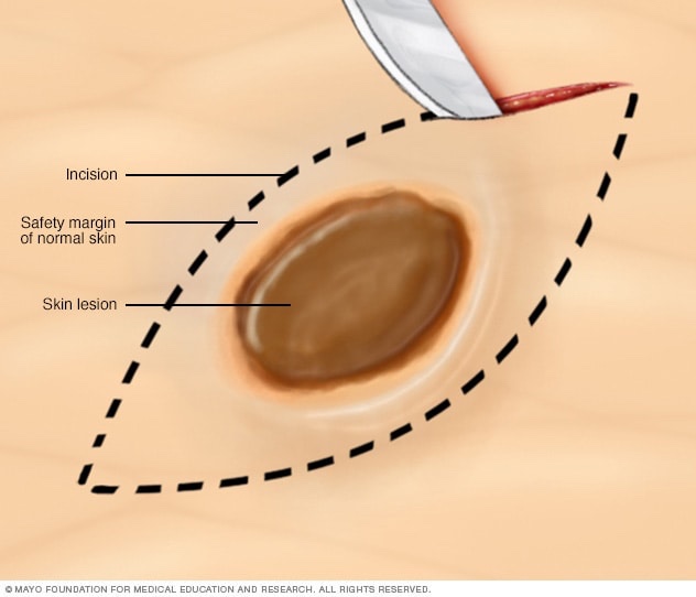
Blog

Two-Stage Total Thyroidectomy for Thyroid Cancer Offers Lower Complication Rates Than One-Stage Surgery

  • Ansart F, de Ponthaud C, Buffet C, et al. One- or two-step total thyroidectomy for cancer indications: a 20-year retrospective study from a referral center. Ann Surg Oncol 2024;32:2329-2334; doi: 10.1245/s10434-024-16707-6. PMID: 39739263.
  • Background:
    • Current guidelines for the management of differentiated thyroid cancer (DTC) recommend consideration of thyroid lobectomy unless there are clear indications to remove the contralateral lobe
    • Lobectomy carries fewer risks, including no risk of postoperative hypoparathyroidism, no risk of bilateral recurrent laryngeal nerve (RLN) injury, and lower risk of requiring thyroid hormone supplementation postoperatively
    • However, up to 20% of patients who undergo lobectomy have unfavorable characteristics on final pathology
    • Patients with high-risk DTC, such as extrathyroidal extension, multiple positive lymph nodes, or multifocal lesions, may require completion thyroidectomy
    • The risks of one-stage versus two-stage total thyroidectomy have not been studied thoroughly
    • This study aimed to compare the postoperative morbidity of patients with thyroid cancer who underwent total thyroidectomy in one stage versus two stages
  • Methods:
    • This retrospective study conducted at a high-volume endocrine surgery center included adult patients who underwent total thyroidectomy for DTC
    • Patients either underwent total thyroidectomy in one stage (TT1) or in two stages—thyroid lobectomy followed by completion thyroidectomy (TT2)
    • The primary outcome was postoperative morbidity within 30 days after surgery, which included hematoma requiring reoperation, wound infection or swelling, temporary or permanent RLN injury resulting in vocal cord immobility, and hypocalcemia
    • Transient hypocalcemia was defined as a postoperative serum calcium level lower than 8 mg/dl within the first 6 months, while permanent hypocalcemia was defined as PTH <15 pg/ml or the need for oral calcium supplementation after 6 months
    • For patients in the TT2 group, postoperative complications were quantified as the sum of those observed after each procedure
  • Results:
    • The study cohort consisted of 5693 patients, 5009 in the TT1 group and 684 in the TT2 group
    • The majority of patients in both groups were female, though the rate was higher in the TT1 group than the TT2 group (79% vs. 69.3%, P<0.001)
    • TT1 patients were significantly older (mean [±SD], 49.9±14.7 years) than TT2 patients (45.4±4.5 years, P<0.001)
    • Patients with papillary thyroid cancer primarily underwent TT1 (92.1%), whereas those with follicular or oncocytic cancers underwent TT1 and TT2 equally (P<0.001)
    • There was no difference in lymph node dissection rates between TT1 and TT2; however, on average, there were more nodes removed (18.1±13.4 vs. 12.2±9.5, P<0.001) and more positive results (29.8% vs. 14.3%, P<0.001) in TT1 patients than TT2
    • Tumors in TT2 patients were significantly larger (14.4±13.8 mm vs. 28.2±18.4 mm, P<0.001) but there was no difference in tumor stage
    • TT2 patients had significantly lower rates of all surgical complications (11.7% vs. 22.6% for TT1), transient hypocalcemia (3.2% vs. 14.5%), and permanent hypocalcemia (1.3% vs. 3.1%) (P<0.01 for all)
    • There were no differences in the rate of hematoma, RLN injury, or wound infection between the two groups
    • After propensity-score matching, TT1 patients experienced higher rates than TT2 patients for overall surgical complications (22% vs. 11.7%), transient hypocalcemia (14.1% vs. 3.2%)(P<0.01 for both), and permanent hypocalcemia (2.9% vs. 1.3%, P = 0.024) and there were no differences in the rates of RLN injury, hematoma, or infection
  • Conclusions:
    • Patients undergoing one-stage total thyroidectomy for thyroid cancer experienced higher rates of postoperative hypocalcemia and overall complications as compared with patients undergoing a two-stage operation
    • However, there were no differences in the rates of RLN injury, hematoma, or infection between the two treatment groups
    • Two-stage total thyroidectomy for thyroid cancer may be performed safely, with similar complication rates to those for one-stage surgery
  • This retrospective study demonstrates the safety of thyroid lobectomy followed by completion thyroidectomy as necessitated by pathology results, supporting the trend toward less aggressive surgical management of small DTCs
  • These results are concordant with both a retrospective study of over 70,000 patients in a national database, which found that completion thyroidectomies are associated with lower rates of complications, and a single-center study that reported that completion thyroidectomy was less likely to result in hypocalcemia and poses no additional risk of RLN injury, hematoma, or permanent hypoparathyroidism
  • Thyroid lobectomy also avoids the potential need for lifelong thyroid hormone replacement, and there is no difference in overall prognosis for DTCs < 4 cm in size
  • With higher complication rates identified in the TT1 group in the present study, there is further evidence that initial management of DTC < 4 cm with thyroid lobectomy may be preferred despite the potential need for completion thyroidectomy
  • Applying these results to practice, there is significant evidence to support offering lobectomy to patients with DTC < 4 cm, with the knowledge that it is both therapeutic and diagnostic but also that this approach may necessitate a completion thyroidectomy
  • Although there are some instances where upfront total thyroidectomy may be desired, initial lobectomy offers a less invasive option with lower complication rates even if completion thyroidectomy is needed
  • Encouragingly, complication rates are low for both total and two-stage thyroidectomy, supporting either surgical approach based on patient or treatment team preference
  • For patients with lower-risk thyroid cancers, lobectomy is a safe initial management choice given the favorable outcomes of two-stage thyroidectomy
  • References:
  • Haugen BR, Sawka AM, Alexander EK, et al. American Thyroid Association guidelines on the management of thyroid nodules and differentiated thyroid cancer task force review and recommendation on the proposed renaming of encapsulated follicular variant papillary thyroid carcinoma without invasion to noninvasive follicular thyroid neoplasm with papillary-like nuclear features. Thyroid 2017;27:481–483; CrossrefPubMed.
  • Ansart F, de Ponthaud C, Buffet C, et al. One- or two-step total thyroidectomy for cancer indications: a 20-year retrospective study from a referral center. Ann Surg Oncol 2024;32:2329-2334; CrossrefPubMed.
  • Brauer PR, Reddy CA, Burkey BB, et al. A national comparison of postoperative outcomes in completion thyroidectomy and total thyroidectomy. Otolaryngol. Head Neck Surg 2021;164(3):566-573; CrossrefPubMed.
  • Dedhia PH, Stoeckl EM, McDow AD, et al. Outcomes after completion thyroidectomy versus total thyroidectomy for differentiated thyroid cancer: a single-center experience. J Surg Oncol 2020;122(4):660-664; Crossref.PubMed.
  • Barney BM, Hitchcock YJ, Sharma P, et al. Overall and cause-specific survival for patients undergoing lobectomy, near-total, or total thyroidectomy for differentiated thyroid cancer. Head Neck 2011;33(5):645-649; Crossref.PubMed.
  • Mendelsohn AH, Elashoff DA, Abemayor E, et al. Surgery for papillary thyroid carcinoma: is lobectomy enough? Arch Otolaryngol Head Neck Surg 2010;136(11):1055; CrossrefPubMed

Thyroid Imaging

  • Thyroid gland imaging studies with radionuclides:
    • Provide both structural and functional information and can be very useful in determining the etiology of biochemical hyperthyroidism:
      • In contrast, thyroidal nuclear imaging is not recommended in the evaluation of a patient with hypothyroidism
  • For nuclear imaging, scans using radioactive iodine isotopes are most preferred:
    • Because these directly reflect the active accumulation (trapping) of iodine by the thyroid follicular cell and covalent attachment (organification) of iodine to thyroglobulin
  • The preferred radionuclide for diagnostic nonthyroid cancer imaging:
    • Is 123I, because this isotope emits only gamma rays that pass through tissue without significant cellular damage:
      • In contrast, 131I emits both gamma rays for imaging, as well as damaging beta particles:
        • So it can be used for the treatment of hyperthyroidism and thyroid cancer:
          • To destroy iodine-avid thyroid tissue
    • For diagnostic imaging:
      • 123I is administered orally:
        • With the measurement of iodine uptake and gamma scintigraphy images:
          • Obtained 4 hours and / or 24 hours later
      • Measured thyroidal uptake depends on the activity of NIS and overall iodine status as determined by the amount of circulating nonradioactive iodine:
        • When there is an excess of nonradioactive iodine:
          • The measured radioactive iodine uptake is reduced due to the competition between radioactive and nonradioactive iodine uptake by the thyroid follicular cells:
            • Sources of excess nonradioactive iodine include kelp, seaweed, seafood, iodine-rich medications and agents (amiodarone, saturated solution of potassium iodide [SSKI], Lugol’s solution, povidone iodine, tincture of iodine, iodoform gauze), and radiographic contrast media used commonly in computed tomography (CT) scans and gallbladder studies
  • An alternate radionuclide is technetium99m pertechnetate (99mTc):
    • Which is administered intravenously
    • Images are obtained much more rapidly than 123I:
      • Usually on the order of 30 to 60 minutes after the administration of the radionuclide tracer
      • Although 99mTc will be trapped by the thyroid follicular cells:
        • There is no iodine moiety for attachment to thyroglobulin, and therefore does not as accurately mimic the thyroidal uptake of iodine as radioiodine nuclides:
          • Thus 123I thyroid scans have 5% to 8% fewer false negative results than 99mTc scans:
            • However, because 99mTc scans are easier, faster, more readily available and less expensive to perform, they have largely replaced 123I scans at some institutions
  • Studies of direct comparison of radioiodine and 99mTc thyroid scans have been highly concordant in patients without nodules and in those with cold nodules:
    • One study reported that of 273 patients with thyroid nodules, only two had increased uptake with pertechnetate and no uptake with radioiodine:
      • However, if the results of the 99mTc scan are not in agreement with the clinical picture, an 123I scan should be performed
  • Although nuclear scans are useful in the differential diagnosis of biochemical hyperthyroidism:
    • Other radiologic modalities (e.g., ultrasonography, CT, and magnetic resonance imaging [MRI]) provide information regarding structural anatomy of the thyroid and provide no functional data
  • The primary role of thyroid ultrasound is in the initial evaluation of thyroid nodules:
    • As recommended by the American Thyroid Association and the American Association of Clinical Endocrinologists
    • Although thyroid ultrasound does not have a role in the initial evaluation of biochemical thyroid dysfunction:
      • It may demonstrate changes that are consistent but are not necessarily diagnostic of chronic lymphocytic thyroiditis, subacute granulomatous thyroiditis, and postpartum thyroiditis
    • Some individuals with subclinical hypothyroidism and sonographic features suggestive of chronic thyroiditis:
      • Are at significant risk for developing overt hypothyroidism requiring thyroid hormone replacement therapy

Diagnosis of Ductal Carcinoma In Situ (DCIS)

  • Clinical Presentation:
    • Before the implementation of routine screening mammography:
      • Most patients with DCIS presented with a:
        • Palpable mass
        • Nipple thickening
        • Nipple discharge
        • Paget disease of the nipple
      • Occasionally, DCIS was an incidental finding:
        • In an otherwise benign breast biopsy specimen
      • In patients with palpable lesions:
        • Up to 25% demonstrated foci of invasive disease
    • Now that screening mammography is more prevalent:
      • The incidence of DCIS has increased dramatically and currently comprises:
        • Approximately 20% of all breast cancers
      • Most cases of DCIS are diagnosed:
        • When the tumor is still clinically occult
      • Patients with abnormalities detected by screening mammography:
        • Should always undergo diagnostic imaging of the contralateral breast:
          • Because 0.5% to 3.0% of patients have:
            • Synchronous occult abnormalities or cancers in the contralateral breast
        • Mammographic images should be compared with previous images, if available, to establish interval changes
  • Mammographic features:
    • On a mammogram, DCIS can present as:
      • Microcalcifications:
        • 80% to 90% of mammographic manifestations
      • A soft-tissue density
      • Both
    • Microcalcifications:
      • Are the most common (80% to 90%) mammographic manifestation of DCIS:
        • Which, in turn, accounts for 80% of all breast carcinomas:
          • Presenting with calcifications
    • Any interval change from a previous mammogram:
      • Is associated with malignancy in 15% to 20% of cases:
        • Most often indicates in situ disease
    • Holland et al. (1990):
      • Described two different classes of microcalcifications:
        • Linear branching-type microcalcifications:
          • Which are more often associated with:
            • High–nuclear-grade lesions
            • Comedo-type lesions
        • Fine, granular calcifications:
          • Which are primarily associated with:
            • Micropapillary or cribriform lesions:
              • Of lower nuclear grade and that do not show necrosis
      • Although the morphology of microcalcifications:
        • Suggests the architectural type of DCIS:
          • It is not always reliable
    • Holland et al:
      • Also demonstrated that the mammographic findings:
        • Significantly underestimated the pathologic extent of disease:
          • Particularly in cases of:
            • Micropapillary DCIS:
              • Lesions were more than 2 cm larger by histologic examination than by mammographic estimation:
                • In 44% of cases of micropapillary lesions, compared with only 12% of cases of the pure comedo subtype
        • However, when magnification views were used in diagnostic mammographic examination:
          • The extent of disease was underestimated in only 14% of cases of micropapillary tumors
        • Hence, magnification views increase the image resolution and are better able to delineate the shape, number, and extent of microcalcifications when compared with mammography alone:
          • And should be used routinely in the evaluation of suspicious mammographic findings
Calcifications seen on diagnostic mammography associated with ductal carcinoma in situ (DCIS). Magnification view is shown to demonstrate calcifications spanning approximately 9 cm.

#Arrangoiz #CancerSurgeon #BreastSurgeon #SurgicalOncologist #BreastCancer #LCIS #DCIS #DuctalCarcinomaInsitu #LobularNeoplasia #LobularCarcinomaInsitu #Surgeon #Teacher #Miami #Mexico #MSMC #MountSinaiMedicalCenter

Breast Radiation and Germline Pathogenic Mutations

  • TP53 is a tumor suppressor gene:
    • That plays a crucial role in:
      • Maintaining genomic stability and regulating cell cycle checkpoints
  • Carriers of a TP53 pathogenic variant:
    • Would be expected to be unable to repair tissue damage from DNA-damaging radiotherapy and be at risk for significant radiotherapy-associated sequelae
  • Outcomes reported in published case reports:
    • Support this recommendation against radiotherapy in women with breast cancer who are carriers of a TP53 pathogenic variants:
      • Due to increased risk of radiation-induced secondary malignancies
  • Mutations in TP53 are considered:
    • An absolute contraindication of radiotherapy:
      • Except in those with a significantly high risk of locoregional recurrence
    • Mastectomy is the recommended therapeutic surgical option
  • The radiotherapy-induced toxicity spectrum in patients with breast cancer carrying ATM pathogenic variants is controversial:
    • Seven studies identified in a systematic review addressed radiotherapy after breast-conserving surgery among women with breast cancer who harbor ATM pathogenic variants:
      • Meyer et al found no significant differences in local relapse-free survival in a cohort of 138 patients with early stage breast cancer treated with breast-conserving surgery, followed by radiotherapy, of whom 20 were found to carry either an ATM truncating or missense variant:
        • They concluded that the results do not support the hypothesis that patients with breast cancer carrying a sequence variant in the ATM gene differentially benefit from postoperative radiotherapy
  • According to the ASCO-ASTRO-SSO Consensus recommendations for carriers of BRCA 1/2 and other germline pathogenic variants,:
    • Women with breast cancer may safely be offered radiotherapy when clinically indicated
    • Data on toxicity rates comparing carriers of ATM pathogenic variants and noncarriers:
      • Do not favor avoiding radiotherapy:
        • Potential absolute risks do not appear to be significant:
          • However, more research is needed
    • A discussion with ATM carriers interested in breast-conserving therapy is encouraged
    • There is no evidence of increased toxicity or contralateral breast cancer events from radiation exposure in BRCA 1/2, CHEK2, CDH1 carriers
  • References:
    • Trombetta MG, Dragun A, Mayr NA, Pierce LJ. ASTRO Radiation Therapy Summary of the ASCO-ASTRO-SSO Guideline on Management of Hereditary Breast Cancer. Pract Radiat Oncol. 2020;10:235-242.
    • Lazzari G, Buono G, Zannino B, Silvano G. Breast cancer adjuvant radiotherapy in BRCA1/2, TP53, ATM genes mutations: Are there solved issues? Breast Cancer (Dove Med Press). 2021;13:299-310.
    • Ferrarini A, Auteri-Kaczmarek A, Pica A, et al. Early occurrence of lung adenocarcinoma and breast cancer after radiotherapy of a chest wall sarcoma in a patient with a de novo germline mutation in TP53. Fam Cancer. 2011;10:187-192.
    • Heymann S, Delaloge S, Rahal A, et al. Radio-induced malignancies after breast cancer postoperative radiotherapy in patients with Li- Fraumeni syndrome. Radiat Oncol. 2010;5:104.
    • Meyer A, John E, Dörk T, Sohn C, Karstens JH, Bremer M. Breast cancer in female carriers of ATM gene alterations: outcome of adjuvant radiotherapy. Radiother Oncol. 2004;72: 319-323.

Diagnostic Thyroid Testing: Serum Thyroglobulin

  • Thyroglobulin (Tg):
    • Is a large glycoprotein that is stored as colloid:
      • The primary storage form of thyroid hormone, in the lumen of thyroid follicles
    • It is continuously secreted into circulation from the thyroid gland:
      • Thereby reflecting the mass of normal and malignant thyroid tissue
  • Higher serum concentrations result from:
    • TSH stimulation and / or injury of thyroid tissue:
      • However, for the individual with an intact thyroid gland:
        • Its clinical value for evaluating thyroid dysfunction or goiter is limited in the era of modern serum thyroid function testing and imaging
        • However, the demonstration of a suppressed serum Tg level in such a patient can be useful in differentiating factitious thyrotoxicosis (from exogenous thyroid hormone ingestion) from excessive endogenous thyroid hormone release of any etiology:
          • In this situation, when thyrotoxicosis is due to ingestion of exogenous thyroid hormone:
            • Normal thyroid hormone production is suppressed and serum Tg levels are decreased
          • In contrast, if excess thyroid hormone is produced from the thyroid:
            • Serum Tg levels are elevated
  • In current clinical practice:
    • The primary use of serum Tg concentrations is as a tumor marker in patients with differentiated thyroid cancer:
      • That is obtained to detect persistent and / or recurrent disease after a total thyroidectomy and radioactive iodine (131I) ablation
  • Most Tg assays have only first-generation functional sensitivity between 0.5 and 1 ng/mL:
    • But the second generation Tg assays are rapidly becoming the standard and have an improved functional sensitivity of 0.05 to 0.1 ng/mL
  • The Tg assay can be made more sensitive to detect persistent or recurrent tumor:
    • After stimulation by TSH:
      • Either endogenously by withholding thyroxine treatment in an athyreotic patient or with administration of recombinant human TSH (rhTSH):
        • The latter of which results in an approximate tenfold increase in basal serum Tg concentrations
  • Detection of persistent and / or recurrent disease in thyroid cancer depends on the performance of Tg immunometric assays:
    • Which currently have suboptimal sensitivity and high interassay variability
  • Virtually all immunometric methods:
    • Will report an undetectable Tg level in euthyroid Tg Ab positive controls:
      • Approximately 25% of patients with differentiated thyroid cancer have a positive serum TgAb titer:
        • Thus when a suspicious lymph node or neck mass is detected in an individual who has undergone a total thyroidectomy:
          • An unmeasurable basal or rhTSH-stimulated Tg in the setting of a positive serum TgAb level:
            • Does not necessarily exclude thyroid cancer recurrence
        • It is reasonable in this relatively uncommon situation to measure Tg instead by Tg Ab-resistant radioimmunoassay (RIA) or liquid chromatography tandem mass spectrometry:
          • Which are available at some specialty endocrine laboratories.
  • When the serum Tg Ab titer is positive:
    • It may also be used as a surrogate marker of tumor persistence / recurrence
  • In one study, a > 50% decrease of Tg Ab levels within the first year after a total thyroidectomy:
    • Was associated with the absence of tumor recurrence / persistence in all patients studied
    • Tumor recurrence / persistence was present in 37% of patients who had any rise of serum Tg Ab within the same period
  • Thus thyroid cancer patients with rising Tg antibody levels:
    • Are at high risk for disease persistence / recurrence and should be evaluated promptly
    • In addition, the sensitivities and absolute values reported by different methods of measuring Tg and TgAb are highly variable:
      • It is essential to always use the same Tg and TgAb method when following an individual over time for tumor persistence/recurrence
  • Finally, the presence of interfering heterophile antibodies (antibodies against the animal-derived antibodies used in the immunometric assay):
    • May rarely result in abnormally high or low serum Tg levels
    • The most common interfering antibodies are HAMAs:
      • Clinically, this should be suspected when an elevated serum Tg level is inappropriate for the clinical situation and does not increase with rhTSH stimulation
      • When heterophile antibody is suspected, the clinician should repeat the test using a commercially available heterophile-blocking tube (HBT) or measure Tg with an RIA assay

Diagnostic Thyroid Testing: Serum Free T4

  • Bioactive free T4 concentrations:
    • Can be estimated using a variety of indirect (analog, immunometric, and two-step labeled hormone assays) or direct methods (equilibrium dialysis, ultrafiltration), and are the most commonly used measurements of circulating thyroid hormone levels
  • Generally, most laboratories estimate free T4 by either the analog immunoassay or a calculated FT4I corrected to thyroid hormone binding capacity:
    • The former is readily available and provides quick results; it does not directly measure the free T4 concentration but is a reliable estimate of FT4 levels in most patients, based on one-step, two-step, or labeled antibody approaches
  • This type of free T4 estimate is especially sensitive to abnormal serum albumin levels and should not be used with conditions such as:
    • Familial dysalbuminemic hyperthyroxinemia, pregnancy, or severe nonthyroidal illness:
      • For example, it is common to have a free T4 lower than the reference range in a euthyroid pregnant woman:
        • Due to the high estrogen state of pregnancy greatly increasing serum TBG concentrations, thus resulting in inaccurate FT4 measurements
  • The serum FT4I measurement is a calculated value that is the product of the total T4 concentrations and a correction factor related to the number of available thyroid hormone binding sites:
    • This correction factor may be called:
      • A thyroid hormone binding ratio (THBR), T3 resin uptake (T3RU), or T3 uptake (T3U)
      • The T3RU is inversely related to the free thyroid hormone binding sites but is now used in only a few laboratories
  • Free T4 by equilibrium dialysis:
    • Is the gold standard and measures the 0.03% of T4 that is biologically active and unbound to protein
    • This assay is available only at reference laboratories and is useful to directly determine free T4 levels when other testing does not provide a clear result
  • Although a TSH-first testing algorithm is suffcient for general screening:
    • Both FT4 and TSH assays are needed for:
      • Diagnosing subclinical thyroid dysfunction, central hypothyroidism, and in the assessment of elderly and hospitalized patients, as well as for accurate assessment of treatment effects
  • Guidelines from multiple thyroid and endocrine societies:
    • Have also endorsed a TSH-first strategy in most clinical scenarios with FT4 testing when clinically indicated or TSH is found to be abnormal
    • Work by Henze et al. goes even further suggesting that a TSH-first strategy can be further perfected by:
      • Widening the TSH reference range from 0.4 to 4.0 mIU/L to 0.2–6.0 mIU/L with minimal impact on case detection
      • They found that only 4.2% of TSH values between 0.2 mIU/L and 0.4 mIU/L would not have led to detection of a high FT4 and equally, only 2.5% of TSH values between 4.0 mIU/L and 6.0 mIU/L were associated with low FT4 level
      • It is likely that this small additional group of patients outside the wider range with abnormal FT4 is clinically unimportant in most cases

Diagnostic Thyroid Testing: Serum Total T4 and Total T3

  • Serum total T4 (TT4) and total T3 (TT3) concentrations:
    • Are a measure of both the bound and free hormone levels of these two hormones
  • TT4 or TT3 levels should be interpreted in the context of the clinical situation:
    • Because many clinical conditions and medications alter the concentrations of thyroid hormone binding proteins and / or compete with the binding of thyroid hormones to the binding proteins:
      • As such, measured TT4 and TT3 levels may be affected, even though the bioactive free levels and thus, the thyroidal status, remain unchanged
  • T3 is the active thyroid hormone:
    • It is primarily useful in the diagnosis and management of patients with hyperthyroidism
    • It occasionally can be used to differentiate stimulation induced thyrotoxicosis / Graves’ disease (TT3 / TT4 ratio > 20) from destruction induced thyrotoxicosis / subacute thyroiditis (TT3 / TT4 ratio < 12):
      • This assessment can be further augmented when TSH is considered as serum levels of TSH are generally suppressed in most untreated Graves’ patients, whereas they usually were not completely suppressed in patients with painless thyroiditis or subacute thyroiditis
    • Measurement of serum TT3 is not usually helpful if hypothyroidism is suspected:
      • Because the activity of 5’deiodinas type 2 enzyme (Dio2):
        • Which converts T4 to the biologically active T3:
          • Increases while serum T4 falls:
            • Thus maintaining normal T3 levels until the overall thyroid hormone levels are very low
  • rT3, which may be elevated during nonthyroidal illness:
    • Is not biologically active:
      • As such, the utility of measuring it and other forms of inactive iodothyronine are limited during the evaluation of thyroid status
  • Finally, the human anti-mouse antibodies (HAMAs) that interfere with TSH testing:
    • Can also interfere with the thyroid hormone assays
    • HAMA positivity:
      • May result in artificially elevated or reduced TT4, TT3, FT4, and FT3 levels
    • Patients who have received therapeutic monoclonal antibody treatment may be at increased risk of develop interfering positive HAMA titers

Diagnostic Thyroid Testing: Serum Thyroid Stimulating Hormone (TSH)

  • Thyroid Stimulating Hormone (TSH):
    • Is produced from the anterior pituitary gland:
      • After stimulation by thyrotropin-releasing hormone (TRH)
  • Thyrotropin-releasing hormone (TRH):
    • Is a modified three-amino acid peptide:
      • Produced by neurons of the paraventricular nucleus:
        • In the hypothalamus
    • TRH signaling from the hypothalamus:
      • Is achieved through a portal venous system:
        • Located in the infundibulum of the pituitary stalk:
          • Which allows communication to the pituitary gland
  • Both TRH and TSH gene expression:
    • Are decreased by excess thyroid hormone levels:
      • Via negative feedback mechanisms (Figure)
    • Both the hypothalamus and pituitary:
      • Have high levels of 5′deiodinases Type 2 enzyme (Dio2):
        • Catalyzes the removal of the 5′ iodine from the outer ring of thyroxine (T4):
          • To create the metabolically active triiodothyronine (T3)
        • So T4 levels are the primary feedback:
          • T4 is converted locally to T3:
            • Which suppresses TRH and TSH gene expression
Schematic Representation of Control of Thyroid-Stimulating Hormone (TSH) Secretion by the Thyrotroph Cells in the Anterior Pituitary Gland.
High concentrations of T3 suppress TSH release, and low concentrations enhance the expression of TSH. Thyrotropin-releasing hormone (TRH) also stimulates TSH release, and its absence results in failure of the thyrotroph to release TSH, resulting in hypothyroidism. TRH is secreted by neurons in the paraventricular nuclei in the hypothalamus and reaches the anterior pituitary via the hypothalamic hypophyseal portal venous system. TSH travels through the circulatory system to stimulate the thyroid and to produce thyroid hormones T4 and T3. By negative feedback inhibition, the circulating thyroid hormones suppress the production and excretion of TRH and TSH to bring the system back into equilibrium to maintain tight control over circulating thyroid hormone levels.
  • Pituitary TSH is secreted in a pulsatile manner:
    • Higher levels at night and lower levels during the day:
      • The inverse of the cortisol cycle
    • Although pulse frequency is increased nocturnally to result in a diurnal variation of TSH concentrations:
      • Levels remain within the reference range
    • Generally, laboratory testing of serum TSH concentrations during daylight hours:
      • Is not substantially affected by the diurnal variation of TSH secretion:
        • But results outside the reference range may occur in euthyroid individuals when drawn outside of these times
  • TSH stimulates the synthesis and release of thyroid hormone from the thyroid gland:
    • TSH production from the anterior pituitary gland is inverse log-linearly regulated by serum thyroid hormone concentrations:
      • When there are small decreases in thyroid hormone levels in circulation:
        • Large increases in serum TSH stimulate thyroid hormone production by the thyroid gland
      • This negative feedback loop between serum TSH and the serum-free thyroid hormones:
        • Is able to maintain circulating thyroid levels within a tight range
  • Serum TSH is the preferred screening test:
    • In the evaluation of thyroid function in the ambulatory patient:
      • Regardless of whether the patient is taking thyroid hormone replacement medication
  • For the healthy patient in an ambulatory setting:
  • The diagnosis of hypothyroidism or hyperthyroidism:
    • May be determined with approximately 98% sensitivity and 92% specificity using serum TSH
    • In addition, TSH has a narrow intraindividual variability of ± 0.5 mIU/L:
      • Such that thyroid dysfunction may be present if there are significant changes in TSH values over time in an individual, even if they remain within the reference range
  • In certain situations, however, such as known or suspected pituitary or hypothalamic dysfunction, recent hyperthyroidism, critical illness, starvation, use of certain medications (dopamine or high-dose glucocorticoids), interference with serum thyroid autoantibodies, and thyroid hormone resistance syndromes:
    • The TSH level is inaccurate for the thyroidal status and should not be used in isolation to determine thyroid function
  • In addition, the presence of interfering heterophile antibodies (antibodies against the animal-derived antibodies used in the immunometric assay):
    • May rarely cause abnormally high or low TSH levels:
      • These conditions should be suspected when the pattern of the TSH levels does not correlate to the clinical presentation or when the peripheral serum hormone levels do not change as expected with elevated or suppressed serum TSH concentrations
  • Serum TSH assays:
    • Have evolved considerably since measurements were first described in the 1960s:
      • When the functional sensitivity was between 1 and 2 mIU/L
    • The commonly used second-generation TSH assays have an improved functional lower limit of 0.10 to 0.20 mIU/L:
      • Which is able to differentiate between euthyroid and hyperthyroid states but does not indicate the degree of hyperthyroidism
    • In contrast, a third-generation TSH assay can detect levels as low as 0.01 to 0.02 mIU/L:
      • This is helpful when there is a challenging pattern of serum thyroid function tests that include an extremely suppressed TSH
    • In the rare instance they are needed, fourth-generation immunochemiluminometric assays are capable of detecting TSH levels in the range of 0.01 to 0.001 mIU/L
  • A serum TSH level measured in an ambulatory population that lies within the reference range:
    • Is generally considered evidence of normal thyroid function and requires no additional testing
    • Reference ranges for serum TSH can vary slightly from one commercial laboratory to another:
      • It should be noted that normal ranges, which are based on the epidemiologic distribution of serum TSH concentrations in healthy populations:
        • From which serum thyroid autoantibody positivity and iodine status (both of which can affect TSH), may be variable
    • If an abnormal screening TSH result is encountered:
      • The circulating thyroid hormone levels should be assessed
        • The specific pattern of tests will allow further insight into whether clinical thyroid dysfunction should be suspected
Evaluation and Management of Hypothyroidism in Individuals With Corresponding Signs and Symptoms.
Evaluation and Management of Hyperthyroidism in Individuals with Corresponding Signs and Symptoms.

Serum Thyroid Autoantibodies

  • Hyperthyroidism and hypothyroidism:
    • Are often the result of autoimmune diseases:
      • In which immunoglobulin G (IgG) antibodies:
        • Such as thyroid peroxidase (TPO Ab, previously known as antimicrosomal antibodies), thyroglobulin (Tg Ab), and the TSH receptor (TSHR Ab):
          • Are formed against thyroid proteins
    • The presence of TPOab is commonly associated with patients with hypothyroidism:
      • But can be present in normal individuals who do not display any obvious symptoms of clinical thyroid disease
    • TPOabs are present in approximately 10% of normal individuals:
      • While it was detected in almost 100% of samples of patients with autoimmune hypothyroidism
  • More than 90% of patients with autoimmune thyroid disease (Hashimoto’s thyroiditis and Graves’ disease):
    • Will have elevated titers of second-generation assays for TPO Ab and Tg Ab
  • When biochemical hypothyroidism is found:
    • Measuring TPO Ab can be helpful:
      • Because its result can provide additional information regarding the etiology of the thyroid dysfunction
  • Median serum TSH concentrations are increased within the reference range:
    • Among those with serum TPO Ab and Tg Ab positivity:
      • Compared with those without TPO Ab titers, and are a predictor for the development of biochemical thyroid dysfunction in euthyroid individuals
  • In individuals with subclinical hypothyroidism:
    • Both serum TPO Ab positivity and sonographic characteristics suggestive of chronic thyroiditis:
      • Are associated with an increased likelihood of progression to overt hypothyroidism
  • The TSHR Abs are a group of immunoglobulins:
    • That produce Graves’ hyperthyroidism
    • Measured as:
      • TSH receptor binding, TRAB (TSH-receptor antibody) or in a functional bioassay, thyroid stimulating immunoglobulin (TSI)
    • TSH receptor antibodies:
      • Are being increasingly recommended for monitoring activity of disease to assess response to therapy in Graves’ patients
    • Less commonly, TSH receptor binding antibodies:
      • Can block the TSH receptor and produce hypothyroidism
  • In the setting of normal serum thyroid function:
    • Thyroid antibodies should generally not be measured except in special circumstances:
      • Such as a history of hyperthyroidism during pregnancy or recurrent miscarriages:
        • In these situations, both the stimulating and inhibiting TSHR Abs can cross the placenta to affect fetal thyroid function and potentially induce fetal goiter
      • Although serum thyroid antibody positivity during pregnancy:
        • Is associated with a higher risk of postpartum subacute thyroiditis:
          • Antibody screening in pregnant women is not currently recommended

Thyroid Physiology in Nonthyroidal Illness (Euthyroid Sick Syndrome)

  • The evaluation of a chronically ill or hospitalized patient with abnormal serum thyroid function tests:
    • Can often be challenging
  • Nonthyroidal illness is not considered a primary thyroid disorder:
    • Its pathophysiology is not completely understood:
      • Although it is known that the elevation of cytokines and hypoxia plays a significant role
    • It is generally recommended to avoid measuring serum thyroid function tests during acute illness:
      • Unless thyroid dysfunction is thought to be a significant contributor to the illness
  • Severe nonthyroidal illness:
    • Is accompanied by significant alterations in thyroid physiology
  • Due to the decreased availability in all of the thyroid binding proteins (thyroxine bindings globulin, transthyretin, albumin):
    • Serum total T4 and total T3 levels are reduced:
      • Whereas free levels are usually normal or slightly low (Figure)
    • Total T3 levels are further decreased:
      • Due to reduced Dio1 activity:
        • Which converts T4 to T3
    • A relatively greater amount of T4 is metabolized to the inactive metabolite:
      • Reverse T3 (rT3):
        • By Dio3
      • Although measurement of rT3 does not reliably distinguish nonthyroidal illness from primary hypothyroidism
      • The degree of serum rT3 elevation, depressed T3 / rT3 ratio, and decreased FT3 and FT4 concentrations:
        • Have been associated with higher mortality among patients in the intensive care unit (ICU):
          • Treatment with T4 or T3, however, does not consistently improve outcome
      • Whether other T4 metabolites, including 3,3′-diiodothyronine (3,3′-T2); 3,5-diiodothyronine (3,5-T2); and 3-iodothyronamine (3-T1AM):
        • Have functional roles in nonthyroidal illness remains unclear
Alternations in Serum Thyroid Hormone Concentrations During Acute Nonthyroidal Illness and Recovery.
The degree of change in hormone concentrations relates to the severity and duration of the illness. Thyroid-stimulating hormone (TSH) may also be suppressed during severe illness and transiently rise moderately above the reference range before returning to normal with recovery. Mortality correlates inversely with the degree of reduction in total T4 concentration. (From Farwell AF. Sick euthyroid syndrome in the intensive care unit. In: Irwin RS, Rippe JM, eds. Intensive Care Medicine. 5th ed. Philadelphia: Lippincott Williams & Wilkins; 2003.)
  • It is important that the diagnosis of primary thyroid dysfunction:
    • Is not established during severe illness based solely on an abnormal serum TSH
  • In nonthyroidal illness:
    • Serum TSH concentrations may be low, normal, or high:
      • Due to the TSH-lowering effects of commonly used medications (glucocorticoids, ipodate, amiodarone, dopamine) in patients managed for a nonthyroidal illness or from a reversible form of acquired central hypothyroidism in severe nonthyroidal illness
  • During the recovery phase of nonthyroidal illness:
    • The TSH may briefly rise above the upper reference range, as suppression of TSH lessens, before it normalizes
    • When possible, thyroid evaluation after recovery from an acute illness is recommended in patients suspected of having intrinsic thyroid disease