Blog

Anatomy and Histology of the Parathyroid Glands

  • Most patients have four parathyroid glands
    • The superior glands usually are dorsal to the RLN at the level of the cricoid cartilage
    • The inferior parathyroid glands are located ventral to the nerve
  • Normal parathyroid glands are gray and semitransparent in newborns:
    • But appear golden yellow to light brown in adults:
      • Parathyroid color depends on cellularity, fat content, and vascularity
    • Moreover, they often are embedded in and sometimes difficult to discern from surrounding fat
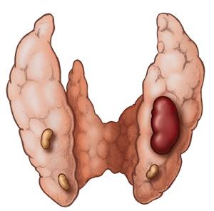
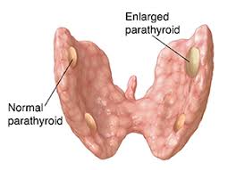
  • There are normally two pairs of parathyroid glands (inferior and superior)
    • The parathyroid gland is oval or bean-shaped (Figure)
    • It typically measures 6 mm × 4 mm × 2 mm
    • They weigh 40 mg to 60 mg
  • Most people have four parathyroid glands:
    • Akerström et al, in a series of 503 autopsies:
      • Identified four parathyroid glands in 84% of the cases
    • Supernumerary glands were found in 13% of the cases:
      • Most commonly in the thymus
    • In the literature, the incidence of supernumerary glands:
      • Is anywhere between 3% and 13%
  • Only in three percent of the cases less than four parathyroid glands are identified
  • The blood supply of the parathyroid glands:
    • Is usually derived from branches of the inferior thyroid artery:
      • Although branches of the superior thyroid artery can supply at least 10% to 45% of the superior parathyroid glands
    • In a study of 354 autopsy specimens, Alverd, observed:
      • That both the superior and inferior parathyroid glands derive their blood supply from the inferior thyroid artery:
        • 86% on the right side and 77% from the left side
    • When the inferior thyroid artery was absent:
      • Both the superior and inferior parathyroid glands were supplied by the superior thyroid artery
    • Branches from the thyroidea ima, and vessels to the trachea, esophagus, larynx, and mediastinum:
      • May also contribute to the irrigation of the parathyroid glands
  • Wang et al., in a study of 160 autopsy specimens:
    • Showed that a low lying inferior parathyroid gland could be identified by following the vascular pedicle of the inferior thyroid artery
  • The parathyroid glands drain ipsilaterally by the:
    • Superior, middle, and inferior thyroid veins
  • The innervation of the parathyroid glands:
    • Occurs via the superior or middle cervical ganglia, or through a plexus in the fascia on the posterior aspect of the thyroid lobe 
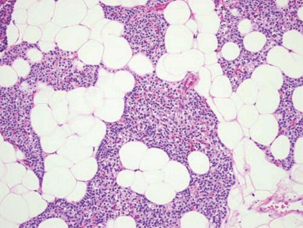
  • Histologically, parathyroid glands are composed of:
    • Chief cells and oxyphil cells arranged in trabeculae, within a stroma composed primarily of adipose cells (Figure)
Normal parathyroid histology showing chief cells interspersed with adipose cells.
  • The parathyroid glands of infants and children:
    • Are composed mainly of chief cells:
      • Which produce parathyroid hormone (PTH)
  • Acidophilic, mitochondria-rich oxyphil cellsL:
    • Are derived from chief cells:
      • Can be seen around puberty:
        • They increase in numbers in adulthood
  • A third group of cells, known as water-clear cells:
    • Also are derived from chief cells
    • Are present in small numbers, and are rich in glycogen
  • Although most oxyphil and water-clear cells retain the ability to secrete PTH:
    • Their functional significance is not known.

Indications for Surgery in Hyperparathyroidism

18767357_298526467261639_5144883419579625143_n

  • All symptomatic patients and most asymptomatic patients should be considered for parathyroidectomy:

    • Patients with nephrolithiasis (kidney stones), fragility fractures, osteoporosis, or evidence of spinal compression fractures are considered symptomatic.

18839633_298526490594970_1808619548355549612_o

18813493_298526493928303_9123677475566775310_nSlide1

  • In asymptomatic patients (usually less than 5% of the cases when an appropriate history is taken), indications for parathyroidectomy include:

    • Patient age younger than 50 years

    • Serum calcium level > 1 mg/dL above the upper limit of normal range,

    • Evidence of renal involvement:

      • Silent nephrolithiasis on imaging

      • Nephrocalcinosis

      • Hypercalciuria defined as a 24-hour urine calcium > 400 mg/dL

      • Impaired renal function defined as glomerular filtration rate < 60 mL/min).

    • Other findings that should prompt consideration for parathyroidectomy in asymptomatic patients include:

      • Neurocognitive dysfunction:

        • Manifesting as anxiety, depression, memory loss, and insomnia, which are multifactorial in cause but might be exacerbated by hypercalcemia

      • Frailty or diminished functional capacity

      • Gastroesophageal reflux

      • Fibromyalgia

      • Cardiovascular disease.

  • It is important to note that asymptomatic patients are often not truly asymptomatic, with insidious symptoms often attributed to other causes when they are related to or exacerbated by the underlying imbalance in calcium homeostasis; thus parathyroidectomy may be indicated even for asymptomatic patients.

Rodrigo Arrangoiz MS, MD, FACS

He is first author on some publications on hyperparathyroidism:

Training:

• General surgery:

• Michigan State University:

• 2004 al 2010

• Surgical Oncology / Head and Neck Surgery / Endocrine Surgery:

• Fox Chase Cancer Center (Filadelfia):

• 2010 al 2012

• Masters in Science (Clinical research for health professionals):

• Drexel University (Filadelfia):

• 2010 al 2012

• Surgical Oncology / Head and Neck Surgery / Endocrine Surgery:

• IFHNOS / Memorial Sloan Kettering Cancer Center:

• 2014 al 2016

Summary of Primary Hyperparathyroidism (PHPT)

  • Primary hyperparathyroidism (PHPT):
    • Is a relatively common disorder:
      • That affects 0.3% of the human population:
        • Most commonly women:
        • 75% of the cases
  • The exact cause of PHPT:
    • Is unknown
  • In 85% to 90% of patients:
    • Only a single adenoma is present
  • Multiple adenomas or hyperplasia:
    • Can be present in up to 15% to 20% of the cases:
      • Doble adenomas:
        • 6% to 9% of the cases
      • Triple adenomas:
        • Less than 0.3% of the cases
      • Hyperplasia:
        • 3% of the cases
  • Patients with PHPT:
    • Can have symptomatic disease:
      • 95% of the cases
    • “Asymptomatic” disease:
      • 5% of the cases
  • Some degree of renal dysfunction:
    • Is present in up to 80% of patients
    • Nephrolithiasis, however, is far less common:
      • With an incidence of approximately 15% to 25%
  • The clinical manifestations of PHPT:
    • Vary widely across patients:
      • But if a detailed history is taken:
        • Many will complain of polydipsia and polyuria:
          • From the calciuresis associated with the disease
  • Although PHPT occurs sporadically in the majority of patients:
    • In a small percentage it is part of a familial syndrome:
      • Multiple endocrine neoplasia type I (MEN-I / Werner Syndrome):
        • Results from a germline mutation in the Menin gene:
          • Located on chromosome 11q12-13
        • Patients with MEN-I are susceptible to the development of:
          • Pancreatic neuroendocrine tumors
          • Pituitary adenomas
          • PHPT
      • MEN-IIA (Sipple Syndrome):
        • Is an autosomal dominantly inherited condition:
          • Caused by a germline mutation on chromosome 11:
            • That is associated with:
              • PHPT
              • Pheochromocytoma
              • Medullary thyroid cancer
      • Patients with familial jaw tumor syndrome:
        • Have a higher risk for the development of:
          • Parathyroid carcinoma
      • Familial hypercalcemic hypocalciuria:
        • Is associated with an:
          • Elevated calcium levels and low urinary excretion of calcium
        • The primary defect is the abnormal sensing of calcium in blood:
          • By the calcium sensing receptor (CaSR):
            • In the the parathyroid glands (chief cells) and the renal tubules:
              • Which causes:
                • Inappropriate secretion of PTH
                • Excessive renal reabsorption of calcium

#Arrangoiz #ParathyroidSurgeon #ParathyroidExpert #Hyerpcalcemia #EndocrineSurgery #HeadandNeckSurgeon #Hyperparathyroidism #PrimaryHyperparathyroidism #Surgeon #Teacher #CancerSurgeon #Miami #Mexico #MSMC #MountSinaiMedicalCenter

Dermatofibrosarcoma Protuberans

  • Dermatofibrosarcoma protuberans:
    • Is an intermediate-grade sarcoma:
      • That presents as a unifocal or multifocal nodular lesion
    • It is a rare, slow-growing, locally aggressive cutaneous sarcoma:
      • With a high rate of local recurrence and low metastatic potential
  • Dermatofibrosarcoma protuberans:
    • Involves the head and neck region in 10% to 20% of cases:
      • With the scalp and supraclavicular fossae the most common sites for involvement (Figure)
  • These slow-growing, locally aggressive tumors have tentacle-like extensions well beyond the visible lesion:
    • Thus the true extent of the disease is often underestimated:
      • Leading to local recurrence in more than 50% of patients
  • Histologically a storiform or fascicular proliferation of spindle cells extends
    from the dermis into the subcutis:
    • With immunohistochemistry showing:
      • CD34 positive staining in most cases
    • Presence of fibrosarcomatous changes and high mitotic rate:
      • May portend a more aggressive course
    • This tumor frequently has a translocation of a fusion protein involving:
      • COL1A1 and PDGFB that functions like PDGFB
  • Wide excision with margins of ≥ 2 cm is
    generally advocated, with adjuvant radiation reserved for larger or recurrent tumors when resection is not feasible
  • Histological subtypes include:
    • Classic DFSP
    • Fibrosarcomatous transformation:
      • Conferring a higher risk of recurrence and metastasis
  • Surgical management is the mainstay of treatment:
    • The National Comprehensive Cancer Network (NCCN), National Cancer Institute, and American Cancer Society recommend:
      • Mohs micrographic surgery (MMS) as the preferred approach due to its ability to achieve complete margin control and minimize tissue loss, especially in anatomically sensitive areas
      • If MMS is unavailable, wide local excision (WLE) with 2 to 4 cm margins down to the investing fascia is acceptable:
        • Achieving negative surgical margins is the most critical factor for reducing recurrence risk; margin width is important, but negative margins are paramount
      • Recent evidence suggests that margins greater than 2 cm to 2.5 cm are associated with significantly lower recurrence rates:
        • But recurrence is rare when negative margins are achieved, regardless of width
      • Routine lymph node dissection is not indicated for DFSP, as the risk of nodal metastasis is extremely low
      • Lymph node dissection should be considered only in cases with clinical or radiologic suspicion of nodal involvement or in tumors with fibrosarcomatous transformation, which carries a higher metastatic risk
      • Sentinel lymph node biopsy is not standard but may be considered in select high-risk cases, such as those with fibrosarcomatous change or lymphovascular invasion, though its utility remains under investigation
  • Adjuvant radiotherapy:
    • Is considered in cases of positive or close margins when further re-excision is not feasible, or for unresectable or recurrent disease
    • The role of adjuvant radiotherapy is supported by its ability to reduce local recurrence in these settings
  • Imatinib, a tyrosine kinase inhibitor:
    • Is indicated for unresectable, recurrent, or metastatic DFSP harboring the t(17;22) translocation (COL1A1-PDGFB fusion)
  • Cytotoxic chemotherapy:
    • Has a limited role and is generally reserved for metastatic disease not amenable to targeted therapy, with inferior outcomes compared to imatinib
  • Surveillance recommendations include:
    • Regular follow-up for early detection of local recurrence:
      • Particularly within the first three years post-resection:
        • As most recurrences occur during this period
      • For patients with negative-margin, non-fibrosarcomatous DFSP:
        • Less intensive follow-up may be appropriate, and some data suggest that ongoing surveillance may not be necessary after negative-margin resection
      • In contrast, patients with fibrosarcomatous transformation should be followed according to soft tissue sarcoma protocols due to higher risk of recurrence and metastasis
  • Areas of ongoing debate include the optimal surgical margin width and the role of sentinel lymph node biopsy, particularly in high-risk subtypes:
    • The consensus remains that complete surgical excision with negative margins is the cornerstone of management, with adjuvant therapies reserved for select cases
  • References:
    • Dermatofibrosarcoma Protuberans: Update on the Diagnosis and Treatment. Hao X, Billings SD, Wu F, et al. Journal of Clinical Medicine. 2020;9(6):E1752. doi:10.3390/jcm9061752.
    • Review of Dermatofibrosarcoma Protuberans. Lim SX, Ramaiya A, Levell NJ, Venables ZC. Clinical and Experimental Dermatology. 2023;48(4):297-302. doi:10.1093/ced/llac111.
      Dermatofibrosarcoma Protuberans: What Is This?. Vitiello GA, Lee AY, Berman RS. The Surgical Clinics of North America. 2022;102(4):657-665. doi:10.1016/j.suc.2022.05.004.
    • Dermatofibrosarcoma Protuberans: An Updated Review of the Literature. Jozwik M, Bednarczuk K, Osierda Z. Cancers. 2024;16(18):3124. doi:10.3390/cancers16183124.
    • Current Patterns of Care and Outcomes for Dermatofibrosarcoma Protuberans: An International Multi-Institutional Collaborative. Winer LK, Akumuo R, Fredette JD, et al. Cancer. 2025;131(1):e35468. doi:10.1002/cncr.35468.
    • Surgical Management of Dermatofibrosarcoma Protuberans. Rust DJ, Kwinta BD, Geskin LJ, et al. Journal of Surgical Oncology. 2023;128(1):87-96. doi:10.1002/jso.27258.
    • Dermatofibrosarcoma Protuberans. Miller SJ, Alam M, Andersen JS, et al. Journal of the National Comprehensive Cancer Network : JNCCN. 2012;10(3):312-8. doi:10.6004/jnccn.2012.0032.
    • Oncological Efficiency of Wide Local Excision in Dermatofibrosarcoma Protuberans. Güç ZG, Güç H, Bütün O, Alacacıoğlu A, Demirdöver C. Journal of Plastic, Reconstructive & Aesthetic Surgery : JPRAS. 2023;77:244-252. doi:10.1016/j.bjps.2022.11.002.
    • Outcome After Surgical Treatment of Dermatofibrosarcoma Protuberans (DFSP): Does It Require Extensive Follow-Up and What Is an Adequate Resection Margin?. Alshaygy I, Mattei JC, Basile G, et al. Annals of Surgical Oncology. 2023;30(5):3106-3113. doi:10.1245/s10434-022-12953-8.
    • Management of Dermatofibrosarcoma Protuberans. Badhey AK, Tikhtman R, Tang AL. Current Opinion in Otolaryngology & Head and Neck Surgery. 2021;29(4):278-282. doi:10.1097/MOO.0000000000000721.
    • Dermatofibrosarcoma Protuberans. Allen A, Ahn C, Sangüeza OP. Dermatologic Clinics. 2019;37(4):483-488. doi:10.1016/j.det.2019.05.006.
Dermatofibrosarcoma Protuberans – It typically presents as a firm, slow-growing plaque or nodule, most commonly on the trunk, followed by the extremities and head and neck.

Parathyroid Gland Imaging

  • Localization studies for Primary Hyperparathyroidism (PHPT):
    • Are used for surgical planning
  • Nuclear medicine parathyroid scan (Sestamibi scan):
    • Is the most commonly employed test
  • There are a number of variations to how this is performed in different centers (e.g., subtraction scans with a second isotope or combined with SPECT [single photon emission CT])
  • Meta-analyses suggest that the sensitivity of this test:
    • Is around 80%
  • However, the range of sensitivities reported in the literature illustrate that the accuracy of this test may be influenced by patients and disease factors, as well as the experience of the practice.
  • More info at:

#Arrangoiz #ParathyroidSurgeon #ParathyroidExpert #Hyperparathyroidism #PrimaryHyperparathyroidism #CancerSurgeon #EndocrineSurgery #Teacher #Surgeon #HeadandNeckSurgeon #SurgicalOncologist #ParathyroidAdenoma #Hypercalcemia #ElevatedCalciumLevels #Miami #MountSinaiMedicalCenter #MSMC #Mexico #Hialeah

Merkel Cell Carcinoma

  • Merkel cell carcinoma
    • Is a neuroendocrine neoplasm of the
      skin
    • It is a rare, aggressive neuroendocrine skin cancer:
      • With a high risk of local recurrence, nodal involvement, and distant metastasis
  • The incidence is rising:
    • Particularly among elderly and immunosuppressed patients, and prognosis remains poor, especially in those with advanced disease or immunosuppression
  • A multidisciplinary approach:
    • Is essential for initial workup and staging
    • Histopathologic confirmation and microstaging of the primary lesion are required
  • For patients with clinically node-negative disease:
    • Sentinel lymph node biopsy (SLNB) is recommended to assess occult nodal involvement:
      • As up to 40% of these patients may harbor microscopic nodal metastases
  • Imaging (e.g., PET/CT):
    • Is indicated for patients with clinically apparent nodal or distant disease
  • The majority of these tumors in North America (80%) are:
    • Caused by infections with Merkel cell polyomavirus (MCV):
      • A double-stranded DNA virus
  • Nearly half of all Merkel cell car-
    cinoma lesions:
    • Occur in the head and neck region:
      • The cheek is the most common site:
        • Followed by the upper neck and nose
  • These lesions typically occur in elderly white persons:
    • They appear as a red to violaceous, smooth, dome-shaped lesion with telangiectasias (Figure)
  • These tumors have a high propensity
    for metastatic spread
    :
    • To regional lymph nodes as well as distant
      sites
  • Histologically they are composed of:
    • Basophilic cells with scant cytoplasm and dark powdery chromatin:
      • They may be morphologically similar to other neuroendocrine carcinomas:
        • Hence metastatic small cell carcinoma, malignant melanoma, or primary neuroendocrine (or “small cell”) carcinoma of the parotid gland may be considerations in the differential diagnosis
    • Immunohistochemical stains:
      • For synaptophysin, chromogranin, and cytokeratin 20 (CK20) (demonstrating a characteristic “dotlike” pattern) or the Merkel cell polyoma virus large T antigen (recognized by the antibody CM2B4) are positive:
        • Whereas thyroid transcription factor-1 (TTF-1) is negative
  • Surgical management:
    • Is the primary treatment for localized MCC
    • The National Comprehensive Cancer Network (NCCN) recommends:
      • Wide local excision (WLE) or Mohs micrographic surgery (MMS) with 1 to 2 cm surgical margins to the investing fascia of muscle when anatomically feasible
      • In anatomically challenging sites (e.g., head and neck):
        • Narrower margins may be acceptable, particularly if adjuvant radiotherapy is planned:
          • Studies indicate that local control is excellent with adjuvant radiotherapy, even when margins are ≤ 1 cm
    • The American Cancer Society aligns with these recommendations, emphasizing the importance of complete excision and individualized margin selection based on tumor location and patient factors
    • Elective neck dissection:
      • Is not routinely recommended for clinically node-negative patients:
        • Instead, SLNB is the preferred method for nodal staging:
          • If SLNB is positive, completion lymph node dissection or nodal radiotherapy may be considered
    • For clinically node-positive disease:
      • Fine needle aspiration or core biopsy is used for confirmation, and management includes lymph node dissection and / or nodal radiotherapy
  • Radiation therapy:
    • Plays a central role in MCC management
    • Adjuvant radiotherapy is generally recommended for most patients:
      • Especially those with high-risk features such as:
        • Lymphovascular invasion
        • Immunosuppression
        • Positive or close margins
        • Large tumor size
    • The recommended dose is 50 to 66 Gy:
      • Tailored to the extent of residual disease and margin status
      • Radiotherapy alone is a definitive option for patients who are not surgical candidates

Clinical appearance of Merkel cell carcinoma
  • National Comprehensive Cancer Network guidelines for HNMCC recommend treatment for localized tumors to include:
    • Surgical excision followed by adjuvant radiotherapy or observation, favoring the use of radiotherapy for patients with HNMCC for its potentially limited ability to achieve 1- to 2-cm margins and the risk of false-negative sentinel lymph node biopsy results
    • Furthermore, MCC is a radiosensitive malignant neoplasm, and postoperative radiotherapy has shown improved outcomes, including increased OS and disease-free survival, compared with surgery alone
  • Chemotherapy is not recommended as adjuvant therapy for localized MCC:
    • As it has not demonstrated a survival benefit and is associated with significant toxicity
    • Its use is generally reserved for select patients with advanced or metastatic disease:
      • Often in the palliative setting
  • Immunotherapy has become the standard of care for advanced or metastatic MCC:
    • Immune checkpoint inhibitors such as avelumab and pembrolizumab are first-line agents:
      • Offering durable responses and improved outcomes compared to traditional chemotheraphy
  • There remain areas of ongoing controversy and research, including the optimal surgical margin size, the precise indications for adjuvant radiotherapy, and the management of high-risk or immunosuppressed patients:
    • The rarity of MCC and lack of prospective randomized trials contribute to variability in practice patterns
  • References:
    • Association Between Surgical Margins Larger Than 1 cm and Overall Survival in Patients With Merkel Cell Carcinoma. Andruska N, Fischer-Valuck BW, Mahapatra L, et al. JAMA Dermatology. 2021;157(5):540-548. doi:10.1001/jamadermatol.2021.0247.
    • Merkel Cell Carcinoma. Lewis DJ, Sobanko JF, Etzkorn JR, et al. Dermatologic Clinics. 2023;41(1):101-115. doi:10.1016/j.det.2022.07.015.
    • Survival of Patients With Head and Neck Merkel Cell Cancer: Findings From the Pan-Canadian Merkel Cell Cancer Collaborative. Nayak AL, Pickett AT, Delisle M, et al. JAMA Network Open. 2023;6(11):e2344127. doi:10.1001/jamanetworkopen.2023.44127.
    • Merkel Cell Carcinoma, Version 1.2018, NCCN Clinical Practice Guidelines in Oncology. Bichakjian CK, Olencki T, Aasi SZ, et al. Journal of the National Comprehensive Cancer Network : JNCCN. 2018;16(6):742-774. doi:10.6004/jnccn.2018.0055.
    • Best Practices in Surgical and Nonsurgical Management of Head and Neck Merkel Cell Carcinoma: An Update. Duarte-Bateman D, Shen A, Bullock T, et al. Molecular Carcinogenesis. 2023;62(1):101-112. doi:10.1002/mc.23483.
    • Overall Survival After Mohs Surgery for Early-Stage Merkel Cell Carcinoma. Cheraghlou S, Doudican NA, Criscito MC, Stevenson ML, Carucci JA. JAMA Dermatology. 2023;159(10):1068-1075. doi:10.1001/jamadermatol.2023.2822.
    • Merkel Cell Carcinoma of the Head and Neck: Epidemiology, Pathogenesis, Current State of Treatment and Future Directions. Yusuf MB, McKenzie G, Rattani A, et al. Cancers. 2021;13(14):3506. doi:10.3390/cancers13143506.
    • Narrow Excision Margins Are Appropriate for Merkel Cell Carcinoma When Combined With Adjuvant Radiation: Analysis of 188 Cases of Localized Disease and Proposed Management Algorithm. Tarabadkar ES, Fu T, Lachance K, et al. Journal of the American Academy of Dermatology. 2021;84(2):340-347. doi:10.1016/j.jaad.2020.07.079.
    • Identifying an Optimal Adjuvant Radiotherapy Dose for Extremity and Trunk Merkel Cell Carcinoma Following Resection: An Analysis of the National Cancer Database. Patel SA, Qureshi MM, Sahni D, Truong MT. JAMA Dermatology. 2017;153(10):1007-1014. doi:10.1001/jamadermatol.2017.2176.
    • Merkel Cell Carcinoma – Current Controversies and Future Directions. Steven N, Lawton P, Poulsen M. Clinical Oncology (Royal College of Radiologists (Great Britain)). 2019;31(11):789-796. doi:10.1016/j.clon.2019.08.012.
Rodrigo Arrangoiz, MD (Oncology Surgeon)

What is Hyperparathyroidism?

  • The overproduction of parathyroid hormone (PTH):
    • Termed hyperparathyroidism (HPT):
      • Can be categorized as:
        • Primary
        • Secondary
        • Tertiary
  • Primary hyperparathyroidism (PHPT) arises from:
    • An unregulated or autonoumous overproduction of PTH from an abnormal parathyroid gland or glands
  • Increased PTH levels may also occur as a:
    • Compensatory response to hypocalcemic states:
      • Resulting from chronic renal failure or
      • Gastrointestinal (GI) malabsorption of calcium
    • This secondary HPT can be reversed by correction of the underlying problem:
      • Kidney transplantation for chronic renal failure
  • However, chronically stimulated parathyroid glands may occasionally become autonomous:
    • Resulting in persistence or recurrence of the hypercalcemia after successful renal transplantation:
      • Resulting in tertiary HPT

#Arrangoiz #ParathyroidSurgeon #ParathyroidExpert #Hyperparathyroidism #PrimaryHyperparathyroidism #CancerSurgeon #EndocrineSurgery #Teacher #Surgeon #HeadandNeckSurgeon #SurgicalOncologist #ParathyroidAdenoma #Hypercalcemia #ElevatedCalciumLevels #Miami #MountSinaiMedicalCenter #MSMC #Mexico #Hialeah

Targeted Therapy for Cutaneous Melanoma

  • Targeted BRAF and MEK dual inhibition (BRAFi /MEKi) in BRAF V600 mutant melanoma:
    • Has been associated with relatively rapid response, high disease control rates, and a survival benefit in stage IV melanoma
    • Unfortunately, this approach has also been associated with limited durability of response, leading some investigators to examine alternate stages of disease for this regimen
  • Long et al. investigated the efficacy of these agents in the adjuvant stage III setting:
    • In a randomized, double-blind, placebo-controlled, phase III trial (COMBI-AD):
      • That randomly assigned patients with completely resected stage III melanoma and BRAF V600 mutations (n = 870) to receive:
        • Oral dabrafenib (150 mg BID) plus trametinib (2 mg once daily) versus placebo in a 1:1 fashion
      • At a median follow-up of 2.8 years:
        • The estimated 3-year RFS was 58% in the combination-therapy group and 39% in the placebo group (HR for relapse or death, 0.47; 95% CI, 0.39 to 0.58; P < .001)
        • In addition, the BRAFi / MEKi group had a significantly higher 3-year OS of 86% versus 77% in the placebo group (HR for death, 0.57; 95% CI, 0.42 to 0.79; P = .0006)
      • Authors noted that this level of improvement did not cross the prespecified interim analysis boundary of P = .000019
      • They concluded that combined dabrafenib plus trametinib in the adjuvant setting resulted in a significantly lower risk of recurrence in patients with stage III melanoma with BRAF V600 mutations and with an acceptable toxicity profile
  • This trial led to the 2018 FDA approval of dabrafenib / trametinib:
    • For use in the adjuvant setting for those with stage III disease and BRAF V600E or V600K mutations

Immune Checkpoint Blockade in Melanoma

  • Immune checkpoint blockade:
    • Has fundamentally changed the management of cutaneous melanoma:
      • Including in the adjuvant setting and for local recurrence after prior therapies
    • These agents, primarily anti–PD-1 antibodies (pembrolizumab, nivolumab) and anti–CTLA-4 (ipilimumab):
      • Work by releasing inhibitory signals on T cells:
        • Thereby enhancing antitumor immunity
      • This mechanism is particularly effective in melanoma:
        • A tumor type with high immunogenicity
  • Historically, adjuvant therapy for resected high-risk melanoma was limited to interferon-α:
    • Which provided only modest benefit and significant toxicity
  • The introduction of immune checkpoint inhibitors:
    • Especially anti–PD-1 agents:
      • Has led to substantial improvements in recurrence-free survival (RFS) and overall survival (OS):
        • With a more favorable toxicity profile compared to ipilimumab or interferon
  • The American Academy of Dermatology and major clinical trials:
    • Have established anti–PD-1 therapy as the standard adjuvant approach for:
      • Resected stage III and select stage IV melanoma
  • Key randomized trials (KEYNOTE-054, CheckMate 238):
    • Demonstrated that adjuvant pembrolizumab or nivolumab significantly improves RFS compared to placebo or ipilimumab:
      • With 3-year RFS rates of 63% to 70% in stage III disease and a reduction in the risk of recurrence or death by 40% to 50%
    • These benefits are observed in both BRAF-mutant and wild-type melanoma:
      • The choice between immunotherapy and BRAF / MEK inhibitors in BRAF-mutant cases:
        • Is individualized based on toxicity profiles and patient preference
  • Despite these advances, local recurrence remains a clinical challenge:
    • Approximately 25% to 30% of patients recur within 1 year of adjuvant anti–PD-1 therapy:
      • With a substantial proportion presenting with locoregional disease
  • Management of local recurrence after prior surgery or intralesional therapy is multidisciplinary:
    • For resectable local recurrences:
      • Surgery remains the mainstay:
        • But systemic therapy is considered for unresectable, multifocal, or high-risk recurrences
    • The effectiveness of immune checkpoint blockade in the setting of local recurrence depends on prior treatment history:
      • Patients who recur during or shortly after adjuvant anti–PD-1 therapy:
        • Have a low likelihood of response to further PD-1 monotherapy:
          • In these cases, escalation to combination immunotherapy (anti–PD-1 plus anti–CTLA-4) or switch to BRAF / MEK inhibitors (for BRAF-mutant melanoma) is recommended, as these approaches have demonstrated higher response rates
      • For patients who recur after a significant interval off adjuvant therapy:
        • Rechallenge with anti–PD-1 may be considered, as some may still respond
        • Retreatment and escalation strategies, including novel combinations, are under investigation, and clinical trial enrollment is encouraged for resistant or recurrent disease
    • Toxicity remains a consideration, with immune-related adverse events:
      • Affecting skin, GI tract, endocrine organs, and other systems:
        • These are generally manageable but require prompt recognition and intervention:
      • Quality of life and patient selection are important, especially in the adjuvant and recurrent settings
  • In summary:
    • Immune checkpoint blockade is the standard of care for adjuvant therapy in high-risk resected cutaneous melanoma and plays a central role in the management of local recurrence, particularly when escalation or combination strategies are employed after prior anti–PD-1 exposure
    • Prospective data on optimal sequencing after adjuvant therapy and in local recurrence are limited, and management should be individualized within a multidisciplinary framework
  • References:
    • Immune Checkpoint Inhibitors in Melanoma. Carlino MS, Larkin J, Long GV. Lancet (London, England). 2021;398(10304):1002-1014. doi:10.1016/S0140-6736(21)01206-X.
    • Neoadjuvant–Adjuvant or Adjuvant-Only Pembrolizumab in Advanced Melanoma. Patel SP, Othus M, Chen Y, et al. The New England Journal of Medicine. 2023;388(9):813-823. doi:10.1056/NEJMoa2211437.
    • Recent Advances in the Treatment of Melanoma. Curti BD, Faries MB. The New England Journal of Medicine. 2021;384(23):2229-2240. doi:10.1056/NEJMra2034861.
    • Guidelines of Care for the Management of Primary Cutaneous Melanoma. Swetter SM, Tsao H, Bichakjian CK, et al. Journal of the American Academy of Dermatology. 2019;80(1):208-250. doi:10.1016/j.jaad.2018.08.055.
    • Adjuvant Pembrolizumab versus Placebo in Resected Stage III Melanoma. Eggermont AMM, Blank CU, Mandala M, et al. The New England Journal of Medicine. 2018;378(19):1789-1801. doi:10.1056/NEJMoa1802357.
    • Novel Adjuvant Options for Cutaneous Melanoma. Dimitriou F, Long GV, Menzies AM. Annals of Oncology : Official Journal of the European Society for Medical Oncology. 2021;32(7):854-865. doi:10.1016/j.annonc.2021.03.198.
    • Monoclonal Antibodies as Adjuvant Therapies for Resected Melanoma. Eljilany I, Garcia JR, Jamal B, Tarhini AA. Expert Opinion on Biological Therapy. 2025;25(5):1-14. doi:10.1080/14712598.2025.2484305.
    • Management of Early Melanoma Recurrence Despite Adjuvant Anti-Pd-1 Antibody Therapy. Owen CN, Shoushtari AN, Chauhan D, et al. Annals of Oncology : Official Journal of the European Society for Medical Oncology. 2020;31(8):1075-1082. doi:10.1016/j.annonc.2020.04.471.
    • Nature and Management of Melanoma Recurrences Following Adjuvant Anti-Pd-1 Based Therapy. Woodford R, McKeown J, Hoeijmakers LL, et al. European Journal of Cancer (Oxford, England : 1990). 2024;212:115055. doi:10.1016/j.ejca.2024.115055.
    • Treatment of Recurrent Melanoma Following Adjuvant Therapy. Malissen N, Grob JJ. American Journal of Clinical Dermatology. 2023;24(3):333-341. doi:10.1007/s40257-023-00762-y.
    • The Concepts of Rechallenge and Retreatment With Immune Checkpoint Blockade in Melanoma Patients. Zaremba A, Eggermont AMM, Robert C, et al. European Journal of Cancer (Oxford, England : 1990). 2021;155:268-280. doi:10.1016/j.ejca.2021.07.002.

KEYNOTE-716 Trial in Melanoma

  • The KEYNOTE-716 trial:
    • Was a randomized, double-blind, phase 3 study:
      • Evaluating adjuvant pembrolizumab (200 mg IV every 3 weeks for up to 1 year) versus placebo:
        • In patients aged ≥ 12 years with completely resected, high-risk stage IIB or IIC cutaneous melanoma (T3b / T4, node-negative)
      • The primary endpoint was:
        • Recurrence-free survival (RFS)
      • Secondary endpoints:
        • Distant metastasis-free survival (DMFS)
        • Safety
    • Patients with recurrence after placebo could cross over to pembrolizumab
  • Key findings:
    • Pembrolizumab significantly reduced the risk of recurrence or death compared to placebo
    • At a median follow-up of 39.4 months, the hazard ratio for RFS was:
      • 0.62 (95% CI, 0.49–0.79)
      • The estimated 36-month RFS was 76.2% for pembrolizumab vs 63.4% for placebo
    • DMFS was also improved:
      • 36-month DMFS:
        • 84.4% vs 74.7%; HR 0.59, 95% CI 0.44–0.79
    • The benefit was consistent across subgroups:
      • Including tumor location and histopathologic features
    • Grade ≥ 3 treatment-related adverse events occurred in 16% of pembrolizumab-treated patients, with no new safety signals on long-term follow-up
    • Overall survival data remain immature, and immune-related toxicity remains a consideration
  • Clinical implications:
    • The KEYNOTE-716 trial established adjuvant pembrolizumab as a standard of care for resected stage IIB / IIC melanoma:
      • Extending the benefit of immune checkpoint blockade to node-negative, high-risk stage II disease
    • The American Society of Clinical Oncology now recommends:
      • Adjuvant pembrolizumab for these patients, with the caveat that the risk of immune-related toxicity must be weighed against the lack of mature overall survival data
  • In context, KEYNOTE-716 complements prior major adjuvant trials:
    • EORTC 18071:
      • Ipilimumab vs placebo, stage III:
        • Improved RFS / OS but high toxicity
    • CheckMate 238:
      • Nivolumab vs ipilimumab, stage III / IV):
        • Superior RFS and safety for nivolumab
    • KEYNOTE-054:
      • Pembrolizumab vs placebo, stage III:
        • improved RFS / DMFS with favorable safety
    • KEYNOTE-716:
      • Is the first to show a significant benefit for adjuvant anti–PD-1 therapy in stage II melanoma:
        • Expanding the population eligible for effective adjuvant immunotherapy
  • References:
    • Pembrolizumab Versus Placebo as Adjuvant Therapy in Completely Resected Stage IIB or IIC Melanoma (KEYNOTE-716): A Randomised, Double-Blind, Phase 3 Trial. Luke JJ, Rutkowski P, Queirolo P, et al. Lancet (London, England). 2022;399(10336):1718-1729. doi:10.1016/S0140-6736(22)00562-1.
    • Pembrolizumab Versus Placebo as Adjuvant Therapy in Resected Stage IIB or IIC Melanoma (KEYNOTE-716): Distant Metastasis-Free Survival Results of a Multicentre, Double-Blind, Randomised, Phase 3 Trial. Long GV, Luke JJ, Khattak MA, et al. The Lancet. Oncology. 2022;23(11):1378-1388. doi:10.1016/S1470-2045(22)00559-9.
    • Pembrolizumab Versus Placebo as Adjuvant Therapy in Resected Stage IIB or IIC Melanoma: Final Analysis of Distant Metastasis-Free Survival in the Phase III KEYNOTE-716 Study. Luke JJ, Ascierto PA, Khattak MA, et al. Journal of Clinical Oncology : Official Journal of the American Society of Clinical Oncology. 2024;42(14):1619-1624. doi:10.1200/JCO.23.02355.
    • Pembrolizumab Versus Placebo as Adjuvant Therapy in Resected Stage IIB or IIC Melanoma: Long-Term Follow-Up, Crossover, and Rechallenge With Pembrolizumab in the Phase III KEYNOTE-716 Study. Luke JJ, Ascierto PA, Khattak MA, et al. European Journal of Cancer (Oxford, England : 1990). 2025;220:115381. doi:10.1016/j.ejca.2025.115381.
    • Systemic Therapy for Melanoma: ASCO Guideline Update. Seth R, Agarwala SS, Messersmith H, et al. Journal of Clinical Oncology : Official Journal of the American Society of Clinical Oncology. 2023;41(30):4794-4820. doi:10.1200/JCO.23.01136.
    • Adjuvant Pembrolizumab in Stage II Melanoma: Outcomes by Primary Tumor Location in the Randomized, Double-Blind, Phase III KEYNOTE-716 Trial. Yoon CH, Ross MI, Gastman BR, et al. Annals of Surgical Oncology. 2025;32(4):2756-2764. doi:10.1245/s10434-024-16642-6.
    • Pembrolizumab Versus Placebo as Adjuvant Therapy in Resected Stage IIB or IIC Melanoma: Outcomes in Histopathologic Subgroups From the Randomized, Double-Blind, Phase 3 KEYNOTE-716 Trial. Schadendorf D, Luke JJ, Ascierto PA, et al. Journal for Immunotherapy of Cancer. 2024;12(3):e007501. doi:10.1136/jitc-2023-007501.