Molecular Genetics of Thyroid Cancer Part 3

  • The second most common type of thyroid cancer:
    • Is follicular thyroid carcinoma (FTC):
      • 4.6% of the cases based on the SEER database between 2010 and 2014
  • Fundamentally all follicular carcinomas are:
    • RAS-like tumors:
      • There profile is different from classic PTC because they do not have BRAF mutations:
        • Most of them have RAS and RAS-like mutations
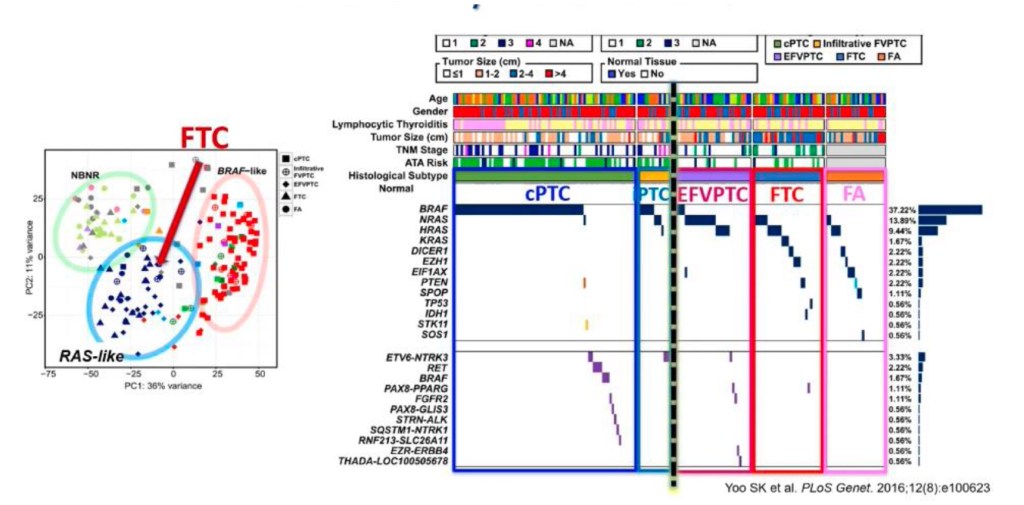
    • Yoo S.K et al, from Korea showed that the genetic profile of follicular carcinomas:
      • Is very similar to follicular adenomas (FA) because they are related tumors:
        • Most FTC originate from a FA and eventually break through the capsule and become carcinomas
    • Encapsulated follicular variant of PTC:
      • Their molecular profile is much closer to a FA and FTC than to classic PTC
    • Infiltrative follicular variant of PTC has a molecular profile:
      • That is more like classic PTC than FTC
    • The biologic difference between
      follicular pattern RAS-like tumors and classic PTC:
      • Is the infiltrative growth pattern (Figure)
    • The difference between these tumors is not only phenotypically based on gross pattern, but also based on biological and clinical differences:
      • Because follicular pattern RAS-like tumors:
        • Retain avidity to radioactive iodine
      • BRAF-like tumors (classic PTC and infiltrative follicular variant of PTC) have the classic features of PTC:
        • They are infiltrative, they spread to lymph nodes first and later to distant sites, and they lose the expression of genes associated with thyroid differentiation
      • RAS-like tumors (FA, FTC, NIFTP, and invasive encapsulated follicular variant of PTC):
        • May or may not have nuclear features of PTC, they are encapsulated, they spread to distant sites (rarely to lymph nodes), and they retain expression of genes associated with thyroid differentiation
  • The RAS genes (HRAS, KRAS and NRAS):
    • Encode for the interconnected G-proteins:
      • That play a critical role in the intracellular transduction of signals arising from cell membrane receptors
  • RAS protein in its inactive state:
    • Is bound to guanosine diphosphate (GDP):
      • Upon activation, it releases GDP and binds guanosine triphosphate (GTP):
        • Thus activating the MAPK and other signaling pathways, such as PI3K/AKT
    • Typically, the activated RAS / GTP protein becomes promptly inactive:
      • Due to its intrinsic GTPase activity and the action of cytoplasmic GTPase-activating proteins:
        • Point mutations in the domains of the RAS gene either increase its affinity for GTP (mutations in codons 12 and 13) or inactivate its autocatalytic GTPase activity (mutation in codon 61):
          • The consequence of this is that the mutant protein becomes permanently switched in the active position and continuously activates its downstream targets
    • Mutations in the RAS genes are believed play
      an early role in the cellular transformation and may predispose to the progression benign tumors to malignant tumors
    • Point mutations involving the specific sites (codons 12, 13 and 61) of the NRAS, HRAS or KRAS genes:
      • Are identified in roughly 10% to 20% of PTC:
        • PTC holding RAS mutation invariably have follicular subtype histology:
          • This mutation also correlates with significantly less prominent nuclear features of PTC, more common
            thyroid encapsulation, and a lower rate of lymph node metastases
        • A few studies have linked RAS
          mutations with PTC:
          • That have a more aggressive behavior, such as a higher frequency of distant metastases
        • Mutations in the RAS gene are not limited to PTC and also found in other benign and malignant thyroid neoplasms, as well as in tumors from other tissues
        • RAS gene mutations are identified in approximately:
          • 20% to 40% of follicular thyroid adenomas (FTA)
          • 40% to 50% of FTC
          • 20% to 40% of anaplastic thyroid carcinoma (ATC)
      • When a FNA cytology is indetermined and the molecular profile identifies a RAS mutation:
        • The risk of malignancy varies
          between the type of mutation:
          • HRAS = 71%
          • NRAS = 63%
          • KRAS = 33%
  • The concept of progression from benign to malignant tumors is supported by the molecular profile shared by these different tumors:
    • More evidence supporting this concept of cancer progression is the similar morphology that these lesions have, along with experimental mouse data showing very similar results
  • Pathologist observing thyroid nodules have detected this step wise progression
  • The nodule in Figure developed from a single cell driven by the RAS mutation, it continued to grow and grow, it eventually forms a capsule, looking microscopically like a benign adenoma (goiter), then it continues to progress, it accumulates more genetic alterations (micro mRNA, and it involves other molecular pathways), it becomes a tumor, it eventually breaks through the capsule, and it will give you invasive encapsulated follicular variant of PTC:
    • If an FNA is performed of area A it would come back as Bethesda II, in area B it would come back as a Bethesda IV, and in area C as Bethesda V:
      • This has changed the practice of cytopathology with molecular
        testing helping us understand better the biological nature of these tumors

Figure: Molecular Changes Precede Histological Changes. The tumor measures 2.5 cm, it has a thin
capsule (black arrows) and shows an area of flat epithelial cells lining the follicles representing a benign thyroid
goiter (tumor area A). Microfollicular areas with well-developed nuclear features of PTC are seen in tumor area
B representing a probable NFTP. Tumor area C has separation artifact with the formation of papillary structures
with nuclear features of PTC. Molecular studies of each section will show NRAS mutation.

Etiology / Pathogenesis of Medullary Thyroid Cancer

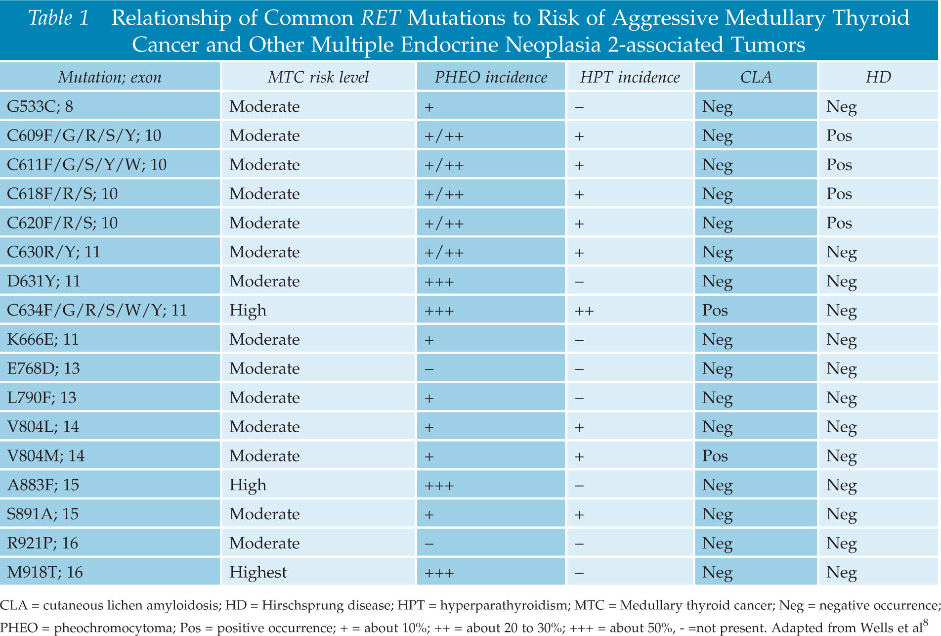
Relationship of Common RET Mutations to Risk of Aggressive MTC

cropped-18403652_10206829497335208_5004404657991480104_n1.jpg

  • What is Head and Neck Surgery?:
    • It is a surgical sub-specialty that deals mainly with benign and malignant tumors of the head and neck region, including:
      • The scalp, facial region, eyes, ears, nose, nasal fossae, paranasal sinuses, oral cavity, pharynx (nasopharynx, oropharynx, hypopharynx), larynx (supraglotic larynx, glottis larynx, subglotic larynx), thyroid gland, parathyroid gland, salivary glands (parotid glands, submandibular glands, sublingual glands, minor salivary glands), soft tissues of the neck, skin of the head and neck region.
        • The head and neck surgeon’s work area:Does not cover tumors or diseases of the brain and other areas of the central nervous system or those of the cervical spine:This is the neurosurgeon field.
    • Among the diagnostic procedures performed by the head and neck surgeon,  are the following:
      • Nasopharyngolaryngoscopy:
        • Performed to examine, evaluate and, possibly perform a biopsy, of oral cavity, pharyngeal and laryngeal lesions.
    • The surgeries most commonly performed by the head and neck surgeon are:
      • Total or near total thyroidectomies
      • Hemithryoidectomies (lobectomies)
      • Comprehensive neck dissections
      • Selective neck dissections
      • Maxillectomies:
        • Total maxillectomy
        • Subtotal maxillectomy
        • Infrastructure maxillectomy
        • Suprastructure maxillectomy
        • Medial maxillectomy
      • Mandibulectomy:
        • Segmental
        • Marginal
      • Tracheostomy
      • Salivary gland surgeries:
        • Parotid gland operations:
          • Limited superficial parotidectomy with identification and preservation of the facial nerve
          • Superficial parotidectomy with identification and preservation of the facial nerve
          • Near total parotidectomy with identification and preservation of the facial nerve
          • Total parotidectomy
        • Submandibular gland resection
        • Sublingual gland resection
      • Resection of tumors of the oral cavity:
        • Glossectomy
        • Resection of the floor of the mouth tumors
      • Resection of tumors of the pharynx
      • Resection of tumors of the larynx
      • Split-thickness skin grafts
      • Full-thickness skin grafts
      • Sentinel lymph node mapping and sentinel lymph node biopsy
      • Resection of malignant skin tumors (BCC, SCC, melanoma) of the head and neck region
  • The formation of the head and neck surgeon includes mastering the following subjects:
    • Surgical Anatomy
    • History and Basic Principles of Head and Neck Surgery
    • Epidemiology, Etiology, and Pathology of Head and Neck Diseases
    • Diagnostic Radiology of the Head and Neck Region
    • Tumors of the Scalp, Skin and Melanoma
    • Eyelids and Orbit
    • Nasal Cavity and Paranasal Sinuses
    • Skull Base and Temporal Bone
    • Lips and Oral Cavity
    • Pharynx and Esophagus
    • Larynx and Trachea
    • Cervical Lymph Nodes
    • Thyroid and Parathyroid Glands
    • Salivary Glands
    • Neurogenic Tumors and Paragangliomas
    • Soft Tissue Tumors
    • Bone Tumors and Odontogenic Lesions
    • Reconstructive Surgery
    • Oncologic Dentistry and Maxillofacial Prosthetics
    • Principles of Radiation Oncology
    • Principles of Chemotherapy
    • Molecular Oncology, Genomics and Immunology
    • Nutrition
    • Biostatistic


  • Rodrigo Arrangoiz MS, MD, FACS a head and neck surgeon / endocrine surgeon / surgical oncologist and is a member of Sociedad Quirúrgica S.C at the America British Cowdray Medical Center in Mexico City:


prof_739_20190417135234

  • Rodrigo Arrangoiz MS, MD, FACS:
    • Is a member of the American Head and Neck Society

img_4750

    • He is a member of the American Thyroid Association:

Unknown

Training:

• General surgery:

• Michigan State University:

• 2004 al 2010

• Surgical Oncology / Head and Neck Surgery / Endocrine Surgery:

• Fox Chase Cancer Center (Filadelfia):

• 2010 al 2012

• Masters in Science (Clinical research for health professionals):

• Drexel University (Filadelfia):

• 2010 al 2012

• Surgical Oncology / Head and Neck Surgery / Endocrine Surgery:

• IFHNOS / Memorial Sloan Kettering Cancer Center:

• 2014 al 2016

#Arrangoiz

#Teacher

#Surgeon

#Cirujano

#ThyroidExpert

#ThyroidSurgeon

#CirujanodeTiroides

#ExpertoenTiroides

#ExpertoenParatiroides

#Paratiroides

#Hiperparatiroidismo

#CancerdeTiroides

#ThyroidCancer

#PapillaryThyroidCancer

#SurgicalOncologist

#CirujanoOncologo

#CancerSurgeon

#CirujanodeCancer

#HeadandNeckSurgeon

#CirugiaEndocrina

#CirujanodeTumoresdeCabezayCuello

#OralCavityCancer

#Melanoma

Medullary Thyroid Cancer Awareness Month

👉Two articles published by a Rodrigo Arrangoiz on medullary thyroid cancer

http://www.remedypublications.com/american-journal-of-otolaryngology-and-head-and-neck-surgery-abstract.php?aid=218

https://s3.amazonaws.com/academia.edu.documents/57472024/final_published_paper.pdf?response-content-disposition=attachment%3B%20filename%3DMedullary_Thyroid_Carcinoma_Literature_R.pdf&X-Amz-Algorithm=AWS4-HMAC-SHA256&X-Amz-Credential=AKIAIWOWYYGZ2Y53UL3A%2F20200302%2Fus-east-1%2Fs3%2Faws4_request&X-Amz-Date=20200302T210556Z&X-Amz-Expires=3600&X-Amz-SignedHeaders=host&X-Amz-Signature=d78d7b605c8a3e8c49d59b7d9545a3f645b1aa4ff1cbda4f57fb4e274bb30849

#Arrangoiz #ThyroidSurgeon

#ThyroidExpert

Molecular Genetics of Thyroid Cancer Part 2

  • Papillary thyroid carcinoma (PTC):
    • Is one of the best molecularly understood cancers:
      • With more that 97% of the driver mutations identified
  • One of the main goals of the cancer genome atlas research network study (TCGA) thyroid project:
    • Was to detect cancer-initiating mutations, i.e., driver mutations:
      • In those cases that lacked the well-known PTC driver mutations (BRAF V600E, point mutations of RAS genes, and gene fusions involving RET and NTRK):
        • These cases are referred to by computational biologists as “dark matter” cases
    • These very important studies under the umbrella of NCI and NIH analyzed 496 PTCs that permitted several analyses:
      • That showed that approximately 75% of all PTC:
        • Developed through the molecular mechanism of point mutation:
        • The most common been the:
          • BRAF V600E mutation
        • The second most common the:
          • RAS mutation
        • The thyroid most common the:
          • TERT mutation – That constituted a small percentage of tumors but was found to be a marker of aggressive disease
      • Roughly 15% of PTC develop through the mechanism of gene fusions:
        • The most common been RET / PTC,
          NTRK 1/3, ALK, BRAF, PAX8 / PPARG
        • The RET / PTC, NTRK 1/3, ALK have very important therapeutic implications for advanced thyroid cancer:
          • Because of the availability of targeted inhibitors with low toxicity and
            high efficacy for the management of these tumors
      • Roughly 7% of PTC develop exclusively from the molecular mechanism of copy number alterations (CNA) (Figure)
  • Roughly 45% to 75% of PTC (29% to 83%) have a mutation in the BRAF gene:
    • Making it the most frequently known genetic
      alteration in PTC
    • Practically all mutations involve:
      • Nucleotide 1799 and result in a valine-to-glutamate substitution at residue 600 (V600E):
        • This point mutation leads to constitutive activation of BRAF kinase:
          • Resulting in a constant phosphorylation of MEK and downstream effectors of the MAPK pathway (Figure)
      • Other mechanisms of BRAF activation in PTC include:
        • K601E point mutation:
          • Small in-frame insertions or deletions surrounding codon 600
        • AKAP9-BRAF rearrangement:
          • Which is more common in PTC associated with radiation exposure
  • BRAF V600E mutation:
    • Is the prevailing mutation in PTC:
      • With classical histology and in the tall cell subtype:
        • But is rare in follicular subtypes tumors
    • In multiple studies, the existence of the BRAF mutation:
      • Has been linked with aggressive tumor biology such as:
        • Advanced stage at presentation
        • Extrathyroidal extension (infiltrative)
        • Recurrence
        • Lymph node or distant metastases
    • BRAF V600E mutation:
      • Is an independent predictor of tumor recurrence:
        • Even in patients with stage I to stage II disease
    • The ability of thyroid cancers to trap
      radioiodine
      :
      • Has been identified to be decreased in tumors with the BRAF V600E mutations and this may lead to treatment failures of the recurrent disease:
        • Which is due to the dysregulation of the function of sodium iodide
          symporter (NIS) and other genes metabolizing iodide in the thyroid follicular cells
    • In thyroid nodules, the V600E BRAF mutation is limited to PTC, poorly differentiated and ATC arising from PTC:
      • Consequently, the identification of the BRAF V600E mutation in FNA cytology of thyroid nodules:
        • Practically confirms the diagnosis of PTC
      • Testing for the BRAF mutation is very useful diagnostically in thyroid FNA samples with indeterminate results:
        • As it can help to determine the diagnosis of PTC in a important portion of these biopsies
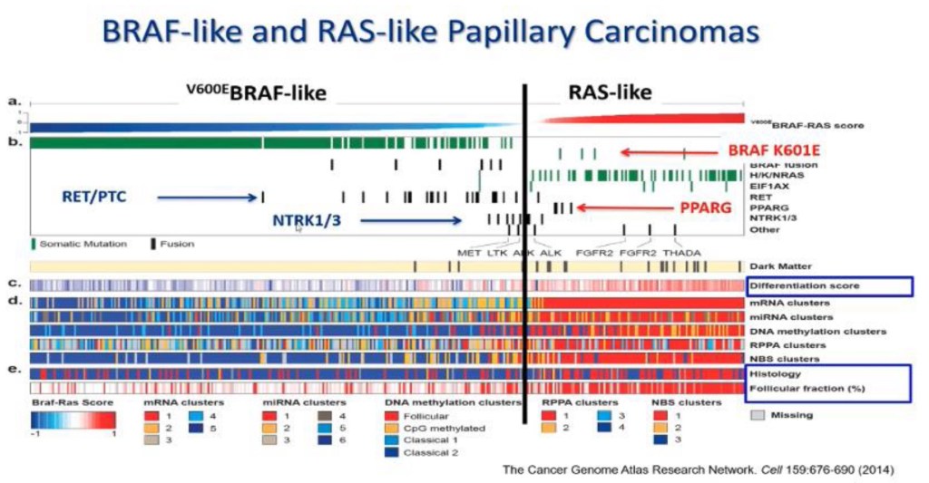
  • Pathologically PTC is one disease and biologically at a minimum PTC has two significantly different groups:
    • Based on multiple type of analysis like gene expression alterations, microRNA alterations, DNA methylation, transcriptomics (Figure)
  • The Cancer Genome Atlas (TCGA) study on PTC highlighted that PTC can be grouped into:
    • BRAFV600E-like and RAS-like tumors:
      • BRAFV600E-like tumors:
        • Also harbor RET fusions
      • RAS-like tumors:
        • Show RAS mutations, the BRAFK601E, and PPARG and THADA fusions
    • The V600E BRAF–like PTC:
      • Are usually classic PTC and tall cell subtype of PTC
    • Clinically when we look at the differentiation score:
      • Which is the expression of genes involved in iodine metabolism and synthesis:
        • BRAF tumors:
          • Loose expression of thyroid differentiation markers
        • RAS-like tumors retain them:
          • Almost at the level of a normal thyroid cell
          • This is important therapeutically because one of the most efficient management options in thyroid cancer is iodine therapy
        • RAS-like tumors:
          • Are usually follicular variant PTC
      • Usually all BRAF mutations are BRAF-like and all RAS mutations are RAS-like:
        • But all other type of molecular alterations can be hidden in either BRAF-like and RAS-like:
          • For example, BRAF K601E mutations are found in RAS-like tumors

Molecular Subtypes of Papillary Thyroid Carcinomas

Graph exemplifying the difference between follicular pattern RAS-like tumors and classic PTC,
which is the infiltrative growth pattern.

Calcitonin

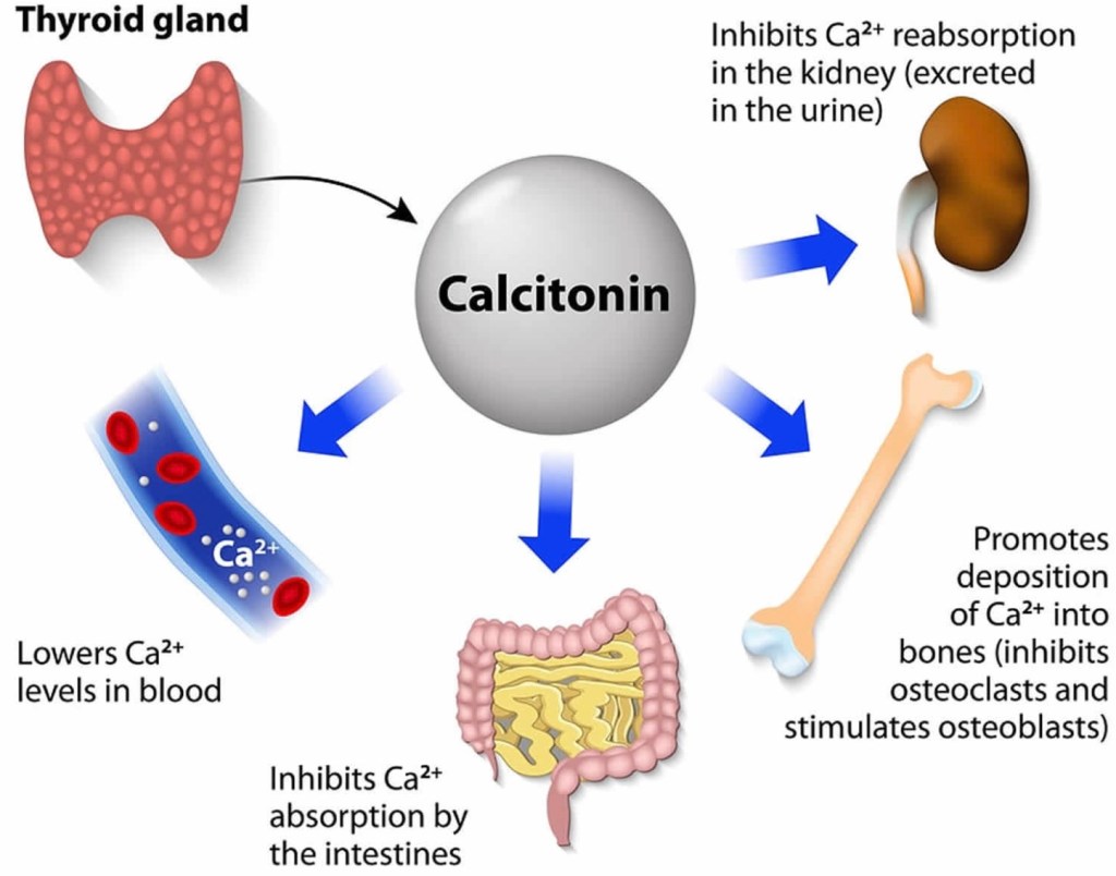
  • Calcitonin:
    • Is produced by the:
      • Parafollicular cells / C-cells of the thyroid gland:
        • Works to oppose the actions of PTH
    • It helps lower ionized calcium levels:
      • Primarily in two ways:
        • First, it inhibits osteoclast-mediated bone resorption
        • Second, it inhibits resorption of calcium and phosphate by the kidney
  • Calcitonin:
    • Has no direct effects on:
      • Intestinal absorption
      • Osteoblast-mediated bone formation
  • It is also used in the treatment of hypercalcemic crisis:
    • 4 IU/kg subcutaneously / intramuscularly
  • It acts quickly (24 to 48 hours):
    • Is more effective when used in combination with glucocorticoids
  • Finally, calcitonin is a useful marker:
    • With regard to surveillance in medullary thyroid carcinoma

#Arrangoiz #Calcitonin #ParathyroidSurgeon #ParathyroidExpert #CalciumHemostasis #Teacher #Surgeon #HeadandNeckSurgeon #CancerSurgeon #MSMC #MountSinaiMedicalCenter #Miami #Mexico

Diagnosis of Medullary Thyroid Carcinoma (MTC)

  • The diagnosis of medullary thyroid cancer (MTC):
    • Is usually made after fine-needle aspiration (FNA) biopsy in a patient who has a solitary thyroid nodule (or a dominant nodule within a multinodular goiter):
      • The sensitivity of FNA is 50% to 80%:
        • Although higher sensitivity can be obtained by the addition of immunohistochemical staining for calcitonin
  • If the clinical suspicion for MTC is high:
    • Patient with diarrhea, flushing, and a thyroid nodule:
      • Calcitonin can be measured in the washout of the FNA biopsy needle:
        • Although this may not be readily available in many commercial laboratories
  • In some cases:
    • The diagnosis of MTC is made after thyroid lobectomy for a suspicious or indeterminate FNA biopsy:
      • Surgical specimens from patients with MTC show:
        • Spindle-shaped and frequently pleomorphic cells without follicle development:
          • Because these cells originate from the calcitonin-producing parafollicular C cells of the thyroid
      • The use of serum calcitonin screening to complement ultrasound and FNA in the routine diagnosis of thyroid nodules is controversial in the United States:
        • Measurement of serum calcitonin has not been a part of the routine evaluation of patients with thyroid nodules in the United States:
          • The high frequency of falsely elevated serum calcitonin values, the inability to confirm the high calcitonin by pentagastrin stimulation in the United States, and the accuracy of FNA biopsy: would argue against a change in this recommendation
        • Further, occasional patients with locoregional metastases or locally invasive MTC will have normal unstimulated serum calcitonin concentrations
  • In some countries (eg, European countries) where pentagastrin is available, however, serum basal and stimulated calcitonin levels are routinely used in the evaluation of thyroid nodules to facilitate the preoperative diagnosis of MTC
  • Differential Diagnosis:
    • The differential diagnosis in a patient presenting with a neck mass:
      • Is extensive and varies with the age of the patient at presentation
    • The majority of these masses represent benign thyroid nodules and cysts
    • Neck masses that are not of thyroidal origin may be from:
      • Congenital
      • Vascular anomaly
      • Inflammatory:
        • Lymph node enlargement
      • Other neoplastic:
        • Primary or metastatic disease disorders
  • In addition to medullary thyroid cancer (MTC):
    • Elevated calcitonin results may also be seen in patients with:
      • Hypercalcemia
      • Hypergastrinemia
      • Neuroendocrine tumors
      • Renal insufficiency
      • Papillary and follicular thyroid carcinomas
      • Goiter
      • Chronic autoimmune thyroiditis
      • Prolonged treatment with:
        • Omeprazole (greater than two to four months)
        • Beta blockers
        • Glucocorticoids:
          • Has been associated with hypercalcitoninemia
  • In addition, the presence of heterophilic antibodies to calcitonin:
    • Can falsely elevate serum calcitonin levels 
  • Elevated carcinoembryonic antigen (CEA) levels can also occur in patients with:
    • Heterophilic antibodies
    • Gastrointestinal tract inflammatory disease
    • Benign lung disease
    • Non-thyroid malignancies
    • Cigarette smoking
  • Evaluation:
    • For patients diagnosed with medullary thyroid cancer (MTC) on the basis of cytologic evaluation of a thyroid nodule:
      • Evaluation should include:
        • Measurement of serum calcitonin, carcinoembryonic antigen (CEA), ultrasonography of the neck (if not already performed), genetic testing for germline RET mutations, and biochemical evaluation for coexisting tumors, especially pheochromocytoma
        • Serum calcitonin and CEA:
          • The serum calcitonin and carcinoembryonic antigen (CEA) concentrations should be measured in patients diagnosed with MTC on the basis of cytologic evaluation of a thyroid nodule
          • These tests can establish that the tumor is capable of hypersecreting the hormones and, if so, the values can be compared with postoperative values
      • Postoperatively:
        • Results may provide a prognostic factor or indicate biochemical cure
  • In a study of 226 patients with MTC:
    • Preoperative serum calcitonin concentrations:
      • Where significantly correlated with tumor size in both the sporadic and familial cases
    • In addition, among 45 patients who had a preoperative serum calcitonin concentration of 50 pg/mL or less:
      • 44 had normal concentrations after surgery:
    • In contrast, only 50 of 120 patients with preoperative serum calcitonin concentrations higher than 50 pg/mL had normal concentrations after surgery
  • In a second study of 224 patients with MTC:
    • 62% of patients without nodal metastases had normal calcitonin postoperatively:
      • 10% of node positive patients had normal postoperative calcitonin levels
  • Assessment of calcitonin and CEA doubling times postoperatively:
    • Provides sensitive markers for progression and aggressiveness of metastatic MTC:
      • Postoperative calcitonin doubling time was a prognostic factor for survival in a study of 65 patients followed for 3 to 30 years:
        • Ten-year survival was: 8%, 37%, and 100% for doubling times:
          • Under six months, between six months and 24 months, and greater than 24 months, respectively
  • Radiologic evaluation:
    • MTC can spread by:
      • Local invasion or metastasis:
        • Within the neck or distantly
    • When MTC is diagnosed by fine-needle aspiration (FNA) biopsy:
      • Ultrasonography of the neck is indicated to look for cervical lymph node involvement
    • For patients with local lymph node metastases on ultrasound or with preoperative serum basal calcitonin > 500 pg/mL (indicating high risk of local or distant metastatic disease):
      • Additional imaging is required to assess for metastatic disease:
        • In this setting, based con literature review I suggest cross-sectional imaging including:
          • Chest computed tomography (CT)
          • Neck CT with IV contast
        • Three-phase contrast-enhanced liver CT or contrast-enhanced liver magnetic resonance imaging (MRI)
        • Axial MRI, and bone scintigraphy:
          • In patients suspected of having skeletal metastases
          • MRI may be superior to other imaging modalities
    • I do not recommend 18-fluoro-2-deoxyglucose positron emission tomography (FDG-PET) imaging or somatostatin receptor imaging:
      • For routine initial screening for metastatic disease:
        • The sensitivity of FDG-PET scanning for detecting metastatic disease is variable:
          • But improves with higher calcitonin levels
          • Sensitivity 78% versus 20% for basal calcitonin value greater than or less than 1000 pg/mL, respectively
      • The use of radionuclide imaging with 111-In-octreotide or 99m-Tc-DMSA:
        • Is not currently recommended for routine initial screening for metastatic disease
        • However, three patients have been described who had regional and distant metastases of MTC detected by somatostatin receptor scintigraphy but not by CT scan
      • How to select patients with a negative CT scan to undergo somatostatin receptor scintigraphy is not clear:
        • Scanning may be more useful in localizing residual or recurrent disease after primary therapy
  • Genetic screening in sporadic MTC:
    • Germline RET testing:
      • In all patients with newly diagnosed C cell hyperplasia or apparently sporadic MTC:
        • Initial germline testing in patients with C cell hyperplasia or apparently sporadic MTC should include:
          • Sequencing of exons 10, 11, and 13 through 16 of the RET gene
        • Sequencing of the remaining exons in the RET gene should be considered in patients with:
          • Clinical features or family history highly suggestive of hereditary medullary syndromes who demonstrate no mutations in exons 10, 11, or 13 through 16
  • While it is possible for clinicians to directly order genetic testing from reference laboratories:
    • It is strongly encourage to have a consultation with genetic counselors who are familiar with both the ethical issues and legal informed consent requirements (which can vary significantly in different regions) that are involved in germline testing
  • When the index patient is positive for a germline mutation:
    • Family members should be offered genetic counseling and genetic screening
  • An important question is what proportion of patients with apparently sporadic MTC have unsuspected germline mutations in the RET proto-oncogene (the underlying defect in MEN2) and, therefore, have heritable disease:
    • Studies of unselected patients with MTC have found, on average:
      • That approximately 6% to 7%  (range 1.5% to 24%) have germline RET mutations
    • In one report, 35 of 482 patients (7.3%) with apparently sporadic MTC had mutations, and in 18 of these 35:
      • Gene carriers were identified in relatives
  • 75% of the familial medullary cases:
    • Had no prior family history:
      • A much higher percentage (approximately 60%) of patients with sporadic MTC have somatic (acquired) mutations in the RET gene within the tumor cells:
        • These mutations are present only in the tumor cells and are not detected by standard genetic testing (ie, using leukocyte DNA)
    • The presence of somatic RET mutations correlate with:
      • Lymph node metastases
      • Persistent disease
      • Lower survival
    • However, in one study:
      • Only mutations in exons 15 and 16 of the RET gene were associated with the worse prognosis:
        • While those in other exons had a more indolent course
  • Since it is unclear how knowledge of a specific somatic (acquired) RET mutation should impact clinical management:
    • I do not routinely test tumor samples
  • Testing for coexisting tumors:
    • Most patients require biochemical evaluation for coexisting tumors (particularly pheochromocytoma and hyperparathyroidism) prior to thyroidectomy:
      • Even when genetic screening is performed preoperatively:
        • The results are rarely known prior to surgery
    • For patients with unknown RET mutational status and for patients who have a germline RET mutation:
      • Serum calcium:
        • To rule out hyperparathyroidism requiring concomitant surgical intervention
      • Plasma fractionated metanephrines:
        • As the initial screen for pheochromocytoma):
          • Normal plasma fractionated metanephrines values exclude a symptomatic catecholamine-secreting neoplasm
          • Mildly elevated values of normetanephrine could be falsely positive in which case additional evaluations including 24-hour urinary fractionated metanephrines, catecholamines, and adrenal imaging may be required to effectively rule in or rule out pheochromocytoma prior to surgery
          • Adrenal imaging should not be performed unless there is biochemical evidence suggesting a possible pheochromocytoma
  • In a patient with negative RET proto-oncogene testing and no family history of MEN2 syndrome:
    • Biochemical testing for coexisting tumors is typically not required
  • What is Head and Neck Surgery?:
    • It is a surgical sub-specialty that deals mainly with benign and malignant tumors of the head and neck region, including:
      • The scalp, facial region, eyes, ears, nose, nasal fossae, paranasal sinuses, oral cavity, pharynx (nasopharynx, oropharynx, hypopharynx), larynx (supraglotic larynx, glottis larynx, subglotic larynx), thyroid gland, parathyroid gland, salivary glands (parotid glands, submandibular glands, sublingual glands, minor salivary glands), soft tissues of the neck, skin of the head and neck region. The head and neck surgeon’s work area:Does not cover tumors or diseases of the brain and other areas of the central nervous system or those of the cervical spine:
        • This is the neurosurgeon field
  • Among the diagnostic procedures performed by the head and neck surgeon,  are the following: Nasopharyngolaryngoscopy:
    • Performed to examine, evaluate and, possibly perform a biopsy, of oral cavity, pharyngeal and laryngeal lesions.
  • The surgeries most commonly performed by the head and neck surgeon are: Total or near total thyroidectomies
  • Hemithryoidectomies (lobectomies)
  • Comprehensive neck dissections
  • Selective neck dissections
  • Maxillectomies: Total maxillectomy
  • Subtotal maxillectomy
  • Infrastructure maxillectomy
  • Suprastructure maxillectomy
  • Medial maxillectomy
  • Mandibulectomy: Segmental
  • Marginal
  • Tracheostomy
  • Salivary gland surgeries: Parotid gland operations: Limited superficial parotidectomy with identification and preservation of the facial nerve
  • Superficial parotidectomy with identification and preservation of the facial nerve
  • Near total parotidectomy with identification and preservation of the facial nerve
  • Total parotidectomy
  • Submandibular gland resection
  • Sublingual gland resection
  • Resection of tumors of the oral cavity: Glossectomy
  • Resection of the floor of the mouth tumors
  • Resection of tumors of the pharynx
  • Resection of tumors of the larynx
  • Split-thickness skin grafts
  • Full-thickness skin grafts
  • Sentinel lymph node mapping and sentinel lymph node biopsy
  • Resection of malignant skin tumors (BCC, SCC, melanoma) of the head and neck region
  • The formation of the head and neck surgeon includes mastering the following subjects: Surgical Anatomy
  • History and Basic Principles of Head and Neck Surgery
  • Epidemiology, Etiology, and Pathology of Head and Neck Diseases
  • Diagnostic Radiology of the Head and Neck Region
  • Tumors of the Scalp, Skin and Melanoma
  • Eyelids and Orbit
  • Nasal Cavity and Paranasal Sinuses
  • Skull Base and Temporal Bone
  • Lips and Oral Cavity
  • Pharynx and Esophagus
  • Larynx and Trachea
  • Cervical Lymph Nodes
  • Thyroid and Parathyroid Glands
  • Salivary Glands
  • Neurogenic Tumors and Paragangliomas
  • Soft Tissue Tumors
  • Bone Tumors and Odontogenic Lesions
  • Reconstructive Surgery
  • Oncologic Dentistry and Maxillofacial Prosthetics
  • Principles of Radiation Oncology
  • Principles of Chemotherapy
  • Molecular Oncology, Genomics and Immunology
  • Nutrition
  • Biostatistic
  • Rodrigo Arrangoiz MS, MD, FACS a head and neck surgeon / endocrine surgeon / complex surgical oncologist and is a member of Mount Sinai Medical Center, in Miami Beach, Florida
  • Rodrigo Arrangoiz MS, MD, FACS:
    • Is a member of the American Head and Neck Society
    • He is a member of the American Thyroid Association
  • Training:
    • General surgery:
      • Michigan State University:
      • 2004 al 2010
  • Surgical Oncology / Head and Neck Surgery / Endocrine Surgery:
    • Fox Chase Cancer Center (Filadelfia):
      • 2010 al 2012
  • Masters in Science (Clinical research for health professionals):
    • Drexel University (Filadelfia):
    • 2010 al 2012
  • Surgical Oncology / Head and Neck Surgery / Endocrine Surgery:
    • IFHNOS / Memorial Sloan Kettering Cancer Center:
      • 2014 al 2016

Molecular Genetics of Thyroid Cancer Part 1

  • Most of the data that has been gathered is associated with somatic mutations:
    • Many of which occur early in the
      transformation process and are crucial for cancer development
  • In thyroid cancer, vital genes are commonly mutated via two separate molecular mechanisms:
    • Point mutations
    • Chromosomal rearrangements
  • Point mutations:
    • Are the result of a single nucleotide change within the DNA chain:
      • Which activates or inactivates a
        protein
  • Chromosomal rearrangements:
    • Represent a large-scale genetic abnormality with breakage and fusion of parts of the same chromosome or different chromosomes
  • The molecular pathogenesis of thyroid cancer:
    • Is relatively simple with two molecular pathways:
      • The mitogen‑activated protein kinase (MAPK) pathway
      • The phosphatidylinositol 3 – kinase – protein kinase B (PI3K-AKT) pathway (Figure)
  • These pathways are activated in most thyroid cancers:
    • Via distinct molecular mechanisms mentioned previously, like:
      • Point mutations (single nucleotide variant):
        • Where a single nucleotide is
          changed:
          • Which activates or inactivates a protein
      • Gene fusions:
        • Were parts of two different genes located in the same or different chromosome are fused to each other:
          • That results in the generation of chimeric protein
      • Copy number alterations (CNA):
        • That have been recently accepted as a driver mutation for oncocytic cell tumors
  • Activation of the MAPK pathway:
    • Is a critical step for tumor initiation
    • The mutated genes that impact
      these pathways encode:
      • The cell-membrane receptor tyrosine kinases:
        • RET and NTRK1
      • Intracellular signal transducers:
        • RAS and BRAF
    • These mutually exclusive mutations:
      • They do not overlap with each other because they activate the same pathway and only one event is sufficient to activate the process for cancer development:
        • Occur in roughly 70% to 75% of patients with PTC and are associated with specific clinical, pathologic, and biological tumor characteristics (Table)
    • In follicular thyroid cancer (FTC), in addition to mutations of RAS:
      • Another common event is PAX8 / PPARG rearrangement
  • Thyroid cancer progression and dedifferentiation involves a number of additional mutations:
    • That affects the PI3K-AKT pathway and other cell signaling pathways

Clinical Presentation of Medullary Thyroid Carcinoma

  • Sporadic MTC (75% of all cases):Typically occurs between the fourth to sixth decade of life:However, patients with hereditary disease:Present earlier
    • Patients with MTC who present with a palpable thyroid nodule:70% will have cervical lymphadenopathy
      • 15% to 20% will have palpable cervical lymphadenopathy.
    • Pain or aching:Is also a common symptom of MTC
    • Local tumor invasion may produce symptoms of:Dysphagia, dyspnea, or dysphonia
    • Patients with extensive metastatic disease frequently develop:Diarrhea:Which may result from:Increased intestinal motility and impaired intestinal water and electrolyte flux:Due to high calcitonin levels
      • About 2% to 4% of patients develop:Cushing syndrome:As a result of ectopic production of ACTH
  • Classic MEN2A is the most common variant of MEN2 (95% of the cases):Most patients carry germline RET mutations in:Codons 609, 611, 618, or 620 of exon 10
      • Codon 634 of exon 11
    • Almost all patients develop MTC:But pheochromocytomas have the highest penetrance in patients with:Codon 634 mutations:52% by age 50
          • 88% by age 77
        • These adrenal tumors:Are almost always benign
          • Are usually multicentric
          • Are associated with diffuse adrenal nodular hyperplasia
      • HPT:Is usually mild and may involve one to four parathyroid glands.
        • RET codon 634 mutations:Are associated with a penetrance of up to 30% for PHPT, whereas it ranges from 2 to 12% for the remaining mutations.
  • Cutaneous lichen amyloidosis (CLA):Can occur sporadically or in an inherited manner:Either separately or in the context of MEN2A
    • The skin lesions of CLA in MEN2A:Are most apparent in the dermatomes corresponding to T2 to T6:Scapular region of the back
    • The classic symptom is:Itching:That worsens in times of stress
        • Improves with sun exposure
    • The lesions are incited by notalgia paresthetica:A sensory neuropathy of the dorsal spinal nerve:May occur prior to MTC
    • CLA is almost exclusively found in patients with:The codon 634 mutations (exon 11)
      • Although cases have also been reported in patients with:Codon 804 mutations
      • Pheochromocytomas and PHPT in CLA patients:Occur at frequencies similar to classic MEN2A
  • In patients with MEN2B:MTC often presents in infancy and is usually highly aggressive
    • Approximately 75% to 90% of MEN2B cases occur in patients:Having de novo RET mutations:Whereas 10% to 25% occur in families with:Known history of MEN2B
    • About 95% of MEN2B individuals:Have codon M918T mutations (exon 13) – Highest risk (HST)The remaining 5% have codon A883F mutations (exon 15) – High risk (H):The MTCs in codon A883F tend to be less aggressive
    • Pheochromocytomas develop in:About 50% of MEN2B patients
    • These individuals also have a typical appearance that includes:Eye abnormalities:Thickened and everted eyelids
        • Mild ptosis
        • Thickened corneal nerves
      • Marfanoid body habitus
      • Long narrow facies
      • Pes cavus
      • Pectus excavatum
      • High-arched palate
      • Scoliosis
      • Slipped capital femoral epiphyses
      • Mucosal neuromas
      • Diffuse ganglioneuromatosis of the aerodigestive tractLeading to bloating, intermittent constipation, diarrhea, and even intestinal obstruction
    • Some patients have atypical MEN2B:Which presents later in life:Around 20 to 30 years of age:These patients have double RET germline mutations:Occurring in tandem on the same allele:Usually V804M and either Y806C, S904C, E805K, or Q781R 


prof_739_20190417135234

  • Rodrigo Arrangoiz MS, MD, FACS:
    • Is a member of the American Head and Neck Society

img_4750

    • He is a member of the American Thyroid Association:

Unknown

Training:

• General surgery:

• Michigan State University:

• 2004 al 2010

• Surgical Oncology / Head and Neck Surgery / Endocrine Surgery:

• Fox Chase Cancer Center (Filadelfia):

• 2010 al 2012

• Masters in Science (Clinical research for health professionals):

• Drexel University (Filadelfia):

• 2010 al 2012

• Surgical Oncology / Head and Neck Surgery / Endocrine Surgery:

• IFHNOS / Memorial Sloan Kettering Cancer Center:

• 2014 al 2016

#Arrangoiz

#Teacher

#Surgeon

#Cirujano

#ThyroidExpert

#ThyroidSurgeon

#CirujanodeTiroides

#ExpertoenTiroides

#ExpertoenParatiroides

#Paratiroides

#Hiperparatiroidismo

#CancerdeTiroides

#ThyroidCancer

#PapillaryThyroidCancer

#SurgicalOncologist

#CirujanoOncologo

#CancerSurgeon

#CirujanodeCancer

#HeadandNeckSurgeon

#CirugiaEndocrina

#CirujanodeTumoresdeCabezayCuello

#OralCavityCancer

#Melanoma

Thyroid Neoplasms

  • The preponderance of thyroid neoplasms:
    • Originate from the thyroid epithelial follicular cells:
      • While 3% to 5% of neoplasms arise from the C cells or parafollicular cells
  • Differentiated thyroid cancer (DTC):
    • Which derives from these follicular cells, includes:
      • Papillary thyroid carcinoma (PTC)
      • Follicular thyroid carcinoma (FTC)
      • Oncocytic cell carcinoma (formerly known as Hürthle Cell Carcinoma / OCC)
      • Poorly differentiated carcinoma
        (insular carcinoma)
      • Anaplastic thyroid carcinoma (ACC / undifferentiated)
    • These thyroid tumors comprise the majority, more than 90% of the cases, of all thyroid neoplasms
    • Of all these subtypes, ATC is the rarest and is characterized by its extremely poor prognosis
    • Likewise, poorly differentiated carcinoma is
      characterized by its aggressive behavior and its unfavorable prognosis
  • Between the year 2010 and 2014:
    • 63,229 patients per year were diagnosed with thyroid cancer:
      • Of these 89.4% had PTC, 4.6% had FTC, 2.0% had OCC, 1.7% had medullary thyroid carcinoma (MTC), and 0.8% had ATC
  • A follicular adenoma:
    • Is a benign tumor (clonal neoplasm):
      • That may serve as a precursor lesion for some follicular carcinomas
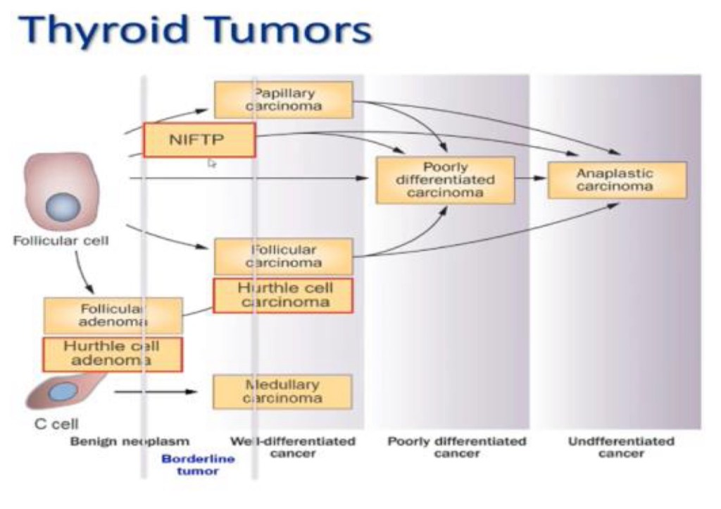
  • Less-differentiated thyroid cancers, namely poorly differentiated carcinomas, and anaplastic carcinomas:
    • Can develop de novo:
      • Although many of them arise through the process of a stepwise dedifferentiation of papillary and follicular carcinomas (Figure)
  • Thyroid nodules:
    • Are a major health problem worldwide
  • Studies have shown that the prevalence of palpable thyroid nodules:
    • Is roughly 5% in women and 1% in men:
      • Living in parts of the world with sufficient iodine
  • In contrast, high-resolution ultrasound:
    • Can detect thyroid nodules in around 19% to 68% of randomly selected people:
      • With higher frequencies in women and the elderly
  • The clinical significance of thyroid nodules lies in the need to exclude thyroid cancer:
    • Which occurs anywhere between 7% and 15% of the cases:
      • Depending on age, gender, radiation exposure history, and family history
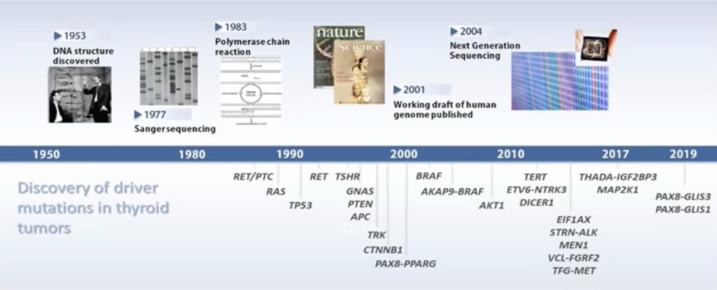
  • The discovery in 1953 of the double helix structure of deoxyribonucleic acid (DNA), by James Watson and Francis Crick:
    • Marked a milestone in the history of science and gave rise to modern molecular genetics that has
      rapidly advanced and has help to identify the driver mutations in in many cancers, including thyroid neoplasms:
      • RET / PTC, RAS, TP53, RET, TSHR, GNAS, PTEN, APC, TRK, CTNNB1, PAX8-PPARG, BRAF, AKAP9-BRAF, AKT1, TERT, ETVS-NTRK3, DICER1, EIFF1AX, STRN-ALK, MEN1, VCL-FGRF2, TGF-MET, THADA-IGF2BP3, MAP2K1, PAX8-GLIS3, PAX8-GLIS1 (Figure)
  • Analogous to other malignant neoplasms, the initiation and progression of thyroid cancer:
    • Occurs through steady accumulation of multiple genetic and epigenetic alterations, including:
      • Activating and inactivating somatic mutations
      • Alteration in gene expression patterns
      • MicroRNA (miRNA) dysregulation
      • Aberrant gene methylation

Medullary Thyroid Carcinoma Part 2

img_3549

  • Diagnosis:The diagnosis of medullary thyroid cancer (MTC):Is usually made after fine-needle aspiration (FNA) biopsy in a patient who has a solitary thyroid nodule (or a dominant nodule within a multinodular goiter):The sensitivity of FNA is 50% to 80%:Although higher sensitivity can be obtained by the addition of immunohistochemical staining for calcitoninIf the clinical suspicion for MTC is high:Patient with diarrhea, flushing, and a thyroid nodule):Calcitonin can be measured in the washout of the FNA biopsy needle:Although this may not be readily available in many commercial laboratoriesIn some cases:The diagnosis of MTC is made after thyroid lobectomy for a suspicious or indeterminate FNA biopsy:Surgical specimens from patients with MTC show:Spindle-shaped and frequently pleomorphic cells without follicle development:Because these cells originate from the calcitonin-producing parafollicular C cells of the thyroidThe use of serum calcitonin screening to complement ultrasound and FNA in the routine diagnosis of thyroid nodules is controversial in the United States:Measurement of serum calcitonin has not been a part of the routine evaluation of patients with thyroid nodules in the United States:The high frequency of falsely elevated serum calcitonin values, the inability to confirm the high calcitonin by pentagastrin stimulation in the United States, and the accuracy of FNA biopsy:Would argue against a change in this recommendation
          • Further, occasional patients with locoregional metastases or locally invasive MTC will have normal unstimulated serum calcitonin concentrations
          • In some countries (eg, European countries) where pentagastrin is available, however, serum basal and stimulated calcitonin levels are routinely used in the evaluation of thyroid nodules to facilitate the preoperative diagnosis of MTC

PastedGraphic-1

  • Differential Diagnosis:The differential diagnosis in a patient presenting with a neck mass:Is extensive and varies with the age of the patient at presentationThe majority of these masses represent benign thyroid nodules and cysts
      • Neck masses that are not of thyroidal origin may be from:Congenital:Vascular anomaly)Inflammatory:Lymph node enlargement)Other neoplastic:Primary or metastatic disease disorders
    • In addition to medullary thyroid cancer (MTC):Elevated calcitonin results may also be seen in patients with:Hypercalcemia
        • Hypergastrinemia
        • Neuroendocrine tumors
        • Renal insufficiency
        • Papillary and follicular thyroid carcinomas
        • Goiter
        • Chronic autoimmune thyroiditis
      • Prolonged treatment with:Omeprazole (greater than two to four months)Beta blockersGlucocorticoids:Has been associated with hypercalcitoninemia
      • In addition, the presence of heterophilic antibodies to calcitonin:Can falsely elevate serum calcitonin levels 
    • Elevated carcinoembryonic antigen (CEA) levels can also occur in patients with:Heterophilic antibodiesGastrointestinal tract inflammatory diseaseBenign lung diseaseNonthyroid malignanciesCigarette smoking
  • Evaluation:For patients diagnosed with medullary thyroid cancer (MTC) on the basis of cytologic evaluation of a thyroid nodule:Evaluation should include:Measurement of serum calcitonin, carcinoembryonic antigen (CEA), ultrasonography of the neck (if not already performed), genetic testing for germline RET mutations, and biochemical evaluation for coexisting tumors, especially pheochromocytoma. Serum calcitonin and CEA:The serum calcitonin and carcinoembryonic antigen (CEA) concentrations should be measured in patients diagnosed with MTC on the basis of cytologic evaluation of a thyroid nodule:
          • These tests can establish that the tumor is capable of hypersecreting the hormones and, if so, the values can be compared with postoperative values
        • Postoperatively:Results may provide a prognostic factor or indicate biochemical cure
      • In a study of 226 patients with MTC:Preoperative serum calcitonin concentrations:Where significantly correlated with tumor size in both the sporadic and familial casesIn addition, among 45 patients who had a preoperative serum calcitonin concentration of 50 pg/mL or less:44 had normal concentrations after surgery:In contrast, only 50 of 120 patients with preoperative serum calcitonin concentrations higher than 50 pg/mL had normal concentrations after surgery
      • In a second study of 224 patients with MTC:62% of patients without nodal metastases had normal calcitonin postoperatively:10% of node positive patients had normal postoperative calcitonin levels
      • Assessment of calcitonin and CEA doubling times postoperatively:Provides sensitive markers for progression and aggressiveness of metastatic MTC:Postoperative calcitonin doubling time was a prognostic factor for survival in a study of 65 patients followed for 3 to 30 years: Ten-year survival was: 8%, 37%, and 100% for doubling times:Under six months, between six months and 24 months, and greater than 24 months, respectively
    • Radiologic evaluation:MTC can spread by:Local invasion or metastasis:Within the neck or distantlyWhen MTC is diagnosed by fine-needle aspiration (FNA) biopsy:Ultrasonography of the neck is indicated to look for cervical lymph node involvementFor patients with local lymph node metastases on ultrasound or with preoperative serum basal calcitonin > 500 pg/mL (indicating high risk of local or distant metastatic disease):Additional imaging is required to assess for metastatic disease:In this setting, based con literature review I suggest cross-sectional imaging including:Chest computed tomography (CT)
            • Neck CT with IV contast
            • Three-phase contrast-enhanced liver CT or contrast-enhanced liver magnetic resonance imaging (MRI)
            • Axial MRI, and bone scintigraphy:In patients suspected of having skeletal metastases:MRI may be superior to other imaging modalities
        • I do not recommend 18-fluoro-2-deoxyglucose positron emission tomography (FDG-PET) imaging or somatostatin receptor imaging:For routine initial screening for metastatic disease:The sensitivity of FDG-PET scanning for detecting metastatic disease is variable:But improves with higher calcitonin levels:Sensitivity 78% versus 20% for basal calcitonin value greater than or less than 1000 pg/mL, respectively)
            • The use of radionuclide imaging with 111-In-octreotide or 99m-Tc-DMSA:Is not currently recommended for routine initial screening for metastatic disease:However, three patients have been described who had regional and distant metastases of MTC detected by somatostatin receptor scintigraphy but not by CT scan
                • How to select patients with a negative CT scan to undergo somatostatin receptor scintigraphy is not clear:Scanning may be more useful in localizing residual or recurrent disease after primary therapy
  • Genetic screening in sporadic MTC:Germline RET testing:In all patients with newly diagnosed C cell hyperplasia or apparently sporadic MTC:Initial germline testing in patients with C cell hyperplasia or apparently sporadic MTC should include:Sequencing of exons 10, 11, and 13 through 16 of the RET gene
          • Sequencing of the remaining exons in the RET gene should be considered in patients with:Clinical features or family history highly suggestive of hereditary medullary syndromes:Who demonstrate no mutations in exons 10, 11, or 13 through 16
          • While it is possible for clinicians to directly order genetic testing from reference laboratories:It is strongly encourage to have a consultation with genetic counselors who are familiar with both the ethical issues and legal informed consent requirements (which can vary significantly in different regions) that are involved in germline testing
    • When the index patient is positive for a germline mutation:Family members should be offered genetic counseling and genetic screening
    • An important question is what proportion of patients with apparently sporadic MTC have unsuspected germline mutations in the RET proto-oncogene (the underlying defect in MEN2) and, therefore, have heritable disease:Studies of unselected patients with MTC have found, on average:That approximately 6% to 7%  (range 1.5% to 24%) have germline RET mutationsIn one report:35 of 482 patients (7.3%) with apparently sporadic MTC had mutations, and in 18 of these 35:Gene carriers were identified in relatives
    • 75% of the familial medullary cases:Had no prior family history:A much higher percentage (approximately 60%) of patients with sporadic MTC have somatic (acquired) mutations in the RET gene within the tumor cells:These mutations are present only in the tumor cells and are not detected by standard genetic testing, ie, using leukocyte DNA
          • The presence of somatic RET mutations correlate with:Lymph node metastasesPersistent diseaseLower survivalHowever, in one study:Only mutations in exons 15 and 16 of the RET gene:Were associated with the worse prognosis:While those in other exons had a more indolent course
        • Since it is unclear how knowledge of a specific somatic (acquired) RET mutation should impact clinical management:I do not routinely test tumor samples
    • Testing for coexisting tumors:Most patients require biochemical evaluation for coexisting tumors (particularly pheochromocytoma and hyperparathyroidism) prior to thyroidectomy:Even when genetic screening is performed preoperatively:The results are rarely known prior to surgery.For patients with unknown RET mutational status and for patients who have a germline RET mutation:Serum calcium:To rule out hyperparathyroidism requiring concomitant surgical interventionPlasma fractionated metanephrines:As the initial screen for pheochromocytoma):Normal plasma fractionated metanephrines values:Exclude a symptomatic catecholamine-secreting neoplasm
            • Mildly elevated values of normetanephrine could be falsely positive:In which case additional evaluations including 24-hour urinary fractionated metanephrines, catecholamines, and adrenal imaging may be required to effectively rule in or rule out pheochromocytoma prior to surgery:Adrenal imaging should not be performed unless there is biochemical evidence suggesting a possible pheochromocytoma. (See 
    • In a patient with negative RET proto-oncogene testing and no family history of MEN2 syndrome:Biochemical testing for coexisting tumors is typically not required


cropped-18403652_10206829497335208_5004404657991480104_n1.jpg

  • What is Head and Neck Surgery?:
    • It is a surgical sub-specialty that deals mainly with benign and malignant tumors of the head and neck region, including:
      • The scalp, facial region, eyes, ears, nose, nasal fossae, paranasal sinuses, oral cavity, pharynx (nasopharynx, oropharynx, hypopharynx), larynx (supraglotic larynx, glottis larynx, subglotic larynx), thyroid gland, parathyroid gland, salivary glands (parotid glands, submandibular glands, sublingual glands, minor salivary glands), soft tissues of the neck, skin of the head and neck region.
        • The head and neck surgeon’s work area:Does not cover tumors or diseases of the brain and other areas of the central nervous system or those of the cervical spine:This is the neurosurgeon field.
    • Among the diagnostic procedures performed by the head and neck surgeon,  are the following:
      • Nasopharyngolaryngoscopy:
        • Performed to examine, evaluate and, possibly perform a biopsy, of oral cavity, pharyngeal and laryngeal lesions.
    • The surgeries most commonly performed by the head and neck surgeon are:
      • Total or near total thyroidectomies
      • Hemithryoidectomies (lobectomies)
      • Comprehensive neck dissections
      • Selective neck dissections
      • Maxillectomies:
        • Total maxillectomy
        • Subtotal maxillectomy
        • Infrastructure maxillectomy
        • Suprastructure maxillectomy
        • Medial maxillectomy
      • Mandibulectomy:
        • Segmental
        • Marginal
      • Tracheostomy
      • Salivary gland surgeries:
        • Parotid gland operations:
          • Limited superficial parotidectomy with identification and preservation of the facial nerve
          • Superficial parotidectomy with identification and preservation of the facial nerve
          • Near total parotidectomy with identification and preservation of the facial nerve
          • Total parotidectomy
        • Submandibular gland resection
        • Sublingual gland resection
      • Resection of tumors of the oral cavity:
        • Glossectomy
        • Resection of the floor of the mouth tumors
      • Resection of tumors of the pharynx
      • Resection of tumors of the larynx
      • Split-thickness skin grafts
      • Full-thickness skin grafts
      • Sentinel lymph node mapping and sentinel lymph node biopsy
      • Resection of malignant skin tumors (BCC, SCC, melanoma) of the head and neck region
  • The formation of the head and neck surgeon includes mastering the following subjects:
    • Surgical Anatomy
    • History and Basic Principles of Head and Neck Surgery
    • Epidemiology, Etiology, and Pathology of Head and Neck Diseases
    • Diagnostic Radiology of the Head and Neck Region
    • Tumors of the Scalp, Skin and Melanoma
    • Eyelids and Orbit
    • Nasal Cavity and Paranasal Sinuses
    • Skull Base and Temporal Bone
    • Lips and Oral Cavity
    • Pharynx and Esophagus
    • Larynx and Trachea
    • Cervical Lymph Nodes
    • Thyroid and Parathyroid Glands
    • Salivary Glands
    • Neurogenic Tumors and Paragangliomas
    • Soft Tissue Tumors
    • Bone Tumors and Odontogenic Lesions
    • Reconstructive Surgery
    • Oncologic Dentistry and Maxillofacial Prosthetics
    • Principles of Radiation Oncology
    • Principles of Chemotherapy
    • Molecular Oncology, Genomics and Immunology
    • Nutrition
    • Biostatistic


  • Rodrigo Arrangoiz MS, MD, FACS a head and neck surgeon / endocrine surgeon / surgical oncologist and is a member of Sociedad Quirúrgica S.C at the America British Cowdray Medical Center in Mexico City:


prof_739_20190417135234

  • Rodrigo Arrangoiz MS, MD, FACS:
    • Is a member of the American Head and Neck Society

img_4750

    • He is a member of the American Thyroid Association:

Unknown

Training:

• General surgery:

• Michigan State University:

• 2004 al 2010

• Surgical Oncology / Head and Neck Surgery / Endocrine Surgery:

• Fox Chase Cancer Center (Filadelfia):

• 2010 al 2012

• Masters in Science (Clinical research for health professionals):

• Drexel University (Filadelfia):

• 2010 al 2012

• Surgical Oncology / Head and Neck Surgery / Endocrine Surgery:

• IFHNOS / Memorial Sloan Kettering Cancer Center:

• 2014 al 2016

#Arrangoiz

#Teacher

#Surgeon

#Cirujano

#ThyroidExpert

#ThyroidSurgeon

#CirujanodeTiroides

#ExpertoenTiroides

#ExpertoenParatiroides

#Paratiroides

#Hiperparatiroidismo

#CancerdeTiroides

#ThyroidCancer

#PapillaryThyroidCancer

#SurgicalOncologist

#CirujanoOncologo

#CancerSurgeon

#CirujanodeCancer

#HeadandNeckSurgeon

#CirugiaEndocrina

#CirujanodeTumoresdeCabezayCuello

#OralCavityCancer

#Melanoma