Adjuvant Radiation Therapy For Ductal Carcinoma In Situ (DCIS)

  • The National Surgical Adjuvant Breast and Bowel Project (NSABP) B-17 phase III randomized clinical trial and three other prospective randomized clinical trials and their follow-up studies:
    • Have demonstrated and continue to show the benefit of radiation therapy (RT) following breast-conserving surgery for patients with DCIS:
      • With a significant 50% reduction in IBTR
  • In a report by Wapnir et al. on long-term outcomes of the NSABP B-17 and B-24 trials:
    • Patients who only received lumpectomy:
      • Had a local recurrence rate of:
        • 19.4% compared with 8.9% in the lumpectomy plus RT group at 15 years of follow-up:
          • Demonstrating a 52% reduction in the risk of IBTR
  • Similarly, European Organization for Research and Treatment of Cancer (EORTC) 10853:
    • Showed a risk reduction of 48% with the addition of radiotherapy to locally excised DCIS
  • The Radiation Therapy Oncology Group (RTOG) 9804 trial:
    • Evaluated the impact of RT after breast-conserving therapy (BCT) in patients with mammographically detected low-risk DCIS as defined by:
      • Size less than 2.5 cm
      • Unicentric
      • Low- and intermediate-grade
      • Margins > 3 mm
    • The primary endpoint of ipsilateral local failure was:
      • 0.9% in the RT arm versus 6.7% in the observation arm
  • The E5194 trial:
    • Included a similar patient population of 665 patients placed into two cohorts:
      • Low- and intermediate-grade DCIS with a tumor size less than 2.5 cm or high-grade DCIS with a tumor size less than 1 cm
    • All patients were treated with BCT yielding greater than 3 mm margins without RT with a median follow-up of 12.3 years
    • These results demonstrated tumor size and cohort to be significant predictors of developing an ipsilateral breast event:
      • 14.4% for the low-risk cohort versus 24.6% for the high-risk cohort
    • This risk continued to increase without plateau through the follow-up period, necessitating further follow-up to determine long-term outcomes
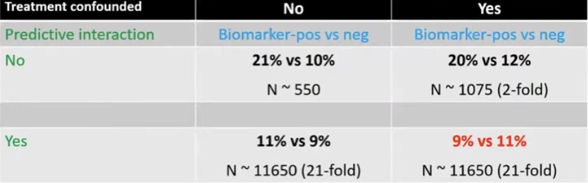
  • Differences in the IBTR rates between these studies:
    • May be due to the effect of tamoxifen use:
      • Which was not controlled in either trial
      • Which is known to reduce the risk of IBTR in combination with RT after BCT as shown by the NSABP B-24 trial
  • Together, the results of both RTOG 9804 and E5194:
    • Support the individualized treatment of DCIS:
      • Based on identification of patients at low risk for IBTR determined by:
        • Pathologic and clinical features that may omit RT
  • A 12-year follow-up study of RTOG 9804 demonstrated:
    • That the 12-year cumulative incidence of local recurrence (LR) was:
      • 2.8% with RT and 11.4% with observation alone
    • The 12-year cumulative incidence of invasive LR was:
      • 1.5% with RT and 5.8% with observation alone
  • References:
    • Wapnir IL, Dignam JJ, Fisher B, Mamounas EP, Anderson SJ, Julian TB, et al. Long-term outcomes of invasive ipsilateral breast tumor recurrences after lumpectomy in NSABP B-17 and B-24 randomized clinical trials for DCIS. J Natl Cancer Inst. 2011;103(6):478-488.
    • EORTC Breast Cancer Cooperative Group; and EORTC Radiotherapy Group; Bijker N, Meijnen P, Peterse JL, et al. Breast-conserving treatment with or without radiotherapy in ductal carcinoma-in-situ: ten-year results of European Organisation for Research and Treatment of Cancer randomized phase III trial 10853—a study by the EORTC Breast Cancer Cooperative Group and EORTC Radiotherapy Group. J Clin Oncol. 2006;24(21):3381-3387.
    • McCormick B, Winter K, Hudis C, Kuerer HM, Rakovitch E, Smith BL, et al. RTOG 9804: a prospective randomized trial for good-risk ductal carcinoma in situ comparing radiotherapy with observation. J Clin Oncol. 2015;33(7):709-715.
    • Solin LJ, Gray R, Hughes L, Wood WC, Lowen MA, Badve SS, et al. Surgical excision without radiation for ductal carcinoma in situ of the breast: 12-year results from the ECOG-ACRIN E5194 Study. J Clin Oncol. 2015;33(33):3938-3944.
    • 5. McCormick B. Randomized trial evaluating radiation following surgical excision for “good risk” DCIS: 12-year report from NRG/RTOG 9804. Int J Radiat Oncol Biol Phys. 2018;102(5):1603.

#Arrangoiz #BreastSurgeon #CancerSurgeon #SurgicalOncologist #BreastCancer #DCIS #RadiationTherapy #Miami #Mexico #Teacher

Occult Primary Breast Cancer

  • Three important conclusions are agreed upon regarding this clinical entity:
    • Prognosis of occult primary breast cancer is the same or slightly better than women with classic stage IIA disease (cT0, cN1, cM0)
    • An exhaustive workup for the non-breast primary:
      • Is usually not fruitful
    • Treatment of the breast in some manner:
      • Decreases the risk of local failure over time
  • Occult primary breast cancer was first recognized by William Halsted:
    • Who described three patients presenting with axillary tumors that were eventually found to represent breast cancer
  • In modern series:
    • Occult breast cancer accounts for:
      • 0.1% to 0.8% of all newly diagnosed breast cancers:
        • The incidence has not decreased with improvements in breast imaging
    • Differential diagnosis:
      • In general:
        • Palpable axillary nodes are more often related to benign rather than malignant disorders:
          • However, when cancer is identified:
            • The most common tumor causing axillary lymphadenopathy is:
              • Breast cancer
  • In several series:
    • The incidence of breast cancer in mixed populations of men and women with metastatic axillary adenopathy:
      • Is 50% or higher:
        • The vast majority are women:
          • Although occult primary breast cancer has been reported in men:
            • It is very rare
  • Other neoplasms that may present with axillary nodal involvement are:
    • Lymphomas
    • Melanomas
    • Sarcomas
    • Thyroid cancers
    • Skin cancers
    • Lung cancers
    • Less often:
      • Uterine, ovarian, sweat gland, or gastric cancers
  • In approximately 30% of cases:
    • The primary site is never identified
  • Initial Diagnostic Workup:
    • Biopsy:
      • The first step in the diagnostic workup of a patient with unexplained axillary adenopathy is:
        • A biopsy
      • Besides standard light microscopic examination of hematoxylin and eosin-stained sections:
        • Other techniques such as:
          • Immunohistochemistry and sometimes electron microscopy can help to narrow the differential diagnosis:
            • Immunohistochemistry:
              • The pathologic examination of a biopsy specimen for an isolated axillary lymph node metastatic adenocarcinoma or poorly differentiated carcinoma in a woman should include immunohistochemical staining for the following markers:
                • Carcinoembryonic antigen (CEA)
                • Cytokeratins 7 and 20
                • Estrogen receptor (ER) and progesterone receptor (PR)
                • Gross cystic disease fluid protein-15:
                  • GCDFP-15:
                    • Is identified by staining with the monoclonal antibody BRST2
                • Mammaglobin
                • Thyroid transcription factor (TTF-1)
                • CA125
              • Men:
                • Should have routine staining for prostate cancer markers as well
          • While none of these markers is sufficiently sensitive or specific to be used alone, certain patterns of expression favor the diagnosis of an occult breast cancer:
            • Positive staining for:
              • CEA, CK7, ER/PR, mammaglobin, CA125, and BRST2
            • Negative staining for:
              • CK20 and TTF-1
            • CEA is a sensitive marker for:
              • Adenocarcinomas of the breast, lung, and gastrointestinal tract:
                • But does not help to distinguish among these sites of origin
            • On the other hand, differential expression of cytokeratins (CKs) can assist in this differentiation:
              • CK20 is a low molecular weight cytokeratin:
                • That is normally expressed in the gastrointestinal epithelium, urothelium, and in Merkel cells
    • CK7 is expressed by tumors of the:
      • Lung, ovary, endometrium, and breast:
        • Not in the lower gastrointestinal tract
      • The pattern of CK20 and CK7 may be particularly helpful in suggesting a primary site:
        • The presence of CK7 and absence of CK20:
          • Favors a diagnosis of breast cancer
      • TTF-1:
        • Is rarely positive in breast cancers:
          • While it is positive in 70% to 80% of non-squamous cancers arising in the lung
      • CA-125:
        • Is commonly positive in ovarian carcinomas:
          • But is positive in about 10% of breast cancers
  • ER /PR:
    • Its presence in an axillary node, particularly in conjunction with other compatible IHC findings:
      • Lends support to a diagnosis of an occult breast primary
    • Although positive staining for ER and / or PR supports a possible diagnosis of breast cancer:
      • These markers are nonspecific and they may also be expressed in:
        • Ovarian, uterine, lung, stomach, thyroid, and hepatobiliary cancers:
          • However:
            • ER/PR staining of an axillary node is compelling evidence of a primary breast cancer
  • Other breast cancer-specific IHC stains are:
    • BRST2 (for GCDFP) and mammaglobin:
      • BRST2 is positive in 65% to 80% of cases:
        • Is relatively specific for breast cancer:
          • Rarely, it is positive in:
            • Skin adnexal tumors, endometrial cancers, and salivary gland tumors
    • Mammaglobin is more sensitive, it is less specific for breast cancer:
      • Gynecologic, lung, urothelial, thyroid, colon and hepatobiliar tumors may stain positive:
        • Both stains are thus typically used together
    • HER2 immunostaining:
      • Is not generally useful for the differential diagnosis of a carcinoma arising in the axillary nodes as it lacks specificity:
        • Furthermore, only 18% to 20% of breast cancers overexpress this protein:
          • However, assay for HER2 overexpression by IHC or fluorescent in situ hybridization (FISH) is a routine component of the evaluation of all breast cancers:
            • As it permits the identification of those women who are most likely to respond to treatments targeting HER2 (eg, the therapeutic monoclonal antibody trastuzumab)
  • Modified radical mastectomy:
    • Has been the traditional surgical treatment for many years
  • Previously, the primary breast cancer was found in the mastectomy specimen:
    • 40% to 80% of the time, but with the advent of much better mammography and with breast MRI, this rate is much lower:
      • However, what was true then and still holds today:
        • Is that no treatment to the breast itself results in an unacceptably high local recurrence rate
      • An alternative to a modified radical mastectomy:
        • Is complete ALND followed by whole-breast irradiation
          • Axillary dissection:
            • Provides local control while also fine tuning staging
          • Theoretically the whole-breast radiation:
            • Should control any subclinical disease in the breast not detected on imaging
            • Primary radiation to the breast, axilla, and supraclavicular area without any surgery of the breast or axilla:
              • Results in higher local and regional recurrence compared to surgery and radiation combined
              • Axillary node dissection and whole-breast irradiation:
                • Has been found to have equivalent survival as a modified radical mastectomy:
                  • A recent meta-analysis of seven studies and more than 240 patients with occult primary breast cancers (0.3% to 0.8% of all breast cancers):
                    • Found 39% were treated with ALND and radiation while 47% had modified radical mastectomy and 15% had ALND alone:
                      • With a mean follow-up of 5 years the study found no difference in local regional recurrence (12.7% vs 9.8%), distant metastasis (7.2% vs 12.7%), or mortality (9.5% vs 17.9%) between ALND and radiation vs modified radical mastectomy (all p>0.16)
                      • ALND with radiation was superior to ALND alone in terms of local regional recurrence (12.7% vs
                        34.3%, p < 0.01) and trended towards improved survival but this was not statistically significant
                        (P=0.09)
  • References:
    • Barton SR, Smith IE, Kirby AM, Ashley S, Walsh G, Parton M. The role of ipsilateral breast
      radiotherapy in management of occult primary breast cancer presenting as axillary
      lymphadenopathy. Eur J Cancer. 2011;47:2099-2106. PMID: http://www.ncbi.nlm.nih.gov/pubmed/21658935
    • Dockery MB, Gray HK, Pierce EH. Surgical significance of isolated axillary adenopathy. Ann
      Surg. 1957;145:104-107. http://www.ncbi.nlm.nih.gov/pubmed/13395289
    • Macedo FI, Eid JJ, Flynn J, Jacobs MJ, Mittal VK. Optimal surgical management for occult
      breast carcinoma: a meta-analysis. Ann Surg Oncol. 2016;23:1838-https://www.ncbi.nlm.nih.gov/pubmed/26832884
    • Rueth NM, Black DM, Limmer AR, et al. Breast conservation in the setting of contemporary multimodality treatment provides excellent outcomes for patients with
      occult primary breast cancer. Ann Surg Oncol. 2015;22:90-95. [epub ahead of
      print]. http://www.ncbi.nlm.nih.gov/pubmed/25249256

Margins after Breast Conserving Surgery in Invasive and Noninvasive Breast Cancer

  • In 2013 the SSO and ASTRO convened a multidisciplinary expert panel to review the available evidence regarding margin width and IBTR in patients with invasive cancer having breast conservation therapy
  • Meta-analysis and secondary data from prospective and retrospective trials led them to conclude that positive margins (defined as ink on invasive cancer):
    • Is associated with at least a 2-fold increase in IBTR
  • Among patients with negative margins:
    • margin width of no ink on tumor represented the optimal margin width to minimize the risk of IBTR:
      • Notably the routine practice of obtaining wider negative margins than no ink on tumor is not indicated
  • While younger age:
    • Is associated with both increased IBTR after breast-conserving therapy as well as increased local chest wall relapse after mastectomy:
      • There is no evidence that increased margin width (over no ink on tumor) nullifies this increased risk of IBTR in younger patients
  • In 2016, margin guidelines related to the treatment of non-invasive breast cancer (DCIS) in the setting of breast conservation therapy were developed by the SSO, ASTRO, and ASCO in a similar manner
  • A consensus statement released by a multidisciplinary panel included the optimal margins for pure ductal carcinoma in situ (DCIS) and mixed tumors (invasive and non-invasive components within the same tumor) in the setting of breast conservation:
    • Results from the meta-analysis showed that a 2 mm margin decreases the risk of IBTR in pure DCIS compared to closer negative margins
      • This differs from the previous margin recommendation for invasive cancer, which remains no ink on tumor
    • However, in the setting of mixed tumors (invasive cancer with a DCIS component) the recommendation for negative margins remains no ink on tumor, as patients with mixed disease are treated as invasive cancer and therefore receive systemic therapy more often than pure DCIS patients:
      • In the setting of DCIS with micro-invasion (no focus of invasive disease larger than 0.1 cm):
        • The multidisciplinary panel recommends a 2 mm margin, as these lesions have similar rates of IBTR as pure DCIS
  • References
    • Moran MS, Schnitt SJ, Giuliano AE, Harris JR, Khan SA, Horton J, et al. Society of Surgical Oncology-American Society for Radiation Oncology consensus guideline on margins for breast-conserving surgery with whole-breast irradiation in stages I and II invasive breast cancer. Int J Radiat Oncol Biol Phys. 2014;88(3):553-564.
    • Morrow M, Van Zee KJ, Solin LJ, Houssami N, Chavez-MacGregor M. et al. Society of Surgical Oncology-American Society for Radiation Oncology-American Society of Clinical Oncology Consensus Guideline on margins for breast-conserving surgery with whole-breast irradiation in ductal carcinoma in situ. J Clin Oncol. 2016;34(33):4040-4046.
#Arrangoiz #Doctor #Surgeon #SurgicalOncologist #CancerSurgeon #BreastSurgeon #MountSinaiMedicalCenter #MSMC #Miami #Mexico #DCIS #Margins #BreastCancer

Eastern Cooperative Oncology Group (ECOG) E5194 Study

  • To investigate the risk of ipsilateral breast events (IBEs) in patients with DCIS treated with local excision without radiation:
    • The ECOG and North Central Cancer Treatment Group (NCCTG):
      • Conducted a prospective trial between 1997 and 2002 known as the E5194 study
      • Patients were stratified into two groups based on grade:
        • Low- and intermediate-grade DCIS measuring 2.5 cm or smaller
        • High-grade DCIS measuring 1 cm or smaller
      • Margin widths of 3 mm or wider were required along with no residual calcifications on postoperative mammograms
      • The low- and intermediate-grade DCIS group:
        • Had a 12-year IBE rate of 14.4%
      • The high-grade DCIS group:
        • Had a 12-year IBE rate of 24.6% (p=0.003)
      • Larger tumor size:
        • Was also found to be statistically significantly associated with developing an IBE (P = .01)
      • The risks of developing an IBE for either DCIS or invasive cancer:
        • Increased over time through 12 years of follow-up:
          • Without plateau
  • Current literature reports a 50% decrease in local recurrence with radiotherapy after surgical excision of DCIS:
    • However, we continue to look for low risk subsets of patients with DCIS:
      • In whom the absolute benefit of radiation therapy of lumpectomy may be small
  • The DCIS score (12-gene signature):
    • Was developed from tissue samples from the E5194 study:
      • With the goal of identifying low and high-risk subsets of patients with DCIS following lumpectomy alone
    • This assay utilized quantitative reverse transcriptase-polymerase chain reaction (RT-PCR):
      • From tumor specimens from 327 patients with DCIS treated with surgical excision without radiation from the E5194 study
    • The DCIS score of 0 to 100 (low less than 39, intermediate 39 to 54, high ≥ 55):
      • Was then designed to predict the recurrence of IBE overall, as well as DCIS or invasive cancer recurrence
      • The DCIS score correlated with 10-year IBE risk of:
        • 10.6% in the low-risk group
        • 26.7% in the intermediate-risk group
        • 25.9% in the high-risk group
      • Young age and larger tumor size:
        • Also were found to be independent predictors of recurrence
      • So, this test may be most beneficial in post-menopausal women with small tumors
  • References:
    • Solin LJ, Gray R, Hughes LL, Wood WC, Lowen MA, Badve SS, et al. Surgical excision without radiation for ductal carcinoma in situ of the breast: 12-year results from the ECOG-ACRIN E5194 Study. J Clin Oncol. 2015;33(33):3938-3944.
    • Solin LJ, Gray R, Baehner FL, Butler SM, Hughes LL, Yoshizawa C, et al. A multigene expression assay to predict local recurrence risk for ductal carcinoma in situ of the breast. J Natl Cancer Inst. 2013;105(10):701-710.

#Arrangoiz #BreastSurgeon #CancerSurgeon #SurgicalOncologist #BreastCancer #DCIS #ECOG5194 #Teacher #Surgeon #Miami #MountSinaiMedicalCenter #Mexico

Idiopathic Granulomatous M. astitis (IGM)

  • Idiopathic granulomatous mastitis (IGM):
    • Is a rare, benign, chronic inflammatory condition of the breast occurring mostly in non-white women of childbearing age
  • In the United States:
    • It’s more commonly seen in the Hispanic population
  • Idiopathic granulomatous mastitis often presents with:
    • A unilateral, hard, discrete tender mass
      • Nonetheless, it may also present with:
        • Multiple indurated inflammatory masses
      • A non-tender mass
        • With pain as a solitary symptom
  • In many cases, a central mass is accompanied by:
    • Multiple simultaneous areas of peripheral infection with abscesses
    • Erythema and ulceration of the overlying skin, and / or fistula formation
  • Pathologic features of IGM include:
    • Non-caseating granulomatous inflammation with necrosis and giant cell formation on a background of neutrophils all surrounding a breast lobule
  • Given the overlap between clinical and radiographic features of IGM and other disease processes:
    • Histopathologic evaluation has emerged as the gold standard for diagnosis of IGM
  • There is no formal consensus regarding the appropriate treatment for patients diagnosed with IGM:
    • The available treatment options include:
      • Expectant management
      • Steroid therapy
      • Immunomodulators such as:
        • Methotrexate and azathioprine
      • Limited or wide surgical excision
      • Antibiotic treatment in the setting of superimposed infection
  • References:
    • Mohammed S., Statz A, Lacross JS, Lassinger BK, Contreras A, Gutierrez C, et al. Granulomatous mastitis: a 10-year experience from a large inner city county hospital. J Surg Res. 2013;184(1):299-303.
    • Pandey TS, Mackinnon JC, Bressler L, Millar A, Marcus EE, Ganschow PS. Idiopathoic granulomatous mastitis – a prospective study of 49 women and treatment outcomes with steroid therapy. Breast J. 2014;20(3):258-266.
    • Oran ES, Gurdal SO, Yankol Y, Öznur M, Calay Z, Tunacı M, et al. Management of idiopathic granulomatous mastitis diagnosed by core biopsy: a retrospective multicenter study. Breast J. 2013;19(4):411-418.
    • Akbulut S, Yilmaz D, Bakir S. Methotrexate in the management of idiopathic granulomatous mastitis: review of 108 published cases and report of four cases. Breast J. 2011;17(6):661-668.

#Arrangoiz #BreastSurgeon #BreastCancer #CancerSurgeon #IGM #IdiopathicGranulomatousMastitis #Miami #Mexico

What is Head and Neck Surgery?:

  • What is Head and Neck Surgery?
    • It is a surgical sub-specialty that deals mainly with benign and malignant tumors of the head and neck region, including:
      • The scalp, facial region, eyes, ears, nose, nasal fossae, paranasal sinuses, oral cavity, pharynx (nasopharynx, oropharynx, hypopharynx), larynx (supraglotic larynx, glottis larynx, subglotic larynx), thyroid gland, parathyroid gland, salivary glands (parotid glands, submandibular glands, sublingual glands, minor salivary glands), soft tissues of the neck, skin of the head and neck region
        • The head and neck surgeon’s work area:
          • Does not cover tumors or diseases of the brain and other areas of the central nervous system or those of the cervical spine:
            • This is the neurosurgeon field
    • Among the diagnostic procedures performed by the head and neck surgeon,  are the following:
      • Nasopharyngolaryngoscopy:
        • Performed to examine, evaluate and, possibly perform a biopsy of the oral cavity, pharynx and larynx
    • The surgeries most commonly performed by the head and neck surgeon are:
      • Total or near total thyroidectomies
      • Hemithryoidectomies (lobectomies)
      • Comprehensive neck dissections
      • Selective neck dissections
      • Maxillectomies:
        • Total maxillectomy
        • Subtotal maxillectomy
        • Infrastructure maxillectomy
        • Suprastructure maxillectomy
        • Medial maxillectomy
      • Mandibulectomy:
        • Segmental
        • Marginal
      • Tracheostomy
      • Salivary gland surgeries:
        • Parotid gland operations:
          • Limited superficial parotidectomy with identification and preservation of the facial nerve
          • Superficial parotidectomy with identification and preservation of the facial nerve
          • Near total parotidectomy with identification and preservation of the facial nerve
          • Total parotidectomy
        • Submandibular gland resection
        • Sublingual gland resection
      • Resection of tumors of the oral cavity:
        • Glossectomy
        • Resection of the floor of the mouth tumors
      • Resection of tumors of the pharynx
      • Resection of tumors of the larynx
      • Split-thickness skin grafts
      • Full-thickness skin grafts
      • Sentinel lymph node mapping and sentinel lymph node biopsy
      • Resection of malignant skin tumors (BCC, SCC, melanoma) of the head and neck region
  • The formation of the head and neck surgeon includes mastering the following subjects:
    • Surgical Anatomy
    • History and Basic Principles of Head and Neck Surgery
    • Epidemiology, Etiology, and Pathology of Head and Neck Diseases
    • Diagnostic Radiology of the Head and Neck Region
    • Tumors of the Scalp, Skin and Melanoma
    • Eyelids and Orbit
    • Nasal Cavity and Paranasal Sinuses
    • Skull Base and Temporal Bone
    • Lips and Oral Cavity
    • Pharynx and Esophagus
    • Larynx and Trachea
    • Cervical Lymph Nodes
    • Thyroid and Parathyroid Glands
    • Salivary Glands
    • Neurogenic Tumors and Paragangliomas
    • Soft Tissue Tumors
    • Bone Tumors and Odontogenic Lesions
    • Reconstructive Surgery
    • Oncologic Dentistry and Maxillofacial Prosthetics
    • Principles of Radiation Oncology
    • Principles of Chemotherapy
    • Molecular Oncology, Genomics and Immunology
    • Nutrition
    • Biostatistic
  • Rodrigo Arrangoiz MS, MD, FACS, FSSO a head and neck surgeon / thyroid surgeon / parathyroid surgeon / complex surgical oncologist
prof_739_20190417135234
  • Rodrigo Arrangoiz MS, MD, FACS, FSSO:
    • Is a member of the American Head and Neck Society
img_4750
  • He is a member of the American Thyroid Association:
Unknown

Training:

• General surgery:

• Michigan State University:

• 2005 al 2010

• Surgical Oncology / Head and Neck Surgery / Endocrine Surgery:

• Fox Chase Cancer Center (Filadelfia):

• 2010 al 2012

• Masters in Science (Clinical research for health professionals):

• Drexel University (Filadelfia):

• 2010 al 2012

• Surgical Oncology / Head and Neck Surgery / Endocrine Surgery:

•International Federation of Head and Neck Societies / Memorial Sloan Kettering Cancer Center:

• 2014 al 2016

#Arrangoiz

#Teacher

#Surgeon

#Cirujano

#ThyroidExpert

#ThyroidSurgeon

#CirujanodeTiroides

#ExpertoenTiroides

#ExpertoenParatiroides

#Paratiroides

#Hiperparatiroidismo

#Hyperparathyroidsim

#CancerdeTiroides

#ThyroidCancer

#PapillaryThyroidCancer

#SurgicalOncologist

#CancerSurgeon

#CirujanodeCancer

#HeadandNeckSurgeon

#CirugiaEndocrina

#EndocrineSurgery

#CirujanodeTumoresdeCabezayCuello

#OralCavityCancer

#Melanoma

#MountSinaiMedicalCenter

#Miami

#MSMC

#Mexico

COMET Trail in Ductal Carcinoma In Situ (DCIS)

  • Key Points:
    • Question:
      • What is the short-term safety of an active monitoring approach vs guideline-concordant care (surgery with or without radiation therapy):
        • For hormone receptor–positive, grade 1 or grade 2 breast ductal carcinoma in situ?
    • Findings:
      • In this prospective randomized clinical trial of:
        • 957 participants
      • The 2-year Kaplan-Meier cumulative rate of ipsilateral invasive cancer was 5.9% in the guideline-concordant care group vs 4.2% in the active monitoring group:
        • A difference of −1.7% (upper limit of the 95% CI, 0.95%):
          • Indicating that active monitoring is not inferior to guideline concordant care
      • Meaning:
        • These data support the short-term safety of active monitoring compared with guideline-concordant care in patients with low-risk ductal carcinoma in situ
  • Abstract:
    • Importance:
      • Active monitoring for low-risk ductal carcinoma in situ (DCIS) of the breast has been proposed as an alternative to guideline-concordant care, but the safety of this approach is unknown
    • Objective :
      • To compare rates of invasive cancer in patients with low-risk DCIS receiving active monitoring vs guideline-concordant care
    • Design, Setting, and Participants:
      • Prospective, randomized noninferiority trial enrolling 995 women aged 40 years or older with a new diagnosis of hormone receptor–positive grade 1 or grade 2 DCIS without invasive cancer at 100 US Alliance Cancer Cooperative Group clinical trial sites from 2017 to 2023
    • Interventions:
      • Participants were randomized to receive active monitoring (follow-up every 6 months with breast imaging and physical examination; n = 484) or guideline-concordant care (surgery with or without radiation therapy; n = 473)
    • Main Outcomes and Measures :
      • The primary outcome was 2-year cumulative risk of ipsilateral invasive cancer diagnosis, according to planned intention-to-treat and per-protocol analyses, with a noninferiority bound of 5%
    • Results:
      • The median age of the 957 participants analyzed was 63.6 (95% CI, 55.5-70.5) years in the guideline-concordant care group and 63.7 (95% CI, 60.0-71.6) years in the active monitoring group
      • Overall, 15.7% of participants were Black and 75.0% were White
      • In this prespecified primary analysis, median follow-up was 36.9 months; 346 patients had surgery for DCIS, 264 in the guideline-concordant care group and 82 in the active monitoring group
      • Forty-six women were diagnosed with invasive cancer, 19 in the active monitoring group and 27 in the guideline-concordant care group
      • The 2-year Kaplan-Meier cumulative rate of ipsilateral invasive cancer was 4.2% in the active monitoring group vs 5.9% in the guideline-concordant care group, a difference of −1.7% (upper limit of the 95% CI, 0.95%):
        • Indicating that active monitoring is not inferior to guideline-concordant care
      • Invasive tumor characteristics did not differ significantly between groups
    • Conclusions and Relevance:
      • Women with low-risk DCIS randomized to active monitoring did not have a higher rate of invasive cancer in the same breast at 2 years compared with those randomized to guideline-concordant care

Mondor Disease, Axillary Web Syndrome, Zuska’s Disease and Fat Necrosis

  • Mondor’s disease:
    • Is a self-limiting superficial thrombophlebitis of the breast
    • The etiology is not always clear:
      • But it has been reported to be associated with:
        • Trauma
        • Core biopsy
        • Breast surgery
        • Cancer
        • Radiation treatment
        • Underwire bras
        • Oral contraceptives
    • It typically presents as a:
      • Vertically oriented, tender cord on the breast:
        • Which becomes more prominent when the arm is raised and can be associated with skin retraction
    • It can be treated with:
      • Warm compresses
      • Aspirin, or non-steroidal anti-inflammatory drugs:
        • It usually resolves within 4 to 6 weeks
  • Axillary web syndrome:
    • Can also present as a palpable cord, but is often found in the axilla:
      • Can extend to involve the medial aspect of the ipsilateral arm down to the antecubital fossa
    • It is associated with limitations in shoulder mobility
  • Fat necrosis:
    • Can cause a palpable mass with skin retraction:
      • But it does not generally conform to the distribution of a superficial vein
  • Zuska’s disease:
    • Is a condition consisting of:
      • Chronic subareolar abscesses and fistulae
    • Caused by squamous metaplasia and keratin plugging of the breast ducts, and it is not related to Mondor’s disease
  • References
    • Pasta V, D’Orazi V, Sottile D, Del Vecchio L, Panunzi A, Urciuoli P. Breast Mondor’s disease: diagnosis and management of six new cases of this underestimated pathology. Phlebology. 2015;30(8):564-568.
    • Salemis NS, Vasilara G, Lagoudianakis E. Mondor’s disease of the breast as a complication of ultrasound-guided core needle biopsy: management and review of the literature. Breast Dis. 2015;35(1):73-76.
    • Shetty MK, Watson AB. Mondor’s disease of the breast: sonographic and mammographic findings. AJR Am J Roentgenol. 2001;177(4):893-896.
    • Tilley A, Thomas-MacLean R, Kwan W. Lymphatic cording or axillary web syndrome after breast cancer surgery. Can J Surg. 2009;52(4):E105-E106.
    • Lannin DR. Twenty-two year experience with recurring subareolar abscess and lactiferous duct fistula treated by a single breast surgeon. Am J Surg. 2004;188(4):407-410.

#Arrangoiz #BreastSurgeon #BreastCancer #CancerSurgeon #SurgicalOncology #Doctor #Surgeon #MondorDisease #AxillaryWebNecrosis #FatNecrosis #ZuskaDisease #MountSinaiMedicalCenter #MSMC #Miami #Mexico

Omitting Radiation Therapy in Ductal Carcinoma In Situ

  • A survey (Jagsi, 2010):
    • Demonstrated that 95% of women with breast cancer and strong indications for post-lumpectomy radiation:
      • Went on to receive it:
        • But rates of post-lumpectomy radiation therapy use have been shown to vary:
          • Depending on the region of the country that the patient lives in
          • The age of the patient
          • The disease being treated (DCIS vs Invasive Disease)
  • Among patients who undergo BCT for DCIS:
    • Only 50% are estimated to receive adjuvant radiation
  • Many patients choose mastectomy over breast-conserving surgery for DCIS:
    • Because they are not able or willing to complete 6 weeks of daily radiation therapy:
      • Secondary to social or health considerations
  • Other patients who are candidates for breast-conserving surgery:
    • Choose to undergo a mastectomy:
      • Because of concerns about postradiation complications
  • Breast-conserving surgery alone (i.e., without radiation therapy):
    • May be sufficient in a select subgroup of patients with DCIS
  • Initial data that supported the use of breast-conserving surgery alone in the treatment of DCIS came from a study by Lagios et al. (1989):
    • In which 79 patients with mammographically detected DCIS underwent margin-negative excision alone
    • After a follow-up time of 124 months:
      • The local recurrence rate was 16% overall, specifically:
        • 33% for the subgroup of patients with high-grade lesions and comedo necrosis versus only 2% for the patients with low- or intermediate-grade lesions
  • The USC / VNPI score can be a helpful tool in clinical decision making:
    • But even though margin width is an independent prognostic factor for recurrence using the USC / VNPI score:
      • It is unlikely that margin width alone can identify the patients with DCIS treated with breast conservation for whom radiation therapy can be safely omitted
  • In a retrospective analysis of 469 patients with DCIS who underwent breast conservation with margins that were at least 10 mm, Silverstein et al. (1999):
    • Did not detect a lower recurrence rate:
      • When postoperative radiation therapy was employed
  • In contrast, even on reanalysis of the NSABP B-17 data:
    • All patient cohorts benefited from radiation therapy:
      • Regardless of the clinical or mammographic tumor characteristics
  • Furthermore, Wong et al. (2003, 2014):
    • Reported the early termination of a prospective single-arm trial conducted at the Dana-Farber / Harvard Cancer Center:
      • In which radiation therapy was omitted in patients with grade 1 to 2 DCIS that was no more than 25 mm and excised with 10 mm or greater margins:
        • At a median follow-up of 3.3 years:
          • The number of local recurrences observed was 2.5% per patient-year:
            • Corresponding to a 5-year rate of 12.5%
  • In 2010, Rudloff and colleagues at Memorial Sloan Kettering Cancer Center:
    • Published a multivariable nomogram:
      • To estimate risk for local recurrence in women with DCIS treated with breast-conserving surgery
    • The nomogram incorporates commonly available factors that have previously been shown to affect risk of ipsilateral breast tumor recurrence, these include:
      • Age at diagnosis
      • Family history
      • Type of patient presentation:
        • Radiologic or clinical
      • Nuclear grade
      • Necrosis, margins
      • Number of excisions
      • Receipt of radiation and / or adjuvant endocrine therapy
    • The nomogram calculates an actual, individualized estimate of absolute risk of ipsilateral breast tumor recurrence at 5 or 10 years:
      • Which can be weighed against the use of available adjuvant treatment options
  • There are two large, prospective, observational studies:
    • Designed to investigate the role of observation versus radiation therapy after breast-conserving therapy in patients with DCIS
    • As mentioned earlier, Wong and colleagues at Harvard:
      • Conducted a single-arm, phase III observational study examining long-term outcomes in women with small (≤ 2.5 cm), low- and intermediate-grade DCIS who were treated with lumpectomy and margins ≥ 1 cm and did not receive adjuvant tamoxifen or radiation:
      • With a median follow-up of 11 years:
        • 13% (19 of 143) of patients experienced local recurrence:
          • Approximately two-third of which were DCIS
    • In the Eastern Cooperative Oncology Group–American College of Radiology Imaging Network (ECOG-ACRIN; formerly known as the Eastern Cooperative Oncology Group) Cancer Research Group E5194 study:
      • Patients with low- or intermediate-grade DCIS smaller than 25 mm (cohort 1), or high-grade DCIS smaller than 10 mm (cohort 2), with excisional margins of at least 3 mm, underwent breast-conserving surgery without radiation therapy:
        • 30% of patients received tamoxifen
      • At 12 years:
        • 14.4% of the participants in cohort 1 experienced an in-breast even while 24.6% of those in cohort 2 experienced an in-breast event (p = 0.0003), and this difference was driven by a statistically significant difference (p = 0.02) in noninvasive recurrence
        • In addition, membership in cohort 2 and larger tumor size were both found to be associated with increased likelihood of recurrence (Solin, 2015)
  • Finally, in patients over 70:
    • There is increasing evidence that radiation therapy:
      • Depending on the aggressiveness of the DCIS and the expected life expectancy of the patient:
        • Can be omitted on a case-by-case basis

#Arrangoiz #BreastSurgeon #CancerSurgeon #SurgicalOncology #Surgeon #Teacher #BreastCancer #BreastExpert #MSMC #MountSinaiMedicalCenter #Miami #Mexico #DCIS #DuctalCarcinomaInSitu #OmissionofRadiation

DCIS: Implications of ER, PR, and HER2 Expression

  • Prognostic role of estrogen receptor (ER) and HER2 in DCIS:
    • In observational studies:
      • ER status – 5 of 26 studies found a statistically significant lower risk of ipsilateral breast tumor recurrence (IBTR) in ER positive cases
      • HER2 status – 10 out of 27 studies reported a significant increase in the risk of recurrences to be associated with HER2 expression
    • Limitations of these observational studies were:
      • Small sample size (events) in the majority of the studies
      • Selection bias
      • Treatment-related confounding:
        • ER expression is inversely associated and HER2 expression is positively associated with:
          • Adverse histologic features in DCIS
        • Therefore, ER negative or HER2 amplification in DCIS:
          • Is more likely to receive adjuvant treatment that ER positive or HER2 negative DCIS when ER or HER2 status is not known:
            • Potentially masking the true association
        • The probability of masking of the true association:
          • Increases greatly if the biomarker also has predictive characteristics
  • How to eliminate treatment-related confounding:
    • The study population should have random treatment allocation:
      • Cohorts from randomized controlled trials
    • Case-control studies matching by treatment:
      • Does not permit investigation of predictive characteristics of the biomarker
    • Multivariable / adjusted analysis:
      • Power remains an issue
  • Biomarker cohort study:
  • UK, Australia and New Zealand DCIS trial Cuzick J et al Lancet Oncol. 2011; Houghton J eta al Lancet 2003):
    • 2X2 randomized trial comparing the effectiveness of radiotherapy and tamoxifen in reducing recurrences in patients with complete locally excised DCIS
    • # of patients 1694
    • The 2X2 factorial design permits investigation pertaining to both adjuvant treatments in DCIS
    • After a median follow-up of 12.7 years, there have been 162 invasive and 197 DCIS events in these patients:
      • 17 unknown
      • Total 376
  • In the study they observed that in patient with ER positive DCIS:
    • They identified areas within the ducts that were ER negative in the same lesion
Multi-clonal ER Expression: On the right side panel you can see an ER negative duct with an ER positive duct adjacent to it.
Multi-clonal ER Expression
  • In these study 11% of patients were identified to have multi-clonal DCIS:
    • Clonal method:
  • Estrogen receptor (ER) expression and recurrence:
    • ER negative (multi-clonal) DCIS is associated with:
      • A five fold increase of in situ ipsilateral breast event
      • A three fold increase in overall ipsilateral breast event
      • Invasive ipsilateral breast event is not statistically increased
mOR; Matched Odds Ratio, IBE: Ipsilateral breast event, I-IBE: Invasive IBE, In situ IBE
  • The results show that the clonal method:
    • Is superior to the standard method in predicting IBE and DCIS-IBE
  • Progesterone receptor (PgR):
    • Was not significantly associated with recurrence in ER positive DCIS
    • It was not an independent predictor in multivariable models
    • Inclusion of PGR did not significantly improve multivariable models
  • HER2 expression and recurrence:
    • HER2 positively was identified in 55% of the cases of DCIS:
      • Compared to invasive breast cancer which is around 15% to 20%
    • The expression of HER2 was associated with a two fold increase in IBE and in situ IBE
Comparison is HER2 3+ vs. 0, 1+, 2+
Univariable Analysis N = 713; Multivariable Analysis N = 612
IBE: Ipsilateral breast event, I-IBE: Invasive IBE, In situ IBE
  • HER2 status (post-ERBB2-reflex test) and recurrence:
    • HER2 status (ERBB2 reflexes) as a predictor of recurrence:
      • Is associated with nearly a three fold increase in IBE and in situ IBE
      • Is associated with an increase risk of I-IBE but it did not reach statistical significance
HER2 status (ERBB2 reflexes) is assigned after ERBB 2 mRNA expression a reflex test.
Comparison of HER2 positive (3+ of IHC 2+ and ERBB2 mRNA expression > 1.1007 vs. HER2 negative (0, 1+ or 2+ with ERBB2 mRNA expression </= 1.1007
  • Radiation therapy benefit:
    • In HER2 positive disease was much larger as compared to HER 2 negative disease (statistically significant difference)
    • In HER2 negative DCIS radiation therapy reduced events by 53% compared to 80% in HER2 positive disease
Kaplan Meir curves showing the difference in benefit of radiation therapy in HER2 positive disease
  • In conclusion:
    • ER is a strong prognostic factor:
      • IBE mOR 3.33 for clinical method
    • HER2 is a strong prognostic factor:
      • IBE HR 2.84 for ERBB2-reflexes status
    • Radiotherapy benefit greater in HER2 positive DCIS:
      • HR 0.21 vs 0.47:
        • En by greater benefit in reducing DCIS-IBE
    • ER and HER2 evaluation should be routinely carried out?
  • Endocrine therapy considerations:
    • NSABP-B24:
      • ER is a predictor of tamoxifen benefit
      • Ipsilateral ER status is not a predictor of contralateral breast cancer (CBC) risk or tamoxifen benefit in preventing CBC
  • Combining the data from the RTOG 9804 trial and the UK/ANZ DCIS trial in the low risk DCIS (< 10 mm):
    • Can we recommend radiation therapy only to ER – negative or HER2 – positive DCIS?
      • The effect size and predictive benefit are an excellent fit to the RTOG 9804 results if 15% to 20% of patients in the trial were HER2 positive (proportion similar to the UK/ANZ DCIS trial)
      • With that proportion of HER2 expression:
        • 15 year cumulative IBE rates (15.1% overall) in the RT arm of the trial would be 9% in HER2 negative (0.6% per years, same as CBC risk) and 26% in HER2 positive disease
#Arrangoiz #Doctor #Surgeon #CancerSurgeon #BreastSurgeon #SurgicalOncologist #MountSinaiMedicalCenter #MSMC #Miami #Mexico #BreastCancer #DCIS