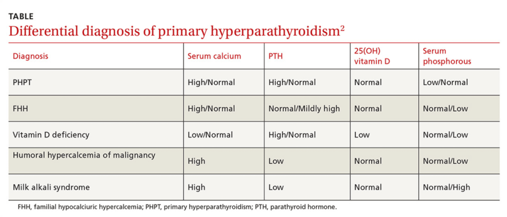
- Primary hyperparathyroidism (PHPT):
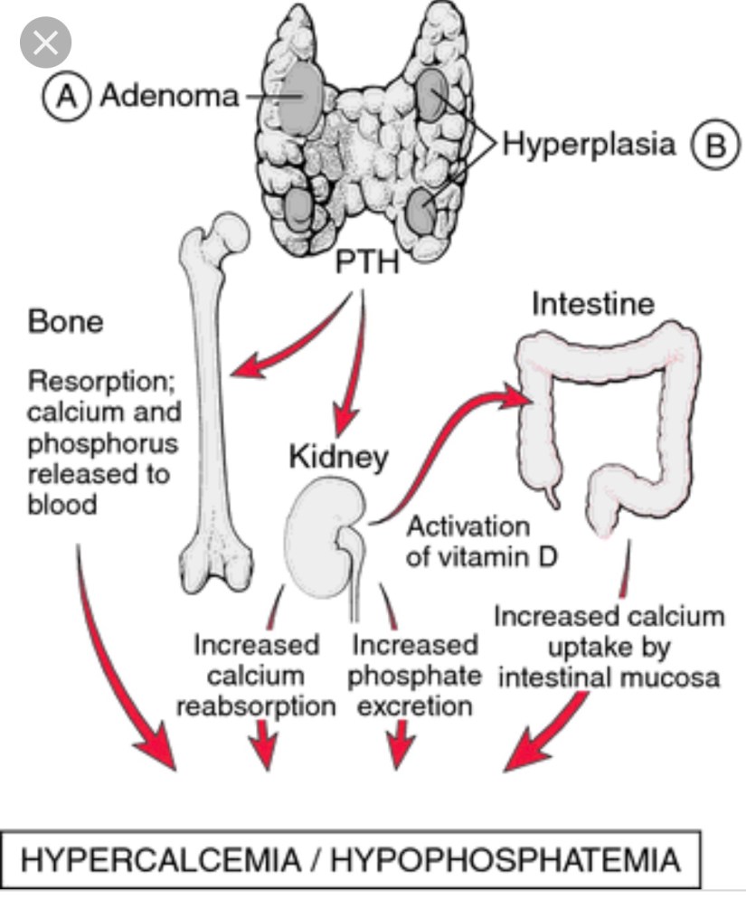
- Is caused by an increased secretion of parathyroid hormone (PTH) by the parathyroid gland(s):
- Which leads to an elevated serum calcium level
- Is caused by an increased secretion of parathyroid hormone (PTH) by the parathyroid gland(s):
- The overproduction of parathyroid hormone (PTH):
- Termed hyperparathyroidism (HPT), can be categorized as:
- Primary
- Secondary
- Tertiary
- Termed hyperparathyroidism (HPT), can be categorized as:
- Primary hyperparathyroidism (PHPT);
- Arises from an unregulated overproduction of PTH from an abnormal parathyroid gland
- Increased PTH levels may also occur as a compensatory response to hypocalcemic states resulting from:
- Chronic renal failure or gastrointestinal (GI) malabsorption of calcium:
- This secondary HPT can be reversed by the correction of the underlying problem:
- For example kidney transplantation for chronic renal failure
- This secondary HPT can be reversed by the correction of the underlying problem:
- Chronic renal failure or gastrointestinal (GI) malabsorption of calcium:
- However, chronically stimulated parathyroid glands:
- May occasionally become autonomous:
- Resulting in the persistence or recurrence of the hypercalcemia after successful renal transplantation:
- Resulting in tertiary HPT
- Resulting in the persistence or recurrence of the hypercalcemia after successful renal transplantation:
- May occasionally become autonomous:
- PHPT is defined as:
- Hypercalcemia or widely fluctuating levels of serum calcium resulting from:
- The inappropriate or autogenous secretion of PTH:
- By one or more parathyroid glands:
- In the absence of a known or recognized stimulus
- By one or more parathyroid glands:
- The inappropriate or autogenous secretion of PTH:
- Hypercalcemia or widely fluctuating levels of serum calcium resulting from:
- The most common cause of hypercalcemia in the outpatient setting is:
- Primary hyperparathyroidism (PHPT):
- With approximately 100,000 new cases per year reported in the United States
- Primary hyperparathyroidism (PHPT):
- Since the advent of routine laboratory testing:
- The prevalence of the disease has increased from:
- 0.1% to 0.4%:
- One to seven cases per 1000 adults
- 0.1% to 0.4%:
- The prevalence of the disease has increased from:
- In a study by Yeh et al:
- The incidence of PHPT fluctuated between:
- 36.3 and 120.2 cases per 100,000 women-years
- 13.4 and 35.6 in 100,000 men-years
- The incidence of PHPT fluctuated between:
- PHPT may present at any age:
- With the vast majority of cases occurring in patients older than 45 years of age
- The mean age at diagnosis has remained between:
- 52 and 56 years
- Women have consistently made up the preponderance of cases:
- With a female-to-male ratio of:
- 3:1 to 4:1
- Based on a population based study from Rochester Minnesota:
- The higher incidence of this could be secondary (hypothetically) to:
- Estrogen deficiency after menopause:
- That reveals underlying HPT
- Estrogen deficiency after menopause:
- The higher incidence of this could be secondary (hypothetically) to:
- With a female-to-male ratio of:
- The precise origin of PHPT is unknown:
- Although exposure to low-dose therapeutic ionizing radiation and familial predisposition account for some cases:
- Irradiation for acne could have accounted for a 2 to 3-fold increase in the incidence of this disease at some point in time, and a 4-fold increase was noted in survivors of the atomic bomb
- Schneider et al., in their study of 2555 patients followed for 50 years, even low doses of radiation exposure during the teenage years:
- Was associated with a slight risk of developing PHPT
- In this study a dose response was documented in people receiving external-beam radiotherapy for benign diseases before their 16th birthday
- The latency period for the development of PHPT after radiation exposure:
- Is longer than that for the development of thyroid tumors, with most cases occurring 30 to 40 years after exposure
- Patients who have been radiated have similar clinical manifestations and serum calcium levels when compared to patients without a history of radiation exposure:
- However, the former tend to have higher PTH levels and a higher incidence of concomitant thyroid neoplasms
- Although exposure to low-dose therapeutic ionizing radiation and familial predisposition account for some cases:
- Certain medications have been implicated in the development of hypercalcemia:
- Lithium therapy has been known to:
- Shift the set point for PTH secretion in parathyroid cells:
- Thereby resulting in elevated PTH levels and mild hypercalcemia
- Shift the set point for PTH secretion in parathyroid cells:
- Lithium stimulates the growth of abnormal parathyroid glands in vitro and also in susceptible patients in vivo
- Unusual metabolic features associated with lithium use include:
- Low urinary calcium excretion
- Normal cyclic AMP excretion
- Lack of calcic nephrolithiasis
- The mechanism probably results from:
- Lithium linking with the calcium sensing receptor on the parathyroid glands resulting in PTH secretion
- Lithium therapy has been known to:
- Elevated serum calcium levels have been associated with thiazide diuretic:
- The overall annual age- and sex-adjusted (to 2000 U.S. whites) incidence was:
- 7.7 (95% CI, 5.9 to 9.5) per 100,000 individuals
- The average 24-hour plasma calcium concentrations are increased with thiazide diuretic use:
- But the mean 24-hour PTH levels remain unchanged in subjects with normal baseline PTH levels and no evidence of hypercalciuria
- Thiazides diuretics have several metabolic effects that may contribute to increased calcium levels:
- A decrease in urine calcium excretion is the most likely cause:
- But in some cases diuretic use has been associates with a metabolic alkalosis:
- That could also increase the total serum calcium levels through a pH-dependent increase in protein-bound calcium
- But in some cases diuretic use has been associates with a metabolic alkalosis:
- Although plasma 1,25 (OH) vitamin D levels are unchanged:
- Increased intestinal calcium absorption in response to thiazide diurectic use:
- Has been noted and could also contribute to an increase in serum calcium
- Increased intestinal calcium absorption in response to thiazide diurectic use:
- One last possible explanation for the elevated serum calcium levels associated with thiazide diuretic use is:
- Hemoconcentration associated with dieresis
- A decrease in urine calcium excretion is the most likely cause:
- The overall annual age- and sex-adjusted (to 2000 U.S. whites) incidence was:
- Numerous genetic abnormalities have been identified in the development of PHPT, including:
- Anomalies in tumor suppressor genes and proto-oncogenes
- Specific DNA mutations in a parathyroid cell:
- May confer a proliferative advantage over normal neighboring cells:
- Thus allowing for clonal growth:
- Large populations of these altered cells containing the same mutation within hyper functioning parathyroid tissue:
- Suggest that such glands are a result of clonal expansion
- Large populations of these altered cells containing the same mutation within hyper functioning parathyroid tissue:
- Thus allowing for clonal growth:
- May confer a proliferative advantage over normal neighboring cells:
- The majority of PHPT cases are:
- Sporadic
- Nonetheless, PHPT also occurs within the spectrum of a number of inherited disorders such as:
- Multiple endocrine neoplasia syndromes (MEN):
- MEN type 1 (Wermer Syndrome)
- MEN type 2A (Sipple Syndrome)
- Isolated familial HPT
- Familial HPT with jaw-tumor syndrome
- Multiple endocrine neoplasia syndromes (MEN):
- All of these are inherited in an:
- Autosomal dominant fashion
- The earliest and most common presentation of MEN type 1 (Wermer Syndrome):
- Is PHPT:
- Develops in approximately 80% to 100% of patients by age 40 years
- These patients also are predisposed to the development of:
- Pancreatic neuroendocrine tumors
- Pituitary adenomas
- Less frequently:
- Skin angiomas
- Lipomas
- Adrenocortical tumors
- Neuroendocrine tumors of the:
- Thymus
- Bronchus
- Stomach
- MEN type 1 has been shown to result from:
- A germline mutation in a tumor suppressor gene:
- Called MEN1 gene:
- Located on chromosome 11q12-13 that encodes Menin:
- A protein that is postulated to interact with the transcription factors JunD and nuclear factor-κB in the nucleus, in addition to replication protein A and other proteins
- Located on chromosome 11q12-13 that encodes Menin:
- Called MEN1 gene:
- A germline mutation in a tumor suppressor gene:
- Pre-symptomatic screening for mutation carriers for MEN type 1:
- Is difficult because generally MEN1 mutations result in a nonfunctional protein and are scattered throughout the translated nine exons of the gene
- MEN1 mutations also have been found in kindred’s initially suspected to represent isolated familial HPT
- Screening for mutation carriers for MEN type 1 has a very high detection rate greater than 94%, and is used in Sweden for patients with:
- PHPT with a first-degree relative with a major endocrine tumor, age of onset is less than 30 years and / or if multiple pancreatic tumors / parathyroid hyperplasia is detected
- Is PHPT:
- Approximately 20% of patients with MEN type 2A (Sipple Syndrome):
- Develop PHPT:
- Which is usually less severe
- MEN type 2A is caused by:
- A germline mutation of the RET proto-oncogene:
- Located on chromosome 10
- A germline mutation of the RET proto-oncogene:
- Genotype and phenotype correlations have been noted in this syndrome:
- In that individuals with mutations at codon 634:
- Are more likely to develop PHPT
- In that individuals with mutations at codon 634:
- Develop PHPT:
- Patients with the familial HPT with jaw-tumor syndrome:
- Have an increased predisposition to:
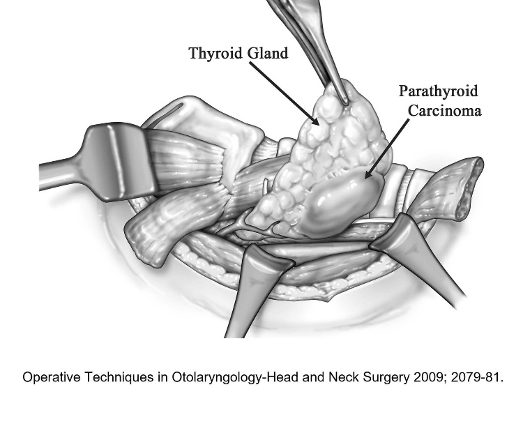
- Parathyroid carcinoma
- This syndrome maps to a tumor suppressor locus HRPT2 (parafibromin):
- On chromosome 1
- Have an increased predisposition to:
- Sporadic parathyroid adenomas and some hyperplastic parathyroid glands:
- Have loss of heterozygosity (LOH) at 11q13:
- The site of the MEN1 gene in approximately 25% to 40% of the cases
- Have loss of heterozygosity (LOH) at 11q13:
- Over expression of PRAD1:
- Which encodes cyclin D1:
- A cell cycle control protein:
- Is found approximately 18% of parathyroid adenomas
- A cell cycle control protein:
- This was proven to result from a rearrangement on chromosome 11:
- That places the PRAD1 gene:
- Under the control of the PTH promoter
- That places the PRAD1 gene:
- Which encodes cyclin D1:
- Other chromosomal regions deleted in parathyroid adenomas and possibly reflecting loss of tumor suppressor genes include:
- 1p
- 6q
- 15q
- Amplified regions suggesting oncogenes have been identified at:
- 16p
- 19p
- RET mutations:
- Are unusual in sporadic parathyroid tumors
- Sporadic parathyroid cancers are characterized by:
- Uniform loss of the tumor suppressor gene RB:
- Which is involved in cell cycle regulation
- 60% have HRPT2 (CDC73) mutations located in chromosome 1:
- Encodes the protein Parafibromin
- These alterations are rare in benign parathyroid tumors;
- May have implications for diagnosis
- The p53 tumor suppressor gene:
- Is also inactivated in a subset (30%) of parathyroid carcinomas
- Uniform loss of the tumor suppressor gene RB:
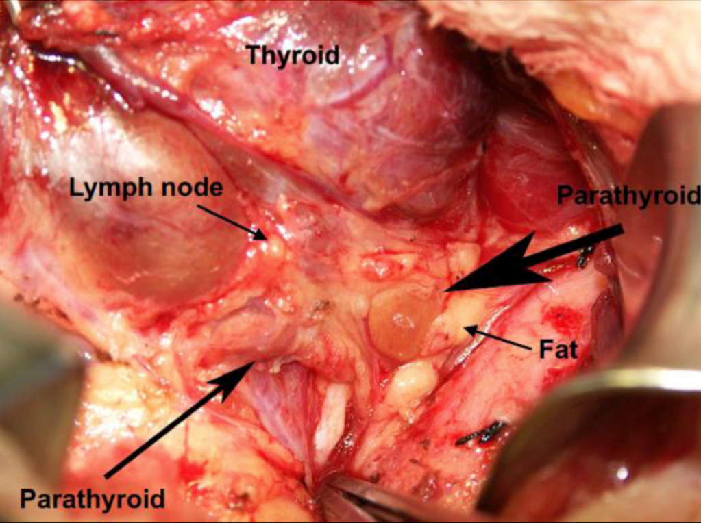
- Single gland adenoma:
- Is the most common cause (75% to 90%) of PHPT
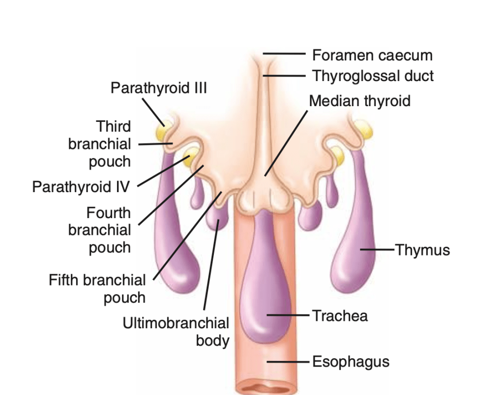
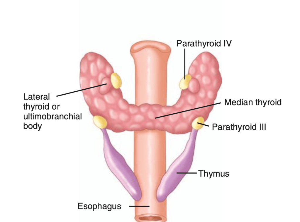
- Lower pole adenomas (in relation to the thyroid):
- Are more common than are upper pole adenomas
- Sizes range from 1 cm to 3 cm:
- The normal parathyroid gland measures approximately 6 mm X 4 mm X 2 mm
- The weight of parathyroid adenomas vary between:
- 553.7 mg +/- 520.5 mg (range, 66-2536):
- The normal weight of a parathyroid gland is:
- Approximately 40 mg to 50 mg
- The normal weight of a parathyroid gland is:
- 553.7 mg +/- 520.5 mg (range, 66-2536):
- Ectopic glands can be present:
- 4% to 16% of cases
- PHPT is caused by multiple adenomas or hyperplasia in:
- 15% to 25% of the cases
- Parathyroid carcinoma as the cause of PHPT:
- Is extremely rare in most parts of the world (~1%)
- Multi-gland adenoma arises in a significant number of patients:
- Double adenomas are seen in approximately:
- 2% to 12% of the cases
- Triple adenomas:
- In less than 1% the cases
- Four adenomas or parathyroid gland hyperplasia:
- In less than 3% to 15% of the cases
- Double adenomas are seen in approximately:
- Most parathyroid adenomas:
- Consist of parathyroid chief cells
- They are usually encapsulated
- In 50% of the cases they are surrounded by normal parathyroid tissue
- Some adenomas, nevertheless, are composed of oxyphil cells:
- These adenomas are usually larger than chief cell adenomas
- Parathyroid adenomas are sometimes located within the thymus:
- They express a parathyroid-specific gene:
- GCMB
- Contrasting with the normal thymus:
- Which does not neither express PTH nor GCMB
- They express a parathyroid-specific gene:
- In a study by Ruda et al:
- 225 patients with PHPT:
- Parathyroid hyperplasia accounted for approximately 6% of cases
- 225 patients with PHPT:
- In parathyroid hyperplasia all four glands are enlarged:
- With the lower glands typically being larger than the upper time
- The glands are usually composed of:
- Chief cells
- Clear cell hyperplasia is very rare and is the only one in which the upper parathyroid glands are larger than the lower ones

#Arrangoiz #ParathyroidSurgeon #ParathyroidExpert #Hyperparathyroidism #PrimaryHyperparathyroidism #CancerSurgeon #EndocrineSurgery #Teacher #Surgeon #HeadandNeckSurgeon #SurgicalOncologist #ParathyroidAdenoma #Hypercalcemia #ElevatedCalciumLevels #Miami #MountSinaiMedicalCenter #MSMC #Mexico #Hialeah