- Type A:
- Adherent to the posterior thyroid parenchyma:
- Posterior to the upper pole of the thyroid:
- But not intrathyroidal
- Posterior to the upper pole of the thyroid:
- Type A glands are in the accepted, expected location of a normal parathyroid gland
- Adherent to the posterior thyroid parenchyma:
- Type B:
- Behind the thyroid parenchyma
- Type B glands are exophytic to the thyroid parenchyma and lie in the tracheoesophageal groove:
- This category includes adenomas in:
- Retroesophageal, retropharyngeal, high lateral pharyngeal, and carotid sheath locations
- This category includes adenomas in:
- A ‘‘B+’’ subcategory can be used to document the location of adenomas above the level of the hyoid bone:
- The ‘‘+’’ is meant to reflect cranial elevation
- Type C:
- Caudal to the thyroid parenchyma:
- In the tracheoesophageal groove
- A type C gland is more inferior than a type B gland on lateral images:
- Located inferior to the inferior pole of the thyroid (closer to the clavicle)
- Caudal to the thyroid parenchyma:
- Type D:
- Directly over the recurrent laryngeal nerve:
- At the level of the inferior thyroid vessels
- The dissection may be difficult:
- Because a type D gland is dangerously close to the recurrent laryngeal nerve
- Directly over the recurrent laryngeal nerve:
- Type E:
- Located in the external aspect of the inferior pole of the thyroid
- A type E gland is in a location that is:
- More superficial in an anterior–posterior plane than the recurrent laryngeal nerve:
- It is the easiest to resect
- More superficial in an anterior–posterior plane than the recurrent laryngeal nerve:
- Type F:
- ‘‘Fallen’’ into the thyrothymic ligament:
- Below the inferior pole of the thyroid in a pretracheal plane
- A type F gland is frequently referred to as an ectopic gland:
- Its resection usually involves:
- Transcervical delivery of the thyrothymic ligament or superior portion of the thymus
- Its resection usually involves:
- ‘‘Fallen’’ into the thyrothymic ligament:
- Type G:
- A gauge, true intrathyroidal gland location
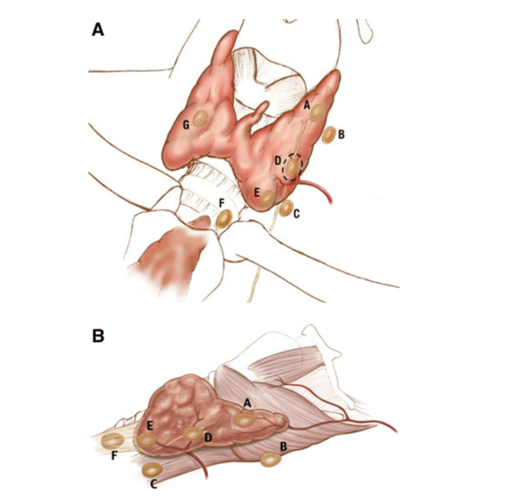
localization of parathyroid adenomas. Anterior view (a); right lateral
view (b) of the superior thyroid pole is oriented to the left. The dotted
circle depicts the region where the recurrent laryngeal nerve is most at
risk
- This nomenclature system has been designed that takes into account the pathologic position of the parathyroid glands (Figure):
- Superior and inferior glands:
- Are defined by the location of the gland’s pedicle and its relationship to the RLN:
- Superior parathyroid glands:
- Anatomically have a vascular pedicle superior and lateral to the RLN (type A through D glands)
- Inferior parathyroid glands:
- Anatomically have a vascular pedicle inferior and medial to the RLN (type D through F glands)
- Superior parathyroid glands:
- Type G glands:
- Represent intrathyroidal parathyroid lesions
- Are defined by the location of the gland’s pedicle and its relationship to the RLN:
- This information not only helps radiologists communicate potential parathyroid lesions of interest to surgeons:
- But also helps surgeons direct their dissection in relation to the RLN
- Superior and inferior glands:
