- Diffusely invasive carcinoma:
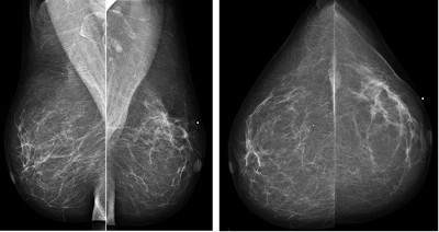
- Has a mammographic appearance of diffuse architectural distortion:
- Usually involving a large area, often larger than a lobe:
- With no central tumor mass and no calcifications:
-
- It sometimes has the appearance of a “spider’s web” as shown in the Image
- Usually involving a large area, often larger than a lobe:
- Has a mammographic appearance of diffuse architectural distortion:
- The diffusely infiltrating cancer:
- Forms concave contours with the surrounding fat in a manner similar to normal fibroglandular tissue (Images)

- The imaging findings of diffusely infiltrating breast cancer are strikingly different:
- From the imaging findings of breast cancers originating either from the terminal ductal lobular units (TDLUs) or the lactiferous ducts:
-
- Suggesting that it may have a different site of origin
- It has been recently proposed that diffusely infiltrating breast cancers:
- May originate from mesenchymal stem cells (progenitors):
- Through a complex process of both:
- Epithelial-mesenchymal transformation and more frequently, mesenchymal-epithelial transformation
- Through a complex process of both:
- May originate from mesenchymal stem cells (progenitors):
- The clinical presentation:
- Is typically a recently detected, extensive, firm lesion:
- Often appearing as an interval cancer following a previous mammogram which was interpreted as normal
- Is typically a recently detected, extensive, firm lesion:
- On clinical breast examination:
- The cancer does not have a distinct tumor mass or focal skin retraction seen in other cancers:
- But rather an indistinct “thickening” and eventually a shrinkage of the breast.
- The cancer does not have a distinct tumor mass or focal skin retraction seen in other cancers:
- In order to make the diagnosis before the development of a palpable mass and a decrease in size of the breast:
- The radiologist and breast surgeon must have a high level of suspicion and a thorough knowledge of the underlying pathophysiology
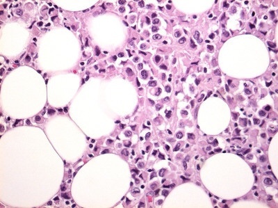
- The subgross (3D) histopathology images show how growth of the mesenchymal tissue:
- Distorts the normal, harmonious connective tissue framework:
- By causing nonuniform thickening of the fine sheets of connective tissue (Images):
- Distorts the normal, harmonious connective tissue framework:

- The predominance of mesenchyme in the diffusely infiltrating breast malignancy:
- Allows it to be imaged with greater sensitivity by ultrasound than by mammography:
-
-
- The thin sheets or veils of tissue reflect the ultrasound waves:
- But are relatively easily penetrated by x-rays
- The thin sheets or veils of tissue reflect the ultrasound waves:
-
-
- The structural / architectural distortion:
-
- While difficult to detect mammographically:
- Is readily detectable on 2-mm thick coronal sections of automated breast ultrasound (Image)
- While difficult to detect mammographically:
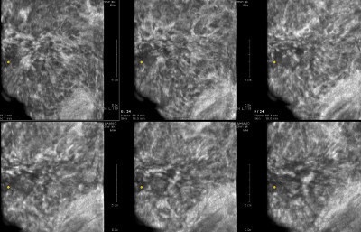
- The 2-mm thick multi-slice series demonstrate the extensive architectural distortion, corresponding to the 3D histology:

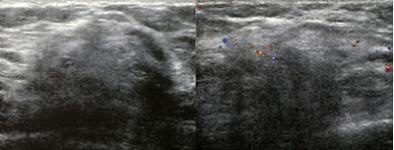
- The hypoechoic changes can also usually be seen on hand held ultrasound, Image:
- The growth pattern and cell type of diffusely invasive breast cancer:
- Is very similar to that of diffuse gastric carcinoma (linitis plastica), and both of these diseases can be associated with:
- A deleterious mutation in the CDH1 gene:
- Which is located on chromosome 16q22 and codes for e-cadherin protein (Image):
- A deleterious mutation in the CDH1 gene:
- Is very similar to that of diffuse gastric carcinoma (linitis plastica), and both of these diseases can be associated with:

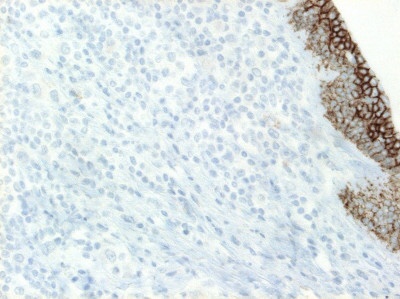
- CDH1 was initially known as a susceptibility gene for diffuse gastric cancer (linitis plastica)
- The histopathologic characteristics of diffuse gastric cancer:
- Show similarities with e-cadherin negative:
- Diffusely infiltrating breast cancer (infiltrating “lobular” carcinoma)
- The neoplastic cells permeate the mucosa and wall as scattered individual signet-ring cells or small clusters of cells in an infiltrative growth pattern
- Show similarities with e-cadherin negative:
- Since there are no TDLUs in the stomach:
- If the similar cells in both conditions associated with CDH1 have a common origin, it could not be a TDLU:
-
- Raising the possibility that they could result from mesenchymal cell transformation in both organs

👉Rodrigo Arrangoiz MS, MD, FACS, FSSO cirujano oncology y cirujano de mamá de en Mount Sinai Medical Center:
-
Es experto en el manejo del cáncer de mama

👉Es miembro de la American Society of Breast Surgeons:

Training:
• General surgery:
• Michigan State University:
• 2004 al 2010

• Surgical Oncology / Head and Neck Surgery / Endocrine Surgery:
• Fox Chase Cancer Center (Filadelfia):
• 2010 al 2012

• Masters in Science (Clinical research for health professionals):
• Drexel University (Filadelfia):
• 2010 al 2012

• Surgical Oncology / Head and Neck Surgery / Endocrine Surgery:
• IFHNOS / Memorial Sloan Kettering Cancer Center:
• 2014 al 2016