- Dermatofibrosarcoma protuberans:
- Is an intermediate-grade sarcoma:
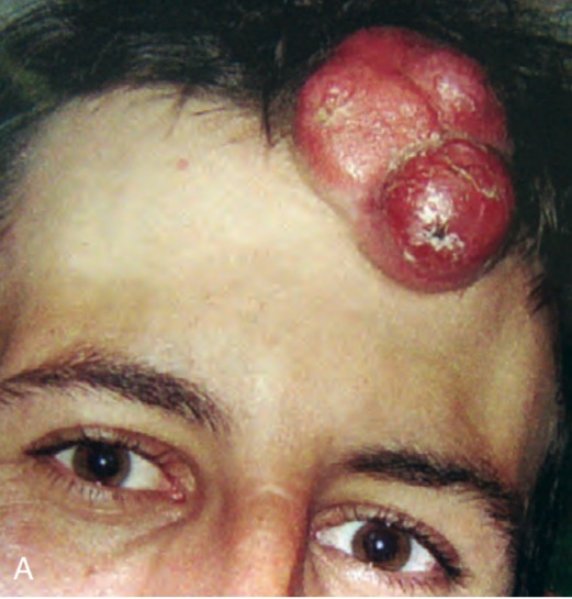
- That presents as a unifocal or multifocal nodular lesion
- It is a rare, slow-growing, locally aggressive cutaneous sarcoma:
- With a high rate of local recurrence and low metastatic potential
- Is an intermediate-grade sarcoma:
- Dermatofibrosarcoma protuberans:
- Involves the head and neck region in 10% to 20% of cases:
- With the scalp and supraclavicular fossae the most common sites for involvement (Figure)
- Involves the head and neck region in 10% to 20% of cases:
- These slow-growing, locally aggressive tumors have tentacle-like extensions well beyond the visible lesion:
- Thus the true extent of the disease is often underestimated:
- Leading to local recurrence in more than 50% of patients
- Thus the true extent of the disease is often underestimated:
- Histologically a storiform or fascicular proliferation of spindle cells extends
from the dermis into the subcutis:- With immunohistochemistry showing:
- CD34 positive staining in most cases
- Presence of fibrosarcomatous changes and high mitotic rate:
- May portend a more aggressive course
- This tumor frequently has a translocation of a fusion protein involving:
- COL1A1 and PDGFB that functions like PDGFB
- With immunohistochemistry showing:
- Wide excision with margins of ≥ 2 cm is
generally advocated, with adjuvant radiation reserved for larger or recurrent tumors when resection is not feasible - Histological subtypes include:
- Classic DFSP
- Fibrosarcomatous transformation:
- Conferring a higher risk of recurrence and metastasis
- Surgical management is the mainstay of treatment:
- The National Comprehensive Cancer Network (NCCN), National Cancer Institute, and American Cancer Society recommend:
- Mohs micrographic surgery (MMS) as the preferred approach due to its ability to achieve complete margin control and minimize tissue loss, especially in anatomically sensitive areas
- If MMS is unavailable, wide local excision (WLE) with 2 to 4 cm margins down to the investing fascia is acceptable:
- Achieving negative surgical margins is the most critical factor for reducing recurrence risk; margin width is important, but negative margins are paramount
- Recent evidence suggests that margins greater than 2 cm to 2.5 cm are associated with significantly lower recurrence rates:
- But recurrence is rare when negative margins are achieved, regardless of width
- Routine lymph node dissection is not indicated for DFSP, as the risk of nodal metastasis is extremely low
- Lymph node dissection should be considered only in cases with clinical or radiologic suspicion of nodal involvement or in tumors with fibrosarcomatous transformation, which carries a higher metastatic risk
- Sentinel lymph node biopsy is not standard but may be considered in select high-risk cases, such as those with fibrosarcomatous change or lymphovascular invasion, though its utility remains under investigation
- The National Comprehensive Cancer Network (NCCN), National Cancer Institute, and American Cancer Society recommend:
- Adjuvant radiotherapy:
- Is considered in cases of positive or close margins when further re-excision is not feasible, or for unresectable or recurrent disease
- The role of adjuvant radiotherapy is supported by its ability to reduce local recurrence in these settings
- Imatinib, a tyrosine kinase inhibitor:
- Is indicated for unresectable, recurrent, or metastatic DFSP harboring the t(17;22) translocation (COL1A1-PDGFB fusion)
- Cytotoxic chemotherapy:
- Has a limited role and is generally reserved for metastatic disease not amenable to targeted therapy, with inferior outcomes compared to imatinib
- Surveillance recommendations include:
- Regular follow-up for early detection of local recurrence:
- Particularly within the first three years post-resection:
- As most recurrences occur during this period
- For patients with negative-margin, non-fibrosarcomatous DFSP:
- Less intensive follow-up may be appropriate, and some data suggest that ongoing surveillance may not be necessary after negative-margin resection
- In contrast, patients with fibrosarcomatous transformation should be followed according to soft tissue sarcoma protocols due to higher risk of recurrence and metastasis
- Particularly within the first three years post-resection:
- Regular follow-up for early detection of local recurrence:
- Areas of ongoing debate include the optimal surgical margin width and the role of sentinel lymph node biopsy, particularly in high-risk subtypes:
- The consensus remains that complete surgical excision with negative margins is the cornerstone of management, with adjuvant therapies reserved for select cases
- References:
- Dermatofibrosarcoma Protuberans: Update on the Diagnosis and Treatment. Hao X, Billings SD, Wu F, et al. Journal of Clinical Medicine. 2020;9(6):E1752. doi:10.3390/jcm9061752.
- Review of Dermatofibrosarcoma Protuberans. Lim SX, Ramaiya A, Levell NJ, Venables ZC. Clinical and Experimental Dermatology. 2023;48(4):297-302. doi:10.1093/ced/llac111.
Dermatofibrosarcoma Protuberans: What Is This?. Vitiello GA, Lee AY, Berman RS. The Surgical Clinics of North America. 2022;102(4):657-665. doi:10.1016/j.suc.2022.05.004. - Dermatofibrosarcoma Protuberans: An Updated Review of the Literature. Jozwik M, Bednarczuk K, Osierda Z. Cancers. 2024;16(18):3124. doi:10.3390/cancers16183124.
- Current Patterns of Care and Outcomes for Dermatofibrosarcoma Protuberans: An International Multi-Institutional Collaborative. Winer LK, Akumuo R, Fredette JD, et al. Cancer. 2025;131(1):e35468. doi:10.1002/cncr.35468.
- Surgical Management of Dermatofibrosarcoma Protuberans. Rust DJ, Kwinta BD, Geskin LJ, et al. Journal of Surgical Oncology. 2023;128(1):87-96. doi:10.1002/jso.27258.
- Dermatofibrosarcoma Protuberans. Miller SJ, Alam M, Andersen JS, et al. Journal of the National Comprehensive Cancer Network : JNCCN. 2012;10(3):312-8. doi:10.6004/jnccn.2012.0032.
- Oncological Efficiency of Wide Local Excision in Dermatofibrosarcoma Protuberans. Güç ZG, Güç H, Bütün O, Alacacıoğlu A, Demirdöver C. Journal of Plastic, Reconstructive & Aesthetic Surgery : JPRAS. 2023;77:244-252. doi:10.1016/j.bjps.2022.11.002.
- Outcome After Surgical Treatment of Dermatofibrosarcoma Protuberans (DFSP): Does It Require Extensive Follow-Up and What Is an Adequate Resection Margin?. Alshaygy I, Mattei JC, Basile G, et al. Annals of Surgical Oncology. 2023;30(5):3106-3113. doi:10.1245/s10434-022-12953-8.
- Management of Dermatofibrosarcoma Protuberans. Badhey AK, Tikhtman R, Tang AL. Current Opinion in Otolaryngology & Head and Neck Surgery. 2021;29(4):278-282. doi:10.1097/MOO.0000000000000721.
- Dermatofibrosarcoma Protuberans. Allen A, Ahn C, Sangüeza OP. Dermatologic Clinics. 2019;37(4):483-488. doi:10.1016/j.det.2019.05.006.