- Merkel cell carcinoma
- Is a neuroendocrine neoplasm of the
skin - It is a rare, aggressive neuroendocrine skin cancer:
- With a high risk of local recurrence, nodal involvement, and distant metastasis
- Is a neuroendocrine neoplasm of the
- The incidence is rising:
- Particularly among elderly and immunosuppressed patients, and prognosis remains poor, especially in those with advanced disease or immunosuppression
- A multidisciplinary approach:
- Is essential for initial workup and staging
- Histopathologic confirmation and microstaging of the primary lesion are required
- For patients with clinically node-negative disease:
- Sentinel lymph node biopsy (SLNB) is recommended to assess occult nodal involvement:
- As up to 40% of these patients may harbor microscopic nodal metastases
- Sentinel lymph node biopsy (SLNB) is recommended to assess occult nodal involvement:
- Imaging (e.g., PET/CT):
- Is indicated for patients with clinically apparent nodal or distant disease
- The majority of these tumors in North America (80%) are:
- Caused by infections with Merkel cell polyomavirus (MCV):
- A double-stranded DNA virus
- Caused by infections with Merkel cell polyomavirus (MCV):
- Nearly half of all Merkel cell car-
cinoma lesions:- Occur in the head and neck region:
- The cheek is the most common site:
- Followed by the upper neck and nose
- The cheek is the most common site:
- Occur in the head and neck region:
- These lesions typically occur in elderly white persons:
- They appear as a red to violaceous, smooth, dome-shaped lesion with telangiectasias (Figure)
- These tumors have a high propensity
for metastatic spread:- To regional lymph nodes as well as distant
sites
- To regional lymph nodes as well as distant
- Histologically they are composed of:
- Basophilic cells with scant cytoplasm and dark powdery chromatin:
- They may be morphologically similar to other neuroendocrine carcinomas:
- Hence metastatic small cell carcinoma, malignant melanoma, or primary neuroendocrine (or “small cell”) carcinoma of the parotid gland may be considerations in the differential diagnosis
- They may be morphologically similar to other neuroendocrine carcinomas:
- Immunohistochemical stains:
- For synaptophysin, chromogranin, and cytokeratin 20 (CK20) (demonstrating a characteristic “dotlike” pattern) or the Merkel cell polyoma virus large T antigen (recognized by the antibody CM2B4) are positive:
- Whereas thyroid transcription factor-1 (TTF-1) is negative
- For synaptophysin, chromogranin, and cytokeratin 20 (CK20) (demonstrating a characteristic “dotlike” pattern) or the Merkel cell polyoma virus large T antigen (recognized by the antibody CM2B4) are positive:
- Basophilic cells with scant cytoplasm and dark powdery chromatin:
- Surgical management:
- Is the primary treatment for localized MCC
- The National Comprehensive Cancer Network (NCCN) recommends:
- Wide local excision (WLE) or Mohs micrographic surgery (MMS) with 1 to 2 cm surgical margins to the investing fascia of muscle when anatomically feasible
- In anatomically challenging sites (e.g., head and neck):
- Narrower margins may be acceptable, particularly if adjuvant radiotherapy is planned:
- Studies indicate that local control is excellent with adjuvant radiotherapy, even when margins are ≤ 1 cm
- Narrower margins may be acceptable, particularly if adjuvant radiotherapy is planned:
- The American Cancer Society aligns with these recommendations, emphasizing the importance of complete excision and individualized margin selection based on tumor location and patient factors
- Elective neck dissection:
- Is not routinely recommended for clinically node-negative patients:
- Instead, SLNB is the preferred method for nodal staging:
- If SLNB is positive, completion lymph node dissection or nodal radiotherapy may be considered
- Instead, SLNB is the preferred method for nodal staging:
- Is not routinely recommended for clinically node-negative patients:
- For clinically node-positive disease:
- Fine needle aspiration or core biopsy is used for confirmation, and management includes lymph node dissection and / or nodal radiotherapy
- Radiation therapy:
- Plays a central role in MCC management
- Adjuvant radiotherapy is generally recommended for most patients:
- Especially those with high-risk features such as:
- Lymphovascular invasion
- Immunosuppression
- Positive or close margins
- Large tumor size
- Especially those with high-risk features such as:
- The recommended dose is 50 to 66 Gy:
- Tailored to the extent of residual disease and margin status
- Radiotherapy alone is a definitive option for patients who are not surgical candidates
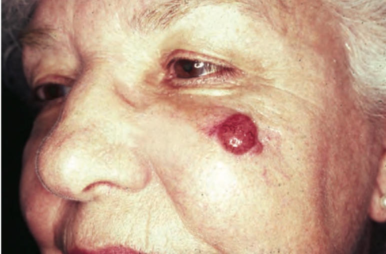
Clinical appearance of Merkel cell carcinoma
- National Comprehensive Cancer Network guidelines for HNMCC recommend treatment for localized tumors to include:
- Surgical excision followed by adjuvant radiotherapy or observation, favoring the use of radiotherapy for patients with HNMCC for its potentially limited ability to achieve 1- to 2-cm margins and the risk of false-negative sentinel lymph node biopsy results
- Furthermore, MCC is a radiosensitive malignant neoplasm, and postoperative radiotherapy has shown improved outcomes, including increased OS and disease-free survival, compared with surgery alone
- Chemotherapy is not recommended as adjuvant therapy for localized MCC:
- As it has not demonstrated a survival benefit and is associated with significant toxicity
- Its use is generally reserved for select patients with advanced or metastatic disease:
- Often in the palliative setting
- Immunotherapy has become the standard of care for advanced or metastatic MCC:
- Immune checkpoint inhibitors such as avelumab and pembrolizumab are first-line agents:
- Offering durable responses and improved outcomes compared to traditional chemotheraphy
- Immune checkpoint inhibitors such as avelumab and pembrolizumab are first-line agents:
- There remain areas of ongoing controversy and research, including the optimal surgical margin size, the precise indications for adjuvant radiotherapy, and the management of high-risk or immunosuppressed patients:
- The rarity of MCC and lack of prospective randomized trials contribute to variability in practice patterns
- References:
- Association Between Surgical Margins Larger Than 1 cm and Overall Survival in Patients With Merkel Cell Carcinoma. Andruska N, Fischer-Valuck BW, Mahapatra L, et al. JAMA Dermatology. 2021;157(5):540-548. doi:10.1001/jamadermatol.2021.0247.
- Merkel Cell Carcinoma. Lewis DJ, Sobanko JF, Etzkorn JR, et al. Dermatologic Clinics. 2023;41(1):101-115. doi:10.1016/j.det.2022.07.015.
- Survival of Patients With Head and Neck Merkel Cell Cancer: Findings From the Pan-Canadian Merkel Cell Cancer Collaborative. Nayak AL, Pickett AT, Delisle M, et al. JAMA Network Open. 2023;6(11):e2344127. doi:10.1001/jamanetworkopen.2023.44127.
- Merkel Cell Carcinoma, Version 1.2018, NCCN Clinical Practice Guidelines in Oncology. Bichakjian CK, Olencki T, Aasi SZ, et al. Journal of the National Comprehensive Cancer Network : JNCCN. 2018;16(6):742-774. doi:10.6004/jnccn.2018.0055.
- Best Practices in Surgical and Nonsurgical Management of Head and Neck Merkel Cell Carcinoma: An Update. Duarte-Bateman D, Shen A, Bullock T, et al. Molecular Carcinogenesis. 2023;62(1):101-112. doi:10.1002/mc.23483.
- Overall Survival After Mohs Surgery for Early-Stage Merkel Cell Carcinoma. Cheraghlou S, Doudican NA, Criscito MC, Stevenson ML, Carucci JA. JAMA Dermatology. 2023;159(10):1068-1075. doi:10.1001/jamadermatol.2023.2822.
- Merkel Cell Carcinoma of the Head and Neck: Epidemiology, Pathogenesis, Current State of Treatment and Future Directions. Yusuf MB, McKenzie G, Rattani A, et al. Cancers. 2021;13(14):3506. doi:10.3390/cancers13143506.
- Narrow Excision Margins Are Appropriate for Merkel Cell Carcinoma When Combined With Adjuvant Radiation: Analysis of 188 Cases of Localized Disease and Proposed Management Algorithm. Tarabadkar ES, Fu T, Lachance K, et al. Journal of the American Academy of Dermatology. 2021;84(2):340-347. doi:10.1016/j.jaad.2020.07.079.
- Identifying an Optimal Adjuvant Radiotherapy Dose for Extremity and Trunk Merkel Cell Carcinoma Following Resection: An Analysis of the National Cancer Database. Patel SA, Qureshi MM, Sahni D, Truong MT. JAMA Dermatology. 2017;153(10):1007-1014. doi:10.1001/jamadermatol.2017.2176.
- Merkel Cell Carcinoma – Current Controversies and Future Directions. Steven N, Lawton P, Poulsen M. Clinical Oncology (Royal College of Radiologists (Great Britain)). 2019;31(11):789-796. doi:10.1016/j.clon.2019.08.012.

